





Introduction
Retroperitoneal fibrosis (RPF) is a rare fibro-inflammatory disorder characterised by the development of fibrous tissue within the retroperitoneum, frequently involving the abdominal aorta, iliac vessels and ureters. Epidemiological data are limited and mainly concern idiopathic RPF, for which the reported annual incidence ranges from approximately 0.1 to 1.3 cases per 100,000 persons, with a prevalence of approximately 1.4 cases per 100,000 inhabitants. Reference Vaglio, Salvarani and Buzio1 , Reference Vaglio and Maritati2 Progressive fibro-inflammatory tissue may encase the ureters, leading to hydronephrosis, obstructive uropathy and renal impairment. Reference Vaglio, Salvarani and Buzio1–Reference Caiafa, Vinuesa and Izquierdo3
Radiation-associated RPF is an uncommon late complication reported after pelvic irradiation and remains poorly documented. Reference Takahashi, Matsushima and Mori4–Reference Shebani, Ikdees and Alazeebi7 Its clinical and radiological features may closely resemble locoregional tumour recurrence, making diagnosis particularly challenging in cancer survivors. Reference Caiafa, Vinuesa and Izquierdo3,Reference Shebani, Ikdees and Alazeebi7 Early recognition is important because prompt urinary decompression and appropriate multidisciplinary management may prevent irreversible renal damage. Reference Caiafa, Vinuesa and Izquierdo3,Reference Jadhav, Kumar and Punatar8
We report a rare case of radiation-associated RPF causing obstructive uropathy following pelvic chemoradiotherapy for rectal adenocarcinoma. This case highlights the importance of considering this delayed radiation toxicity in patients presenting with unexplained renal dysfunction after pelvic irradiation and discusses its implications for radiotherapy practice.
Case
A 59-year-old man with a history of gout was diagnosed in November 2021 with moderately differentiated rectal adenocarcinoma after presenting with chronic diarrhoea and intermittent rectal bleeding. Staging investigations, including pelvic magnetic resonance imaging (MRI) and thoraco-abdominopelvic computed tomography (CT), demonstrated a cT3N0 middle rectal tumour without distant metastases.
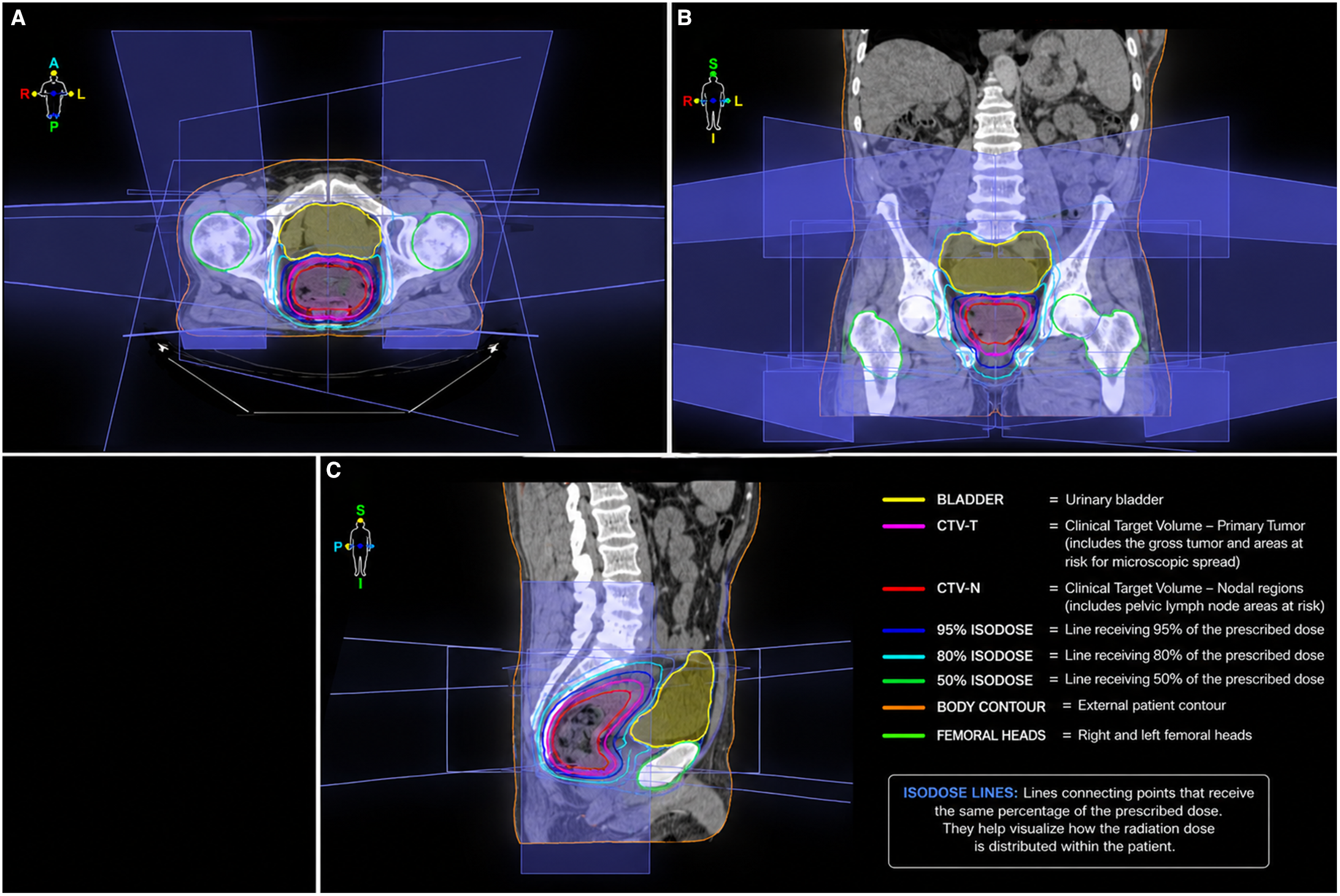
The patient was treated according to a modified PRODIGE-23-like total neoadjuvant strategy. Because of treatment scheduling constraints, two cycles of XELOX were administered before six biweekly administrations of FOLFIRINOX, followed by pelvic chemoradiotherapy with concurrent capecitabine. Pelvic radiotherapy was delivered using a three-dimensional conformal radiotherapy (3D-CRT) technique. A total dose of 46 Gy was delivered to the pelvis in 23 fractions, followed by a 4 Gy boost in two fractions to the primary tumour, with concurrent capecitabine. The treatment plan consisted of four radiation fields: one anterior, one posterior and two opposed lateral fields (Figure 1). Patient positioning was verified before each fraction using daily two-dimensional (2D) planar kV imaging. Treatment was completed in June 2022 with good overall tolerance. The patient subsequently underwent curative surgical resection in September 2022, and pathological examination confirmed pT3N0 disease. Adjuvant capecitabine was subsequently administered.
Three-dimensional conformal radiotherapy treatment plan of our patient. (A) Axial dose distribution and radiation field arrangement. (B) Coronal dose distribution and target volume coverage. (C) Sagittal dose distribution and target volume coverage.

Figure 1. Long description
Panel A: An axial view of a radiotherapy treatment plan showing dose distribution and radiation field arrangement. The image includes labeled regions for bladder, clinical target volume for primary tumor, clinical target volume for nodal regions, and various isodose lines indicating different percentages of the prescribed dose. Panel B: A coronal view of the treatment plan showing dose distribution and target volume coverage. The same labeled regions and isodose lines are visible. Panel C: A sagittal view of the treatment plan showing dose distribution and target volume coverage with the same labeled regions and isodose lines.
Before initiation of systemic chemotherapy, the patient’s baseline serum creatinine was 12.5 mg/L on 7 December 2021. During routine follow-up in May 2023, serum creatinine increased to 17 mg/L. Eleven months after completion of pelvic radiotherapy, surveillance CT demonstrated retroperitoneal soft tissue surrounding the aortic bifurcation, consistent with probable radiation-associated RPF, without significant urinary tract obstruction. Serum creatinine subsequently increased to 18.2 mg/L in August 2023 and decreased to 16.3 mg/L in September 2023. Multidisciplinary assessment involving oncology, nephrology and urology was undertaken. Pelvic MRI performed in September 2023 showed no evidence of local tumour recurrence.
In March 2024, the patient presented with acute kidney injury. CT imaging demonstrated severe right ureterohydronephrosis caused by progression of the retroperitoneal fibrotic process. Percutaneous right nephrostomy was performed, with subsequent clinical improvement.
To exclude recurrent malignancy, positron emission tomography (PET) was performed in July 2024. No abnormal hypermetabolic activity suggestive of locoregional recurrence or metastatic disease was identified. Persistent non-hypermetabolic retroperitoneal soft tissue encasing the major vessels was consistent with a benign fibro-inflammatory process rather than tumour recurrence.
Follow-up CT in November 2024 demonstrated stable RPF with minimal residual right hydronephrosis and no evidence of recurrent cancer. At the most recent follow-up, the nephrostomy had been removed, and the patient remained asymptomatic. The chronological radiological evolution of the disease is summarised in Table 1.
Radiological timeline of retroperitoneal fibrosis progression

Table 1. Long description
A table summarizing the radiological timeline of retroperitoneal fibrosis progression. The table has four rows and three columns. The columns are labeled Date, Imaging modality, and Main findings. Row 1: Date, May 2023; Imaging modality, Computed tomography; Main findings, Retroperitoneal fibrosis surrounding aortic bifurcation. Row 2: Date, March 2024; Imaging modality, Computed tomography; Main findings, Major right hydronephrosis secondary to retroperitoneal fibrosis. Row 3: Date, July 2024; Imaging modality, Positron emission tomography; Main findings, No hypermetabolic recurrence; persistent fibrotic tissue. Row 4: Date, November 2024; Imaging modality, Computed tomography; Main findings, Stable fibrosis with minimal residual hydronephrosis.
Discussion
Radiation-associated RPF is an uncommon but potentially serious late complication of pelvic radiotherapy. Reference Takahashi, Matsushima and Mori4–Reference Shebani, Ikdees and Alazeebi7 Radiation-induced fibrosis has been associated with persistent activation of transforming growth factor-β1 (TGF-β1), which plays a central role in fibroblast activation and extracellular matrix deposition. Reference Martin, Lefaix and Delanian9 Progressive fibro-inflammatory tissue may encase the ureters, causing hydronephrosis, obstructive uropathy and renal impairment. Reference Vaglio, Salvarani and Buzio1–Reference Caiafa, Vinuesa and Izquierdo3 In this case, the temporal relationship with pelvic irradiation, characteristic retroperitoneal distribution and subsequent ureteral obstruction support radiation exposure as a plausible contributing factor. However, a multifactorial aetiology cannot be excluded.
The principal diagnostic challenge is distinguishing benign fibro-inflammatory changes from locoregional tumour recurrence, as both may present with retroperitoneal soft-tissue thickening and ureteric obstruction on imaging. Reference Caiafa, Vinuesa and Izquierdo3,Reference Shebani, Ikdees and Alazeebi7 In our patient, the absence of hypermetabolic activity on PET, together with stable findings on serial CT and MRI, supported a benign fibro-inflammatory process rather than recurrent malignancy. Multimodal imaging therefore guided management and helped avoid unnecessary invasive investigations.
Management of radiation-associated RPF is not standardised because of its rarity and limited evidence. Reference Takahashi, Matsushima and Mori4–Reference Shebani, Ikdees and Alazeebi7 Relief of urinary obstruction remains the priority in patients with hydronephrosis or acute kidney injury. Reference Jadhav, Kumar and Punatar8 In our patient, percutaneous nephrostomy resulted in clinical improvement and subsequent radiological stability.
Causality should nevertheless be interpreted cautiously. The patient’s history of gout and the previously reported association between hyperuricaemia and RPF suggest a possible underlying fibro-inflammatory predisposition. Reference Cullen10 Previous systemic chemotherapy and surgery may also have contributed. In addition, the absence of histopathological confirmation and serum IgG4 assessment precluded definitive exclusion of alternative inflammatory or fibro-inflammatory causes. The condition is therefore best described as radiation-associated or probable radiation-related rather than definitively radiation-induced.
Only a small number of radiation-associated RPF cases have been reported following radiotherapy. Reference Takahashi, Matsushima and Mori4–Reference Shebani, Ikdees and Alazeebi7 This case highlights the importance of considering this complication in patients presenting with unexplained renal dysfunction or obstructive uropathy after pelvic irradiation. Early multidisciplinary assessment, multimodal imaging to exclude recurrence and prompt urinary decompression may help prevent irreversible renal impairment. Reference Caiafa, Vinuesa and Izquierdo3,Reference Jadhav, Kumar and Punatar8
Data availability statement
De-identified data are available from the corresponding author upon reasonable request, subject to institutional and ethical requirements.
Acknowledgements
None.
Author contributions
IB drafted the manuscript and performed the literature review. LD and RA contributed to patient management and data collection. SS, MB, and AMA critically revised the manuscript. All authors read and approved the final manuscript.
Financial support
This research received no specific grant from any funding agency, commercial or not-for-profit sectors.
Competing interests
The authors declare no conflicts of interest.
Ethical standards
The authors confirm that all procedures performed in this study were conducted in accordance with institutional ethical standards and the principles of the Declaration of Helsinki.
Ethics and consent for publication
Ethical approval was not required according to institutional policy. Written informed consent for publication was obtained from the patient.


 Open access

