Introduction
Hypopharyngeal diverticula, also known as pharyngoesophageal diverticula, were first recognised in the medical literature over 200 years ago.Reference Johnson and Postma 1 To date, four types of diverticula have been described: Zenker's diverticulum, Killian–Jamieson diverticulum, Laimer's diverticulum and traction diverticulum. Zenker's diverticula are the most common, occurring in an estimated 0.01–0.11 per cent of the population.Reference Watemberg, Laudau and Avrahami 2 There are no reliable data on the epidemiology of the more rare diverticula, although Killian–Jamieson diverticula are believed to occur in 0.025 per cent of the population.
Killian–Jamieson diverticula, first described by Ekberg and Nylander in 1983,Reference Ekberg and Nylander 3 are rare oesophageal diverticula, and are commonly misdiagnosed as Zenker's diverticula. These two types of diverticula are diagnosed using radiological studies and endoscopy. Differentiation between the two is important, as surgical management is different.
Reports of Killian–Jamieson diverticula are limited, with less than 30 cases described in the literature since 1983.Reference Siow, Mahendran and Hardin 4 Simultaneously occurring Zenker's diverticulum and Killian–Jamieson diverticulum has only been described once previously in the literature.Reference Boysen, Aasen, Lotveit and Bakka 5
We report the case of a 69-year-old man who presented with a 2-year history of dysphagia, regurgitation and cough. Radiological studies confirmed the presence of concurrent Zenker's diverticulum and Killian–Jamieson diverticulum, and an oesophageal web. Following surgical management, the patient remains well and is asymptomatic. We review the literature on Killian–Jamieson diverticula and the surgical options available.
Case report
A 69-year-old man presented to the out-patient department with a 2-year history of worsening dysphagia, regurgitation and cough. His symptoms were worse on lying down. Past medical history included hypertension, inguinal hernia repair and benign prostatic hypertrophy. There was no history of oesophageal reflux or previous cervical surgery. His regular medications included losartan potassium (100 mg once daily), lercanidipine hydrochloride (20 mg once daily), doxazosin (2 mg once daily), dutasteride/tamsulosin (0.5 mg/0.4 mg once daily), and simvastatin (40 mg at night).
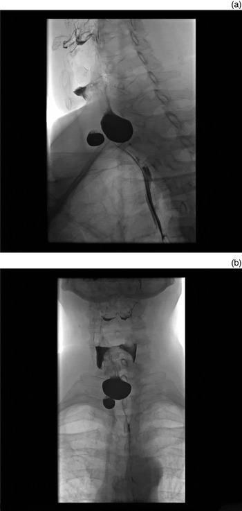
Barium studies demonstrated simultaneous Killian–Jamieson diverticulum and a larger Zenker's diverticulum in the cervical oesophagus (Figure 1a and b). Oesophageal motility appeared normal. A computed tomography (CT) scan of the neck confirmed the presence of diverticula, one at the sixth cervical vertebra (C6) to the first thoracic vertebra (T1) level, and a more anteriorly placed diverticulum at T1. An oesophageal web was present at the T1–2 level.
Barium swallow study images (a & b) demonstrating simultaneous Killian–Jamieson diverticulum and a larger Zenker's diverticulum in the cervical oesophagus.

Endoscopic stapling of the diverticulum was attempted initially, but was unsuccessful because of difficult access. Three months later, open excision of the pouch via a right partial utility incision was performed. Closure of the pharynx was performed with a 3/0 size vicryl suture. Post-operatively, the patient was fed nasogastrically for 7 days.
A water contrast study on day 7 confirmed successful resection of the Killian–Jamieson diverticulum with an intact surgical site, and normal oral intake was resumed. The biopsy demonstrated fibro-adipose tissue with a squamous-lined cystic space consistent with a pharyngoesophageal diverticulum.
At follow up, the patient remained symptomatic. Three months after the Killian–Jamieson diverticulum had been resected, the patient underwent an open excision of the Zenker's diverticulum using a 35 mm endopath stapler device via a left collar incision. The biopsy confirmed the diagnosis of diverticulum, with no evidence of dysplasia or malignancy. The patient's post-operative recovery was good, and two months after resection he was asymptomatic.
Discussion
The physiological mechanism of swallowing is complex, involving a coordinated series of events from the oral cavity down to the oesophagus. The upper oesophageal sphincter (UOS) is a 2.5 cm area extending from the distal pharynx to the proximal oesophagus. It plays an important role in the swallowing process. The cricopharyngeus muscle contributes to the function of the UOS, and is located between the inferior constrictor muscle and the cervical oesophagus. At rest, the UOS is under tonic contraction, preventing aerophagia and gastric reflux. During swallowing, the UOS relaxes, allowing food to pass from the hypopharynx into the proximal oesophagus. With ageing, the cricopharyngeus muscle can become hypertonic, leading to abnormalities in the normal passage of food. Early symptoms include cough and globus sensation, with the symptoms progressing to regurgitation, aspiration and the development of diverticula.Reference Huntley, Spiegel and Boon 6
Pharyngoesophageal diverticula can be classified pathophysiologically (into pulsion or traction diverticula), by composition (true or false diverticula) and anatomically (e.g. hypopharyngeal, mid-thoracic or epiphrenic diverticula). Pulsion diverticula, the most common type, herniate through a weak point in the outer muscular wall because of raised intraluminal pressure. They are considered false diverticula, as biopsy demonstrates the presence of only mucosa and submucosa in the diverticula.
Anatomically, there are three main areas of weakness in the pharyngeal musculature and it is at these points that pulsion diverticula most commonly occur. Zenker's diverticula are pulsion diverticula that arise in Killian's triangle, a space between the inferior pharyngeal constrictor muscle and the cricopharyngeus muscle.Reference Bizzotto, Iacopini, Landi and Costamagna 7 It is widely accepted that the aetiology of Zenker's diverticula is ‘increased intraluminal pressure in the oropharynx during swallowing, against an inadequate relaxation of the cricopharyngeal muscle, and subsequent incomplete opening of the UOS, causing the protrusion of the mucosa through an area of relative weakness’.Reference Bizzotto, Iacopini, Landi and Costamagna 7 A link between gastroesophageal reflux and Zenker's diverticula has been postulated, but never proven.Reference Ferreira, Simmons and Baron 8 Zenker's diverticula protrude posteriorly into the retropharyngeal space and usually extend to the left. Larger diverticula bulge laterally into the visceral space.
First described in 1983,Reference Ekberg and Nylander 3 a Killian–Jamieson diverticulum, also known as a ‘lateral cervical oesophageal diverticulum’ or ‘lateral diverticulum from the pharyngoesophageal junction area’, is an outpouching from the pharyngoesophageal junction through an anterolateral muscular gap – the Killian–Jamieson space. This is located inferior to the transverse fibres of the cricopharyngeus muscle and superior to the longitudinal muscle fibres of the proximal oesophagus in a lateral position behind the cricothyroid joint region. Killian–Jamieson diverticula are false diverticula.Reference Chea, Siow, Khor and Nik Azim 9 The aetiology is unclear, but is likely related to some form of cricopharyngeal muscle dysfunction, possibly dyscoordination of the pharyngeal constrictors, cricopharyngeal spasm, failure of sphincter relaxation or premature contraction.Reference Rodgers, Armstrong and Dana 10 They are not related to reflux oesophagitis.
Laimer's diverticula are rare, with only four cases described in the literature.Reference Boysen, Aasen, Lotveit and Bakka 5 , Reference Kumoi, Ohtsuki and Teramoto 11 , Reference Nguyen, Moslemi, Rawashdeh, Meyer, Garagozlo and Golestani 12 , Reference Kobayashi, Ito, Sawada, Deguchi and Lin 13 These arise inferior to cricopharyngeus in Laimer's triangle at the posterior aspect of the oesophagus, an area covered only by the circular layer of the oesophageal muscle.Reference Schunke, Schulte, Schumacher, Ross and Lamperti 14 A Laimer's diverticulum can be differentiated from a Zenker's diverticulum, as a Laimer's diverticulum: originates below the cricopharyngeus muscle, is broad based, occurs in a younger population and is a full thickness true diverticulum.Reference Nguyen, Moslemi, Rawashdeh, Meyer, Garagozlo and Golestani 12 Given the low incidence of this type of diverticula, the exact aetiology is unclear. The four reported cases suggest a pulsion mechanism secondary to an intrinsic oesophageal dysmotility disorder.
Traction diverticula form as a result of inflammation (resolution of a previous oesophageal perforation or lymphadenitis), which causes scarring or tethering of the hypopharyngeal or oesophageal musculature. Previous anterior cervical spine surgery has also been associated with the development of traction diverticula.Reference Goffart, Lennelle, Morceau and Bovene 15 Although there are less than 20 reported cases, some authors believe the incidence will rise as more surgeons adopt the anterior cervical approach.Reference Allis, Grant and Davidson 16 Traction diverticula are true diverticula as they contain mucosa, submucosa and outer muscular layers.Reference Johnson and Postma 1
Clinical presentation
Zenker's diverticula typically present in elderly individuals, with a 1.5-fold male predominance,Reference Bizzotto, Iacopini, Landi and Costamagna 7 and they are more frequent in northern Europe.Reference Klockars, Sihvo and Maekitie 17 Classical symptoms include: progressive dysphagia (for solids and liquids), regurgitation of undigested food debris due to food entrapment in the diverticulum, pharyngeal stasis of secretion, chronic cough, chronic aspiration, halitosis, globus sensation, hoarseness, whistling and cervical borborygmi.Reference Siddiq, Sood and Strachen 18 Reported complications include: aspiration pneumonia, poor absorption of oral medication, malnourishment, weight loss, diverticulitis, peptic ulceration, bleeding, iatrogenic perforation during endoscopy, fistulae, vocal fold paralysis and malignancy.Reference Ferreira, Simmons and Baron 8 Malignancy is probably due to chronic inflammation. They occur in approximately 0.5 per cent of patientsReference van Overbeek 19 and should be considered in patients with increased symptom severity.Reference Bradley, Kochaar and Quraishi 20
Patients with Killian–Jamieson diverticula tend to be older than those with Zenker's diverticula (72 years vs 66 years),Reference Rubesin and Levine 21 but may present with similar symptoms, mainly dysphagia, cough and epigastric pain. The majority, however, are asymptomatic or have symptoms attributable to abnormal pharyngeal motility. Of note, patients with Killian–Jamieson diverticula are very unlikely to develop aspiration pneumonia, as the closed cricopharyngeus above the diverticula prevents the reflux of retained food into the hypopharynx.Reference Rubesin and Levine 21 Complications of an untreated Killian–Jamieson diverticulum include perforation,Reference Nutter and Ball 22 infection,Reference Kitazawa, Koide, Saito, Kamimura, Uehara and Miyagawa 23 obstruction, ulceration, bezoar formation, fistula formation,Reference Braghetto, Cardemil, Schwartz, Valladares, Rencoret and Estay 24 iatrogenic perforation and malignancy.
Laimer's diverticula are so rare that precise symptomology is hard to determine. In the available cases, cough, dysphagia and regurgitation of food shortly after eating appear to be the prominent symptoms. No long-term complications have been reported.
Typical symptoms of traction diverticula include dysphagia, postural regurgitation, belching, retrosternal pain, gastroesophageal reflux and epigastric pain.Reference Ballehaninna, Shaw and Brichkov 25 Pulmonary complications are not uncommon, with symptoms ranging from a mild nocturnal cough to life-threatening aspiration pneumonia. It is important in these patients to identify any underlying cause, including tuberculosis, histoplasmosis, healed oesophageal perforation, past history of cervical spine surgery and any other inflammatory condition.Reference Johnson and Postma 1
Diagnosis
Killian–Jamieson diverticula are commonly misdiagnosed as Zenker's diverticula, and accurate differentiation relies mainly on barium contrast oesophagography and axial neck CT. During barium studies, it is important to examine both the anteroposterior and lateral radiographs, with the patient in both the supine and erect positions, in order to identify the relationship of the diverticulum to the cricopharyngeus muscle. The opening of a Zenker's diverticulum is shown radiographically directly above the cricopharyngeal bar, with the sac of the diverticulum lying posterior to the oesophagus on lateral images and in the midline on frontal images.Reference Johnson and Postma 1 The opening of a Killian–Jamieson diverticulum is located just below the cricopharyngeus, with the diverticular sac lying lateral to the oesophagus on frontal images and overlapping the anterior wall of the cervical oesophagus on lateral images.Reference Johnson and Postma 1 Zenker's diverticula tend to be larger than Killian–Jamieson diverticula (2.5 cm vs 1.4 cm) and are more likely to retain barium. Zenker's diverticula usually protrude to the left side, with only 10 per cent occurring on the right. Killian–Jamieson diverticula tend to be left-sided, although there have been reports of right-sided Killian–Jamieson diverticula,Reference Kim, Hwang, Lee, Lee, Kim and Chee 26 bilateral Killian–Jamieson diverticulaReference Rubesin and Levine 21 , Reference Boisvert, Bethune, Acton and Klassen 27 and one report of a Killian–Jamieson diverticulum in association with a Zenker's diverticulum.Reference Boysen, Aasen, Lotveit and Bakka 5 Our patient, who had a right-sided Killian–Jamieson diverticulum associated with a Zenker's diverticulum, is very unusual.
Barium studies are also useful in the investigation of Laimer's and traction diverticula, but the exact nature of the diverticulum is usually confirmed at surgery. Both types may be misdiagnosed as a Killian–Jamieson diverticulum.
An axial CT scan can localise the origin of the diverticulum more precisely. A CT scan demonstrates a laterally projecting sac of a Killian–Jamieson diverticulum originating below the level of the cricoid cartilage. Zenker's diverticula arise at a higher level and are more posteriorly orientated.Reference Johnson and Postma 1
Whilst ultrasound scanning does not form part of the diagnostic investigation of pharyngoesophageal diverticula, a number of case reports have highlighted the importance of carefully examining thyroid nodules, as Killian–Jamieson diverticula can mimic these nodules. Kim et al. reviewed 13 cases of Killian–Jamieson diverticula mimicking thyroid nodules.Reference Kim, Lee, Jang, Bae, Lee and Kim 28 Twelve of 13 patients were referred for aspiration of a thyroid nodule and the other patient was referred following an increase in size of a pre-existing nodule. All patients were asymptomatic. Ninety-two per cent of the diverticula were located in the posterior aspect of the left thyroid lobe and the other was located in the right lobe. The study determined that Killian–Jamieson diverticula have specific characteristics on ultrasound scanning: (1) a heterogeneous internal echo with strong echogenic foci caused by air bubbles or food particles; (2) a hypoechoic rim with or without a multi-layered pattern suggesting that the gastrointestinal tract is the origin of the lesion; (3) an irregular boundary of the posterior wall at the posterior portion of the thyroid gland; and (4) changes in the internal echo consistent with changes in the contents of the diverticulum after swallowing. As ultrasound is commonly used in the assessment of thyroid nodules, it is important to recognise the possibility that Killian–Jamieson diverticula may mimic these nodules, as the diagnosis and management of these two conditions is very different.
Management
The management of Zenker's diverticula is well documented, and involves endoscopic or open surgical procedures. A recent systematic review on the treatment of Zenker's diverticula concluded that open surgical procedures are preferable in younger patients and in those with anatomy unsuitable for endoscopy.Reference Verdonck and Morton 29 Flexible endoscopic procedures have been described as an alternative management in patients with co-morbidities that make prolonged general anaesthesia undesirable;Reference Mulder, den Hartog, Robijn and Thies 30 , Reference Case and Baron 31 however, the authors do not have experience of this technique.
The optimal management of Killian–Jamieson diverticula remains unknown because of the limited number of reported cases. Treatment options include open or endoscopic surgery, with diverticulotomy, diverticulectomy with oesophagomyotomy, or diverticulopexy.
Unlike Zenker's diverticula, the site of origin of Killian–Jamieson diverticula is below the level of the cricopharyngeus muscle and is directly adjacent to the entry point of the recurrent laryngeal nerve (RLN) into the larynx. Rodgers et al. reviewed the management of Killian–Jamieson diverticula in 2000 and concluded that an open surgical procedure reduced the possibility of RLN damage.Reference Rodgers, Armstrong and Dana 10 Two reports published in 2008 documented the successful treatment of Killian–Jamieson diverticula endoscopically.Reference Lee, Chung, Park, Lee, Lee and Park 32 , Reference Tang, Tang, Chen and Myers 33 Tang et al. used a needle-knife and pure coagulation current with a setting of 25 W to perform a distal vertical diverticulotomy.Reference Tang, Tang, Chen and Myers 33 There were no complications and the patient remained asymptomatic at follow up. Lee et al. performed flexible endoscopic diverticulotomy, using an isolated-tip needle-knife papillotome to dissect the tissue bridge of the diverticulum, and a flexible overtube with a modified distal end to visualise the tissue bridge and protect the surrounding tissue during dissection.Reference Lee, Chung, Park, Lee, Lee and Park 32 There were no complications and the patient made a good recovery. Neither case report mentions visualisation of the RLN or post-operative laryngoscopy to confirm that the RLN had not been damaged.
In 2016, Chang et al. reported two cases of Killian–Jamieson diverticula managed with the use of a rigid laryngoscope and a point-cutting carbon dioxide (CO2) laser.Reference Chang, Chi and Lee 34 Prior to surgery, serious consideration was given to the prevention of RLN damage. Three main factors were deemed important: prevention of oesophageal perforation, prevention of thermal injury to the RLN and the location of the most appropriate area for laser cutting. In order to avoid oesophageal perforation, no laser energy was applied beyond the longitudinal oesophageal fibres. Studies have shown that thermal injury to the RLN occurs when surface temperatures exceed 47–58 °C.Reference Tang, Tang, Chen and Myers 33 , Reference Beriat, Akmansu, Ezerarslan, Dogan, Han and Saglam 35 , Reference Xu and Pollock 36 Using the CO2 laser, Chang et al. maintained a surface temperature of 43 °C, minimising the risk of thermal damage.Reference Chang, Chi and Lee 34 The septum of the diverticulum was cut at the midpoint. Both operative and hospitalisation times were reduced, and both patients quickly resumed normal oral intake post-operatively.
Whilst these case reports describe successful endoscopic diverticulotomy, this technique is associated with an increased risk of RLN damage.Reference Rodgers, Armstrong and Dana 10 , Reference Tang, Tang, Chen and Myers 33 The base of the diverticulum lies posterior to the nerve, and the narrow operating field, combined with the use of ultrasonic shears, electrocautery or a stapling device, increase the risk of nerve damage. Open diverticulectomy, preferably using a transcervical approach, allows easier dissection of the RLN and easy transection of the diverticular base using an endoscopic stapling device.Reference Siow, Mahendran and Hardin 4 Since 2010, there have been six reported cases advocating an open surgical approach to Killian–Jamieson diverticula,Reference Siow, Mahendran and Hardin 4 , Reference Chea, Siow, Khor and Nik Azim 9 , Reference Kitazawa, Koide, Saito, Kamimura, Uehara and Miyagawa 23 , Reference Kim, Hwang, Lee, Lee, Kim and Chee 26 , Reference Boisvert, Bethune, Acton and Klassen 27 , Reference Undavia, Anand and Jacobson 37 with all but one patientReference Chea, Siow, Khor and Nik Azim 9 undergoing transcervical diverticulectomy. The remaining patient underwent transcervical diverticulopexy. Boisvert et al. reported a case of bilateral Killian–Jamieson diverticula.Reference Boisvert, Bethune, Acton and Klassen 27 Although they undertook open diverticulectomies on the basis of potential RLN damage, no mention was made of RLN visualisation or post-operative assessment for RLN damage. Kitazawa et al. reported the only case to date of cellulitis secondary to a Killian–Jamieson diverticulum.Reference Kitazawa, Koide, Saito, Kamimura, Uehara and Miyagawa 23 At surgery, the RLN was found to be adherent to the base of the diverticulum, and was carefully dissected from the diverticulum and corded by a thin string to preserve it. There was no reported RLN damage.
Chea et al. were unable to perform diverticulectomy because it was technically difficult to apply the stapling device across the entire neck of the diverticulum, as the lower edge extended into the thoracic inlet.Reference Chea, Siow, Khor and Nik Azim 9 At surgery, they identified and preserved the RLN. No post-operative complications were reported, and the authors considered diverticulopexy a feasible alternative to diverticulectomy in patients with complex anatomy. This procedure, however, is more commonly used in patients who would not tolerate prolonged anaesthesia; the management of a Killian–Jamieson diverticulum in this way has not been reported elsewhere. Kim et al. treated a right-sided Killian–Jamieson diverticulum with open diverticulotomy, but did not report identification of the RLN, despite the diverticulum's adherence to adjacent structures.Reference Kim, Hwang, Lee, Lee, Kim and Chee 26 Undavia et al.Reference Undavia, Anand and Jacobson 37 and Siow et al.Reference Siow, Mahendran and Hardin 4 recommended an open surgical approach to Killian–Jamieson diverticula. Both cases were reported as left-sided Killian–Jamieson diverticula, with adherent RLNs. Careful dissection avoided overt damage to the RLN, but only Undavia et al.Reference Undavia, Anand and Jacobson 37 confirmed normal vocal fold function post-operatively.
Although there are limited data on the management of Killian–Jamieson diverticula, the existing literature supports an open surgical approach, primarily because of the risk to the RLN. Diverticulectomy reduces the risk of recurrence, and is preferable in younger patients who have fewer co-morbidities.Reference Siow, Mahendran and Hardin 4 , Reference Kitazawa, Koide, Saito, Kamimura, Uehara and Miyagawa 23 , Reference Boisvert, Bethune, Acton and Klassen 27 , Reference Undavia, Anand and Jacobson 37 Diverticulopexy may be used as an alternative in older patients, or in those with challenging anatomy. Endoscopic surgery requires further evaluation.
Conclusion
Killian–Jamieson diverticula are rare. Thorough examination of barium studies is needed to differentiate Killian–Jamieson diverticula from the more common Zenker's diverticula. Surgical management is necessary because of the list of complications if left untreated. Although the number of cases is small, open transcervical excision (rather than endoscopic surgery) appears to be the most appropriate management.
Our patient is unusual. He had a right-sided Killian–Jamieson diverticulum associated with a Zenker's diverticulum. His management demonstrates clearly the difficulties in approaching a Killian–Jamieson diverticulum endoscopically. The patient's anatomy made such an approach impossible. Open surgery made identification of both pouches much easier. The RLN was not specifically identified at surgery, which we would recommend, but the patient has no clinical evidence of RLN damage.