Introduction
Posterior glottic stenosis is defined as a narrowing or fixation in the larynx as a result of scarring of the posterior commissure, interarytenoid space or cricoarytenoid joints. The disorder is most commonly a consequence of intubation. In particular, in the posterior commissure, the endotracheal tube can abrade the mucosa causing scarring and inflammation. Depending on the nature of the stenosis, patients suffer from various degrees of airway obstruction and dysphonia.
Posterior glottic stenosis can be diagnosed by either indirect or direct laryngoscopy and radiographic studies, but can be elusive. The condition can also be diagnosed by endolaryngeal microsurgery.
Reconstruction of the dynamic larynx after scarring of the posterior glottis can be a difficult task, especially using endolaryngeal microsurgery. Posterior glottic stenosis is easily disrupted by a vertical midline incision, but it will recur if the denuded posterior commissure is not covered by a mucous membrane.Reference Montgomery1
This report describes the use of a posteriorly based mucosal flap, obtained from the postcricoid region using endoscopic laryngeal microsurgery, to cover the denuded lesion in order to prevent scar re-formation.
Patients and methods
A six-year-old girl presented complaining of increasing inspiratory stridor. Three months earlier, she had undergone surgery for lateral curvature. She had been post-operatively intubated for 17 days and had shown inspiratory stridor after extubation.
Laryngoscopy revealed the presence of an adhesion at the posterior commissure which severely limited abduction of the bilateral vocal folds and arytenoids. In addition, it showed the true vocal folds to be immobile bilaterally, with an approximately 2-mm gap between the vocal processes.
Initially, endolaryngeal microsurgery was performed following tracheal fenestration. The scar tissue on the posterior commissure were identified. The passive mobility of the arytenoids and vocal folds was restricted bilaterally. The scar tissue, including the transverse arytenoid muscle, was resected using CO2 laser. As a result, the posterior glottal airway was sufficiently widened.
Post-operatively, however, the posterior commissure and interarytenoid lesion both gradually narrowed. The stenosis recurred, more severely this time. The true vocal folds were seen to be completely immobile bilaterally, with no gap between the vocal processes upon inspiration.
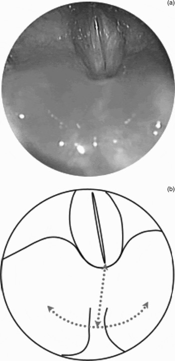
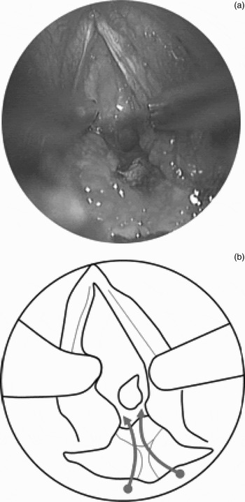
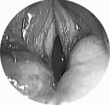
Three months later, endolaryngeal microsurgery was performed again. The scar tissue of the posterior commissure and the interarytenoid lesion was identified (Figure 1a). Using endoscopic microscissors, a transverse incision was made extending from the top of one arytenoid to the other. The scar tissue of the posterior glottis and interarytenoid notch was disrupted and separated by a vertical midline incision (Figure 1b). The interarytenoid muscle scarring and the fibrotic tissue nearby were excised as needed. As a result, free passive motility of the arytenoids was restored bilaterally, without the need to divide the cricoarytenoid joints (Figure 2a). A posteriorly based mucosal flap was submucousally elevated from the postcricoid region. The corniculate cartilages (above the arytenoid cartilages) were debulked bilaterally with CO2 laser. The mucosal flap was extended to approximate to the posterior commissure mucosa. The mucosal flap was sutured to the inferior subglottic mucosa by two 4–0 polyglactin absorbable sutures (Figures 2b and 3).
(a) Endolaryngeal, microsurgical view during second procedure, showing posterior glottic stenosis, interarytenoid scarring and a completely closed airway. (b) A diagram of the same surgical view, with dashed lines indicate planned incision lines.

(a) Endoscopic view of the larynx showing the incision used to separate the arytenoids (the incision is held open by two probes for demonstration purposes). Subglottic stenosis was identified. (b) Schematic drawing of same surgical view; arrows indicate the application of the posteriorly based mucosal flap obtained from the postcricoid region, which was submucosally elevated and then sutured to the inferior subglottic mucosa by two sutures.

Endoscopic view of the larynx showing attachment of the mucosal flap to the inferior subglottic mucosa by two sutures.

Although subglottic stenosis was observed during the two procedures, no specific surgical treatment for this condition was undertaken.
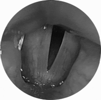
Three months after surgery, the patient's respiratory and phonatory function was satisfactory (Figure 4).
Endoscopic view of the larynx three months after the procedure, showing successful opening of the airway in inspiration.

Discussion
Bogdasarian and OlsonReference Bogdasarian and Olson2 have proposed a classification system for posterior glottic stenosis, involving four categories. Patients with a type one stenosis have normal interarytenoid mucosa (i.e. posterior commissure) and are candidates for simple scar excision, because the normal mucosa serves to prevent scar re-formation between the arytenoids. Patients with types two or three stenosis have mobility in one or both cricoarytenoid joints. Patients with type four stenosis have bilateral cricoarytenoid fixation, and airway surgery must be performed in order to widen the airway; however, this does not reconstruct a dynamic larynx.
In the current case, pre-operative fibreoptic observation of the bilateral arytenoids showed limited abduction and adduction. Judging from the slight passive rotation of the arytenoids and the presence of scar tissue only on the posterior commissure, the case was diagnosed as type two posterior glottic stenosis at the first microsurgical procedure. However, during the second microsurgical procedure, when not only scarring of the interarytenoid mucosa but also bilateral vocal fold fixation was identified, the patient's posterior glottic stenosis was diagnosed as type four, based on the above system.
Previously described methods for treatment of patients with posterior glottic stenosis have included arytenoidectomy, cordectomy,Reference Gaboriau, Laccourreye, Laccourreye and Brasnu3 placement of a keel,Reference Langman, Lee and Dedo4 microtrapdoor flap,Reference Dedo and Sooy5 open scar excision with mucosal flap advancement,Reference Montgomery1 open placement of a posterior cricoid cartilage graft,Reference Rimell and Dohar6, Reference Berkowitz7 and laser lysis with or without botulinum toxin injection.Reference Nathan, Yin and Stucker8 However, simple lysis of the interarytenoid scar is typically followed by re-formation of the scar. Thus, various methods of preventing scar re-formation have been used and discussed, e.g. keel placement and the use of intervening tissue between the separated arytenoids. MontgomeryReference Montgomery1 has reported that advancement of a vascularised mucosal flap required an open procedure via a midline laryngo-fissure. Using the open approach, decannulation rates of 70 per cent have been reported; higher rates have been reported with additional procedures, such as a cordectomy.Reference Hoasjoe, Franklin, Aarstad, Day and Stuker9 However, these procedures can cause scarring of the anterior commissure, and the patient's hoarseness can worsen post-operatively. The method used in the current patient is similar to the endoscopic postcricoid advancement flap described by Goldberg.Reference Goldberg10 This author reported that this flap represented a less invasive means of placing vascularised tissue into the interarytenoid space, in order to prevent re-formation of interarytenoid scar tissue. The procedure combines the advantages of endoscopic surgery with those gained by open placement of vascularised mucosa at the desired location, to prevent re-formation of scar tissue; as a result, tissue trauma is reduced in comparison with an open neck approach.
However, when a procedure is performed with a small flap, the flap contains less vascularised tissue and may not cover the whole exposed surface of the interarytenoid space. As a result, there is a possibility of scar recurrence in the interarytenoid space, and vocal fold mobility can be easily compromised. In the current technique, a flap raised from the wide postcricoid mucosa adequately covers the space exposed by removing the interarytenoid scar, and has better circulation than a small flap.
In the current patient, subglottic stenosis was identified during surgery, but surgical treatment was not performed due the possibility that the stenosis could re-form more severely after such treatment. Furthermore, a tracheal stent (commonly used in open procedures for subglottic stenosis) can cause granulation tissue formation, ulceration and re-stenosis. In the current patient, as a result of not treating the subglottic stenosis, the subglottic airway was sufficient to enable adequate post-operative speech and respiration.
Conclusion
A posteriorly based mucosal flap obtained endoscopically from the postcricoid region is a useful adjunct to other treatment options for posterior glottic stenosis. The highly vascularised tissue is placed endoscopically in the posterior glottis to prevent scar re-formation. This procedure minimises trauma through the use of an endoscopic approach (compared with use of a laryngofissure and laryngeal stent), and it can be performed with available laryngeal microsurgery equipment by surgeons familiar with laryngeal microsurgery.