INTRODUCTION
Neurocysticercosis (NCC) is a severe and specific infection, caused by the ingestion of Taenia solium eggs, and the consequent invasion and development of the larval stages at the central nervous system. The disease is endemic in areas with deficient sanitary conditions mainly in developing countries of America, Africa and Asia (Ndimubanzi et al. Reference Ndimubanzi, Carabin, Budke, Nguyen, Qian, Rainwater, Dickey, Reynolds and Stoner2010; Del Brutto, Reference Del Brutto2012; Martins-Melo et al. Reference Martins-Melo, Ramos, Cavalcanti, Alencar and Heukelbach2016). In these endemic areas, NCC is responsible for about 30% of acquired epilepsy cases (Carpio and Romo, Reference Carpio and Romo2015; Del Brutto and García, Reference Del Brutto and García2015; Gonzales et al. Reference Gonzales, Rivera and Garcia2016).
Southern region of the USA is seen as an endemic area, due to the border with Mexico and the consequent increased immigration from highly endemic regions. In addition, the spread of parasitic diseases can be highlighted in other countries from North America and European Union because of Latin Americans migration (Arriola et al. Reference Arriola, Gonzalez, Gomez-Puerta, Lopez-Urbina, Garcia and Gilman2014; O'keefe et al. Reference O'keefe, Eberhard, Shafir, Wilkins, Ash and Sorvillo2014; Gabriël et al. Reference Gabriël, Johansen, Pozio, Smit, Devleesschauwer, Allepuz, Papadopoulos, Van Der Giessen and Dorny2015; Rizvi et al. Reference Rizvi, Saleh, Frimpong, Mohiy, Ahmed, Edwards and Ahmed2016).
NCC, although a life-threatening disease in severe cases, is generally an acute or long-lasting infection affecting the quality of the patient life and social environment. The disease has socioeconomic importance because majority of patients are at productive ages and frequently become unable to work soon after the onset of symptoms (Madrazo, Reference Madrazo2013).
Diagnosis of NCC is based on clinical, epidemiological and laboratory findings, and it can also be accurately performed using neuroimaging techniques such as computed tomography (CT) and magnetic resonance imaging (Del Brutto et al. Reference Del Brutto, Lama, Zambrano and Del Brutto2014). Unfortunately, these methods are expensive and usually inaccessible at the poorest endemic areas, and therefore the development of other protocols is also needed. The immunological tests most commonly used to detect antibodies against T. solium are the enzyme-linked immunosorbent assay (ELISA) and immunoblotting using cerebrospinal fluid (CSF) and/or serum (Costa et al. Reference Costa, Ferreira, Makino and Camargo1982; Barcelos et al. Reference Barcelos, Mineo, Oliveira-Silva, Ferreira, Moura, Biondi and Costa-Cruz2001; Husain et al. Reference Husain, Shukla, Kumar, Husain, Chaturvedi, Agarwal and Gupta2008), but these tests have problems related to the complex composition of total antigenic preparations and also with the cross-reactions with samples from patients infected with other parasitic diseases.
To overcome problems related with antigen preparation and cross-reactivity there are efforts to develop the use of fractionated heterologous antigens, such as those derived from Taenia saginata metacestodes. The use of this heterologous antigen for NCC immunodiagnosis is important where the collection of T. solium metacestodes to prepare homologous antigen is difficult (Oliveira et al. Reference Oliveira, Machado, Cabral and Costa-Cruz2007, Reference Oliveira, Machado, Gonçalves-Pires, Moura and Costa-Cruz2009; Coral-Almeida et al. Reference Coral-Almeida, Gabriël, Abatih, Praet, Benitez and Dorny2015). Diagnostic performance of heterologous antigens was improved resulting in higher diagnostic indexes through fractionation using other chromatographic techniques (Nunes et al. Reference Nunes, Ribeiro, Manhani and Costa-Cruz2010, Reference Nunes, Gonzaga, Ribeiro, Cunha-Junior and Costa-Cruz2013). Moreover there is a need to use proteomic tools to identify proteins with potential applicability in NCC diagnosis.
In light of these previous reports, we developed a three stage strategy to obtain a protein biomarker as new diagnostic tool for NCC diagnosis in serum. First, we used gel filtration chromatographic technique to fractionate total saline extract (SE) from T. saginata metacestodes obtaining the gel filtration fraction (GFF). Subsequently, the diagnostic potential of the obtained fraction was evaluated by ELISA and immunoblotting. Finally, proteins from the GFF with diagnostic potential were identified through mass spectrometry (MS) and B cell epitopes were predicted using the structure of the identified proteins.
MATERIAL AND METHODS
Ethical statement
This study was conducted according to the ethical guidelines of the Brazilian Health Ministry after being approved by the Research Ethics Committee of the Universidade Federal de Uberlândia, Minas Gerais state, Brazil under the number 382/11.
Serum samples
It was used a panel of serum samples from immunocompetent individuals that were well characterized parasitologically. Serum samples were collected from 160 subjects attended at the following laboratories: (a) Laboratório de Análises Clínicas do Hospital de Clínicas de Uberlândia (Groups 1 and 2); (b) Laboratório de Diagnóstico de Parasitoses of the Universidade Federal de Uberlândia (Group 3); (c) Laboratório de Helmintos Parasitos de Vertebrados do Instituto Oswaldo Cruz, Rio de Janeiro state, Brazil (Group 2); and (d) Laboratório de Genômica Estrutural e Funcional of the Universidade Federal do Rio Grande do Sul, Rio Grande do Sul state, Brazil (Group 2).
Group 1 (G1) consisted of 45 patients with definitive diagnosis of NCC, according to Del Brutto (Reference Del Brutto2012), as follows: (a) all patients presented at least one type of clinical manifestation suggestive of NCC such as: epilepsy, cephalea, dizziness, dementia, faintness, hydrocephalus and no signs or symptoms of cysticercosis in other organs; (b) all patients came from or lived in a cysticercosis endemic area; (c) for immunodiagnosis, cerebrospinal fluid and/or serum samples were positive in an ELISA test for the detection of anti-Taenia IgG; (d) they presented evidence of the parasite in neuroimaging, with the following characteristics: active NCC (n = 24), where metacestodes were viable or in early degeneration, and inactive NCC (n = 21), where metacestodes were completely degenerated, according to Sotelo et al. (Reference Sotelo, Guerrero and Rubio1985).
Group 2 (G2) consisted of 65 patients with other parasitic infections: Ascaris lumbricoides (n = 5), Entamoeba histolytica/dispar (n = 2), Enterobius vermicularis (n = 6), E. granulosus (n = 8), Giardia lamblia (n = 8), hookworm (n = 7), Hymenolepis nana (n = 4), Schistosoma mansoni (n = 4), Strongyloides stercoralis (n = 8), Taenia sp. (n = 5) and Trichuris trichiura (n = 4). Co-infected patients were also tested: hookworm + A. lumbricoides + T. trichiura (n = 1), hookworm + A. lumbricoides (n = 1), Entamoeba histolytica/dispar + G. lamblia (n = 1) and H. nana + E. vermicularis (n = 1). At least three fecal samples were obtained and tested by the parasitological method of Ritchie (Reference Ritchie1948). Patients infected with Echinococcus were considered positive after a positive diagnosis by ultrasound, CT and serology tests and confirmation by parasitological examination of cystic fluid after surgery. All cases underwent surgery for hydatid disease. Serum samples from patients infected with Echinococcus granulosus were received from the Laboratório de Helmintos Parasitos de Vertebrados do Instituto Oswaldo Cruz, Rio de Janeiro state, Brazil and from the Laboratório de Genômica Estrutural e Funcional of the Universidade Federal do Rio Grande do Sul, Rio Grande do Sul state, Brazil.
Group 3 (G3) comprised 50 apparently healthy individuals, based on their clinical presentation. Although they came from a cysticercosis endemic area, all volunteers from this group did not present evidence of household contact with T. solium infection or a history of taeniasis or cysticercosis. In addition, these individuals had three fecal samples negatively tested by the parasitological methods of Baermann–Moraes (Baermann, Reference Baermann1917; Moraes, Reference Moraes1948) based on the positive larval thermo-hydrotropism and Lutz method (Lutz, Reference Lutz1919), a gravity sedimentation technique.
Taenia saginata metacestodes obtention
Taenia saginata metacestodes were obtained from naturally infected cattle at the Triângulo Mineiro region, Minas Gerais state, Brazil. Infected cattle came from abattoirs and were slaughtered in accordance with the inspection technique recommended by the Federal Inspection Service. Taenia saginata metacestodes were obtained by dissecting muscles and only those in the vesicular stage were collected, washed in saline solution (0·15 M NaCl) four times and stored at −20 °C.
Total SE from T. saginata metacestodes preparation
SE was obtained by using 50 T. saginata metacestodes resuspended in phosphate-buffered saline (PBS) containing a protease inhibitors cocktail: 100 µL mL−1 of complete ULTRA extract (Tablets, Mini, EASYpac, Roche, Alemanha) and disrupted by five cycles of freezing, maceration and thawing/sonication until no integral metacestodes were observed. After a 2-h incubation period at 4 °C, under constant shaking, the suspension was centrifuged (12400 × g , 30 min, 4 °C) and the supernatant was assayed according to the procedure of Bradford (Reference Bradford1976) for protein quantification and stored at −20 °C.
Fractionation procedure and antigen analysis
Gel filtration fraction (GFF) was obtained by gel filtration chromatography from the SE and developed in column (glass, size 1·5 × 50 cm2, max volume 89 mL, cross-sectional area 1·77 cm2, Bio-Rad Laboratories, USA) containing resin (Sephacryl S-100 High Resolution, GE Healthcare, USA). A minimum protein concentration of 6000 µg mL−1 of SE (4 mL) was loaded and column was previously equilibrated with five times the resin volume (80 mL) of PBS (4–8 °C). After centrifugation and addition of protease inhibitor cocktail [composed of substances such as benzethonium chloride, α-toluenesulphonyl fluoride, disodium dihydrogen ethylenediaminetetraacetate and 4-(2-Aminoethyl) benzenesulfonyl fluoride hydrochloride, Bestatin, [(2S,3R)-3-amino-2-hydroxy-4-phenylbutanoyl]-L-leucine hydrochloride, pepstatin, Leupeptin hemisulphate 2-oxiranecarboxylic acid, Trypsin inhibitor (pancreatic basic)], SE was applied to the column until total influx, then 4 mL of PBS (4–8 °C) was pumped to prevent mixing chamber formation. Aliquots (1 mL) were collected automatically (Model 2110, Bio-Rad, USA). The final volume collected corresponded to two times the resin volume (160 mL). Aliquots were monitored for the presence of protein according to Bradford (Reference Bradford1976). The chromatogram was generated, and peak from tubes 22–26 was defined as GFF. After use, the resin was washed with PBS and stored (4–8 °C) in PBS with sodium azide (0·1%).
Electrophoretic analysis
Total SE and the collected fractions were analysed by sodium dodecyl sulphate polyacrylamide gel electrophoresis 12% (SDS–PAGE) under non reducing conditions, as described by Laemmli (Reference Laemmli1970) and GFF was selected considering preliminary ELISA results. Proteins were visualized in SDS–PAGE after Coomassie Brilliant Blue staining (Dong et al. Reference Dong, Wang, Wang and Zhang2011).
Analysis was performed using a graphical method to generate protein lane profile plot of each antigenic fraction, by Image J version 1.44 software (National Institutes of Health, Bethesda, USA); and then peaks related to bands were compared with protein standard marker (Real Biotech, RECOM™ Blue Wide Range Prestain Marker, Taiwan) to estimate their relative molecular weight.
ELISA to detect anti-Taenia IgG
ELISA using SE and GFF was carried out according to Nunes et al. (Reference Nunes, Gonzaga, Ribeiro, Cunha-Junior and Costa-Cruz2013). Low-affinity polystyrene microplates (Interlab, Brazil) were coated with antigenic preparations (10 µg mL−1) in carbonate bicarbonate buffer, pH 9·6 and incubated overnight at 4 °C. Then, plates were washed once, for 5 min with PBS containing 0·05% Tween 20 (PBS-T). Serum samples were diluted 1:200 in PBS-T containing 3% of skimmed milk (PBS-TM) and incubated for 45 min at 37 °C. After three washes with PBS-T, 5 min each, the immunoenzymatic conjugate (peroxidase-goat anti-human IgG, Fc specific; Sigma) was added diluted 1:2000 in PBS-T and incubated for 45 min at 37 °C. Subsequently, another step of washing was performed and the assay was developed by adding the enzyme substrate consisting of hydrogen peroxide and orthophenylenediamine (OPD) in 0·1 M citrate phosphate (Na2HPO4) buffer pH 5·5 for 15 min. Reaction was interrupted by adding 25 µL per well of 2 N H2SO4. Absorbance was determined at 492 nm in an ELISA reader (TP Reader, Thermo Plate, Brazil).
Immunoblotting
For membrane preparation SE and GFF were submitted to SDS–PAGE 12% and gels were transferred using a Trans-Blot SD Semy Dry Electrophoretic Transfer Cell (Bio-Rad, Hercules, CA, USA) to nitrocellulose membranes (0·20 µm, Bio-Rad), as described by Towbin et al. (Reference Towbin, Staehelin and Gordon1979). Membranes were cut into strips and immunoblotting was performed as described by Nunes et al. (Reference Nunes, Gonzaga, Ribeiro, Cunha-Junior and Costa-Cruz2013), with modifications. Strips containing SE and GFF were blocked with 2·5% PBS-TM for 2 h at room temperature and washed once, for 5 min with PBS-TM 1%. After, samples [a pool from the most reactive samples in ELISA from G1 (n = 5) and G3 (n = 5)] diluted 1:25 in 1% PBS-TM were added and incubated overnight at 4 °C. Then, after washing five times with PBS-T 0·05%, 5 min each, strips were incubated for 2 h at room temperature with peroxidase-labelled goat anti-human IgG conjugate (whole molecule, Sigma) diluted at 1:1500 in PBS-TM 1%. Subsequently, another step of washing was performed using PBS-T 0·05% and the assay was developed by adding hydrogen peroxide and 3·3′-diaminobenzidine tetra hydrochloride (Sigma) for 3 min. Reaction was stopped by washing strips with water.
MS and protein identification
After confirmation by the immunological tests (ELISA and immunobloting) the band of interest at GFF was elected and excised manually with a clean scalpel from the gel, previously stained with Coomassie Brilliant Blue, for further characterization through MS. All materials were previously washed with methanol to reduce contaminants. The excised gel band was washed with acetonitrile and stored (−20 °C) until processing. Sample was dried by vacuum centrifugation, and digested with trypsin. Analyses were carried out in the Laboratório de Espectrometria de Massas of the Centro de Facilidades de Apoio à Pesquisa (CEFAP) of the Universidade de São Paulo (USP), São Paulo state, Brazil. Sample was applied to an EASY nanoLC II-LTQ Orbitrap Velos-ETD (Thermo Scientific, Waltham, Massachusetts, EUA) coupled to nano liquid chromatography (EASY-nanoLC II). Peptides were loaded in a trap column packed with C-18 resin (reverse-phase high-performance liquid chromatography; RP-HPLC) and sequentially eluted using a gradient from acetonitrile for 45 min, separated and analysed (LTQ-OrbitrapVelos ETD). Data search was conducted in the National Center for Biotechnology Information (NCBI) database. Search was done using Taenia genus considering identified proteins with the highest score, coverage rate and unique peptides.
Prediction of linear B-cell epitopes
The 3D models of the proteins were constructed using Swiss-Model software (http://swissmodel.expasy.org/) and UCSF Chimera (Resource for Biocomputing, Visualization, and Informatics at the University of California, CA, USA). As templates we used tertiary structures of other proteins to construct the models of Taenia identified proteins. The prediction of linear B-cell epitopes was performed by BepiPred platform (http://tools.immuneepitope.org/bcell/).
Statistical analyses
Analyses were performed using GraphPad software package 5·0 (GraphPad Software Inc., USA). Cut-off points were established using a two-graph receiver operating characteristic curve (TG-ROC) (Greiner et al. Reference Greiner, Sohr and Göbel1995) based on ELISA results from positive (G1) and negative samples (G2 and G3). ELISA reactivity index (RI) was obtained by the ratio between OD and cutoff. RI greater than the optimum point of reaction for each extract were considered positive (RI > 1). Sensitivity (Se) and specificity (Sp) were calculated according to Youden (Reference Youden1950). ROC curves were built to describe the tests (Martinez et al. Reference Martinez, Louzada-Neto and Pereira2003). The area under ROC curve (AUC), an index of diagnostic accuracy, was calculated (Hanley and McNeil, Reference Hanley and McNeil1982) and compared (Hanley and McNeil, Reference Hanley and McNeil1983). Likelihood ratios (LR), an efficiency diagnostic parameter (Greiner et al. Reference Greiner, Sohr and Göbel1995) were calculated: LR + indicates how likely patients with NCC are to have a positive test result compared with other patients; and LR− define the probability of having a negative test for patients with NCC. Probability (P) values of <0·0001 were regarded as significant and 95% confidence intervals (CI) were provided for Se, Sp and AUC.
RESULTS
Diagnostic performance of SE and GFF through ELISA
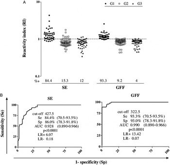
Figure 1A shows ELISA reactivity of the tested serum samples using both antigenic preparations (SE and GFF). Positivity rates for SE and GFF were, respectively: 84·4% (38/45) and 93·3% (42/45) at G1; 15·3% (10/65) and 9·2% (6/65) at G2 and 12% (6/50) and 4% (2/50) at G3. Cross-reactivity in G2 using SE was due to samples from patients infected with hookworm (2/7), S. mansoni (1/4), S. stercoralis (2/8), G. lamblia (2/8), E. histolytica/dispar (1/2), H. nana + E. vermicularis (1/1) and E. granulosus (1/8). When testing GFF cross-reactive samples were from patients with hookworm (1/7), S. stercoralis (1/8), E. histolytica/dispar (2/2), Taenia sp. (1/1) and E. granulosus (1/8).
ELISA using total saline extract (SE) from T. saginata metacestodes and gel filtration fraction (GFF) for detection of IgG anti-Taenia in serum samples from 45 patients with NCC (G1), 65 patients with other parasitic infections (G2), and 50 healthy individuals (G3) RI for each extract were considered positive (RI > 1) (A). Receiver operating characteristic curves (ROC) indicating reaction cut-off point, sensitivity (Se), specificity (Sp), area under curve (AUC), likelihood ratios (LR+ and LR−) and AUC comparison according to Hanley and McNeil (Reference Hanley and McNeil1983), using total SE from T. saginata metacestodes and GFF (B), 95% CI for Se, Sp and AUC are shown in brackets.

GFF reached the best diagnostic parameters (Fig. 1B). Comparing ROC curves, GFF was the most efficient to distinguish patients with NCC (G1) from control groups (G2 and G3) reaching a high AUC value (AUC = 0·990, z = −3·6; P = 0·0003). The LR + values for SE (LR + = 6·07) indicated only a moderate probability of a true positive NCC case, while for GFF this index indicated a high diagnostic value (LR + = 13·42). This efficiency of GFF can be corroborated by analysing LR− value (LR− = 0·07), this parameter virtually exclude the chance of the infection (Jaeschke et al. Reference Jaeschke, Guyatt and Sacket1994).
Electrophoretic profile and Immunoblotting
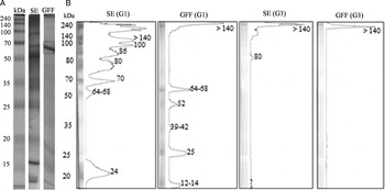
Proteins with molecular weight varying from 64 to 68 kDa, showing high concentration bands, were observed at GFF (Fig. 2A). Considering the promising results obtained with ELISA tests using GFF, immunoblotting was performed and high molecular weight bands (⩾70 kDa) were eliminated for NCC diagnosis in agreement with Barcelos et al. (Reference Barcelos, Moura, Costa, Ferreira and Costa-Cruz2007). Figure 2B showed the immunoblotting reactivity profile when using SE and GFF. For SE with G1 pool, the following immunodominant proteins were recognized: 24, 64–68, 70, 80, 86, 100 and >140 kDa. Observing GFF there was the recognition of the following bands: 12–14, 25, 39–42, 52, 64–68 and >140 kDa. There was no recognition of bands related to NCC diagnosis with G3 pool for both antigens. Special attention should be paid to the 64–68 kDa band, an important marker for NCC diagnosis that clearly appeared at GFF.
Electrophoretic profiles of the total saline extract (SE) from T. saginata metacestodes and gel filtration fraction (GFF) obtained after gel filtration chromatography (SDS–PAGE) (A). Immunoblotting to detect anti-Taenia sp. IgG antibodies in pool of serum samples from patients with NCC (G1 pool) and healthy individuals (G3 pool). Peaks of intensity, indicative of the bands detected, were generated by ImageJ version 1.44. Molecular weight standard in kilodaltons (kDa) (B).

Protein identification
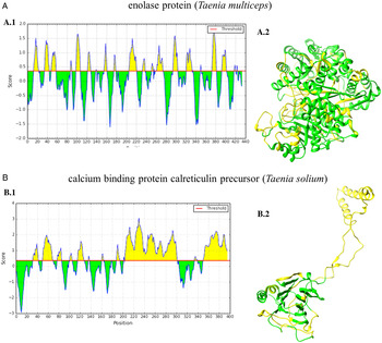
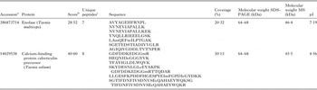
Figure 3A and B shows proteins of diagnostic interest for NCC by MS. In GFF the band with 64–68 kDa (the same molecular weight of a known marker for NCC diagnosis) was selected. Table 1 shows the identified proteins according to score, unique peptides and coverage.
Protein identification: enolase (Taenia multiceps) (A) and calcium-binding protein calreticulin precursor (T. solium) (B) through MS and in silico analysis by predicting linear B-cell epitopes (BepiPred). Amino acid residues are coloured in yellow in the graph when they reach scores above the threshold, where the X-axis correspond to the positions of the residues in the sequence and Y-axis score (A.1 and B.1). Marking of antigenic determinants (yellow) predicted for B-cells. 3D modelling of the proteins showed the B-cell epitopes (yellow) and not predicted (green) (A.2 and B.2).

Protein characterization of fraction obtained by gel filtration on S-100 resin of the saline extract (SE) from T. saginata metacestodes identified by MS and subjected to search in the database for the genus Taenia

pI, isoelectric point.
a Protein Data Bank National Center for Biotechnology Information (NCBI; http://blast.ncbi.nlm.nih.gov/).
b Search platforms attribute scores reflecting similarity between the experimental fragmentation spectrum (measured) and the theoretical spectra of peptides from a given database (Granholm and Käll, Reference Granholm and Käll2011).
c Peptide one is defined as a peptide, regardless of its length, which exists only on a protein of interest a proteome, even this peptide appears more than once in the same protein (Zhao and Lin, Reference Zhao and Lin2010).
Considering the absence of the whole sequence from T. solium enolase and the conserved structure of this protein at Taenia genus we used the estructure of Taenia multiceps to perform in silico analyses for B-cell epitopes prediction (Fig. 3A.1, A.2, B.1 and B.2). Using enolase (T. multiceps) 18 epitopes were predicted. For the calcium binding protein calreticulin precursor (T. solium) 10 epitopes were predicted. B-cell epitopes are shown on three-dimensional (3D) modelling in yellow (Fig. 3A.2 and B.2).
DISCUSSION
The development of a highly specific, sensitive and reliable assay to detect the presence of anti-Taenia antibodies is important to improve the diagnosis of human NCC. Thus, efforts have been done to develop new antigenic fractions and other antigenic sources as heterologous ones.
Taenia saginata heterologous antigens showed to be useful in NCC diagnosis through ELISA and immunoblotting due to the cross-reactivity with T. solium antigens confirming the presence of common antigenic determinants (Espíndola et al. Reference Espíndola, Iha, Fernandes, Takayanagui, Machado, Livramento, Mendes-Maia, Peralta and Vaz2005). Taenia saginata metacestodes can be obtained in considerable quantities, due to the habitual diet in many countries, mainly based on beef consumption (Morakote et al. Reference Morakote, Nawacharoen, Sukonthasun, Thammasonthi and Khamboonruang1992; Oliveira et al. Reference Oliveira, Machado, Cabral and Costa-Cruz2007, Reference Oliveira, Machado, Gonçalves-Pires, Moura and Costa-Cruz2009).
Immunological tests are considered important methods to confirm clinical findings and to give diagnostic help when clinical manifestations are non-specific, as happens in NCC. ELISA is a valuable tool to detect many parasitic infections and provides the possibility for the simultaneous processing of many samples, reducing the cost for the analyses. This test is also quantitative and its results are not subjected to individual interpretation. However, appropriate antigens, easy producible, and highly sensitive and specific are required (Zimic et al. Reference Zimic, Pajuelo, Rueda, López, Arana, Castillo, Calderón, Rodriguez, Sheen, Vinetz, Gonzales, García and Gilman2009).
Cross-reactions between crude antigens and epitopes from other helminths are the major challenge in the diagnostic tests. A simple fractionation is a viable tool to increase positive reactions minimizing cross-reactivity (Gonçalves et al. Reference Gonçalves, Machado, Oliveira, Rezende, Mineo and Costa-Cruz2010; Nunes et al. Reference Nunes, Ribeiro, Manhani and Costa-Cruz2010, Reference Nunes, Gonzaga, Ribeiro, Cunha-Junior and Costa-Cruz2013), maintaining a certain antigenic complexity that leads to an accurate diagnosis, important for patient management, understanding the epidemiology of the disease and establishing preventive measures. Our study shows that even using a simple chromatography procedure we can easily fractionate SE obtaining an antigenic fraction enriched with proteins with diagnostic potential that can be readily used improving specificity and sensitivity ELISA indexes to detect NCC in serum samples. Several studies have demonstrated the usefulness of the partial fractionation to obtain antigenic fractions with increased diagnostic parameters using other chromatographic approaches. Fractionation techniques [detergent Triton X-114 (Machado et al. Reference Machado, Santiago, Mineo and Costa-Cruz2007; Gonçalves et al. Reference Gonçalves, Machado, Oliveira, Rezende, Mineo and Costa-Cruz2010), affinity for concanavalin A and jacalin (Nunes et al. Reference Nunes, Ribeiro, Manhani and Costa-Cruz2010; Oliveira et al. Reference Oliveira, Machado, Mineo and Costa-Cruz2010), ion-exchange chromatography with carboxymethyl sepharose and diethylaminoethyl sepharose (DEAE)] (Nunes et al. Reference Nunes, Gonzaga, Ribeiro, Cunha-Junior and Costa-Cruz2013; Ribeiro et al. Reference Ribeiro, Nunes, Gonzaga, Cunha-Júnior and Costa-Cruz2014) lead to antigenic fractions with high potential in NCC immunodiagnosis using T. saginata or T. solium metacestodes.
A previous study successfully used the homologous antigen from T. solium metacestodes to detect porcine cysticercosis, the band of 14 kDa was obtained from crude cyst fluid of T. solium metacestodes by two-step chromatography: the first step gel filtration (Sephacryl S-300) and the second using anion exchange column (Mono Q HR 5/5) on HPLC (Assana et al. Reference Assana, Kanobana, Tume, Zoli, Nguekam, Geerts, Berkvens and Dorny2007). In our study, GFF was obtained after a single-step chromatography and this fraction showed higher diagnostic parameters if compared with SE. The major advantages of using gel filtration chromatography are the minimal cost, simple and easy extraction and high efficiency. The GFF revealed antigenic components with 64–68 kDa, other studies showed the importance of these antigenic markers. Shiguekawa et al. (Reference Shiguekawa, Mineo, Moura and Costa-Cruz2000) found that the 47, 52, 64–68 kDa were immunodominant when serum samples were tested in immunoblotting. Using another chromatography technique and the heterologous antigen from T. saginata metacestodes, Nunes et al. (Reference Nunes, Gonzaga, Ribeiro, Cunha-Junior and Costa-Cruz2013) obtained the DEAE S2 fraction with bands with 39 to 42, 47–52 and 64–68 kDa with potential applicability in active NCC diagnosis (Nunes et al. Reference Nunes, Gonzaga, Ribeiro, Cunha-Junior and Costa-Cruz2013); however, these proteins were not identified yet.
Recent advances in MS and proteomics have shown promising results for the diagnosis of parasitic infections (Vasoo and Pritt, Reference Vasoo and Pritt2013), since these tools are helpful for antigen identification and characterization supporting the development of accurate diagnostic tests. The prediction of B-cell epitopes was performed at the structure of the identified proteins (enolase and calreticulin), and exposed regions of these molecules could be antibody targets.
Enolase is a multifunctional glycolytic enzyme, that catalyse the reversible dehydration of 2-phospho-D-glycerate to phosphoenolpyruvate and plasminogen receptor on the surface of several pathogens (Ramajo-Hernández et al. Reference Ramajo-Hernández, Pérez-Sánchez, Ramajo-Martín and Oleaga2007; Cayir et al. Reference Cayir, Erdemir, Ozkan, Topuzogullari, Bolat, Akat and Turgut-Balik2014). Furthermore, the protein can also induce host-protective immunity and is considered a potential vaccine candidate being evaluated in several helminths (Yang et al. Reference Yang, Qiu, Xia, Yao, Fu, Yuan, Feng and Lin2010; Chen et al. Reference Chen, Yuan, Xu, Zhou, Zhang, Zhang, Wang, Yan, Lin and Zhu2012). Calreticulin is a highly conserved multifunctional protein found mainly at the lumen of the endoplasmic reticulum in almost all eukaryotic cells and is involved in the homoeostasis of intracellular Ca2+, regulating diverse cellular functions, such as secretion, contraction–relaxation, adhesion, protein synthesis, differentiation, organogenesis and gene expression. This protein is also important regulating T. solium development at the tissues, especially during germ cell development and embryogenesis (Mendlovic et al. Reference Mendlovic, Ostoa-Saloma, Solís, Martínez-Ocaña, Flisser and Laclette2004).
In conclusion, we developed a rapid, simple, sensitive and specific fraction to detect specific antibodies against Taenia sp. infection. Proteins that were identified here are composed of immunodominant peptides suggesting the potential applicability in human NCC diagnosis with implications in clinical practice and as screening tool.
ACKNOWLEDGEMENTS
Dra. Rosângela Rodrigues e Silva of the Instituto Oswaldo Cruz, Rio de Janeiro state, Brazil and Dr. Henrique Bunselmeyer Ferreira of the Universidade Federal do Rio Grande do Sul, Rio Grande do Sul state, Brazil, by donation of serum samples from patients infected with Echinococcus granulosus.
FINANCIAL SUPPORT
This research was supported by the Fundação Coordenação de Aperfeiçoamento de Pessoal de Nível Superior (CAPES-3200601004 M-8) (to D. S. N.), Fundação de Amparo à Pesquisa do Estado de Minas Gerais (FAPEMIG-CBB-PPM-00396-13) (to J. M. C. C), Rede de Pesquisa em Doenças Infecciosas Humanas e Animais do Estado de Minas Gerais (CDS-RED-00013-14) (to J. M. C. C and J. P. C. J), Conselho Nacional de Desenvolvimento Científico e Tecnológico (CNPq-302426/2012-4) (to J. M. C. C) and by the Universidade Federal de Uberlândia (UFU), (to D. S. N, H. T. G., V. S. R., J. P. C. J. and J. M. C. C.), Brazil.
CONFLICT OF INTEREST
The authors declare to have no conflict of interest.