





Introduction
Protozoa of the genus Cryptosporidium are epicellular parasites that infect the gastrointestinal and respiratory tracts of a wide range of vertebrates (O’Donoghue, Reference O’Donoghue1995). Currently, 48 species of Cryptosporidium and a large number of genotypes that differ in tissue and host specificity have been validly described (Zikmundová et al., Reference Zikmundová, Holubová, Fenclová, Sak, Rost, Gomulkiewicz, Konečný, McEvoy, Xiao and Kváč2025). Within the genus Cryptosporidium, we distinguish 2 phylogenetic groups: species that infect the stomach of their hosts and species that infect the intestine, whereby within this group we can further distinguish species and genotypes that parasitize the small intestine, caecum, large intestine or cloaca (Fayer and Xiao, Reference Fayer and Xiao1997). In terms of host specificity, most species and genotypes are narrowly host-specific, infecting only a limited number of phylogenetically closely related hosts (Ryan et al., Reference Ryan, Fayer and Xiao2014; Feng et al., Reference Feng, Ryan and Xiao2018). The second, less numerous group, comprising mainly C. parvum, the species with the widest host range, as well as C. ubiquitum and C. baileyi, represents cryptosporidia infectious to a wide range of hosts across different orders of mammals or birds.
This group also includes C. muris, a species that primarily parasitizes rodents (Iseki et al., Reference Iseki, Maekawa, Moriya, Uni and Takada1989; Aydin and Ozkul, Reference Aydin and Ozkul1996; Chalmers et al., Reference Chalmers, Sturdee, Bull, Miller and Wright1997; Torres et al., Reference Torres, Gracenea, Gomez, Arrizabalaga and Gonzalez-moreno2000), but which has also been detected in humans, wood mice, bank voles, Syrian hamsters, desert hamsters, squirrels, Siberian chipmunks, marsupials, Bactrian camels, reticulated giraffe, mountain goats, lambs, rabbits, ringed seals, hyraxes, cynomolgus monkeys, dogs and cats (Iseki et al., Reference Iseki, Maekawa, Moriya, Uni and Takada1989; Aydin and Ozkul, Reference Aydin and Ozkul1996; Katsumata et al., Reference Katsumata, Hosea, Ranuh, Uga, Yanagi and Kohno2000; Hůrková et al., Reference Hůrková, Hajdušek and Modrý2003; Kváč et al., Reference Kváč, Havrdová, Hlasková, Daňková, Kanděra, Ježková, Vítovec, Sak, Ortega, Xiao, Modrý, Chelladurai, Prantlová and McEvoy2016).
Cats are among the hosts in which infection has been documented under both experimental and natural conditions. Iseki et al. demonstrated that C. muris is infectious to cats and causes long-lasting and massive infection (Iseki et al., Reference Iseki, Maekawa, Moriya, Uni and Takada1989). Although it could be assumed that C. muris infection in cats should be common, given that rodents – the natural hosts of C. muris (Tyzzer, Reference Tyzzer1910; Xiao et al., Reference Xiao, Fayer, Ryan and Upton2004) – are their typical prey, the number of described natural cases of infection in cats is very low (Table 1). To date, the detailed biological characteristics of C. muris isolates obtained from cats remain unknown. Most studies have determined the species based on partial sequences of the small subunit (SSU) rRNA (Ryan et al., Reference Ryan, Feng, Fayer and Xiao2021). Only Pavlásek and Ryan (Reference Pavlásek and Ryan2007) demonstrated the infectivity of an isolate from a cat to mice, but without further details. This study aims to describe the course of chronic, natural infection of cats with Cryptosporidium muris, including molecular genotyping of the isolate and a description of its biological properties, including oocyst morphology, infectivity for model hosts and oocyst viability.
Overview of published reports of Cryptosporidium muris infection in cats

Table 1 Long description
Columns list year of detection, type of infection, number of cats examined, diagnostic method, evidence of oocyst infectivity, sampling strategy, and reference. In 1989, an experimental infection examined 3 cats, used microscopy, reported infectivity evidence as yes in mice, and used repeated sampling, reference Iseki et al., 1989. In 2006, a natural infection examined 1 cat, used PCR, reported no infectivity evidence, and used one shot sampling, reference Santín et al., 2006. In 2007, the type of infection cell contains 1, the number of cats examined cell is blank, diagnostic method lists microscopy and PCR, infectivity evidence is yes in mice, and sampling strategy is repeated sampling, reference Pavlásek and Ryan, 2007. In 2011, the type of infection cell contains 1, the number of cats examined cell is blank, diagnostic method lists histology and PCR, infectivity evidence is no, and sampling strategy is repeated sampling, reference FitzGerald et al., 2011. In 2015 and 2025, the type of infection cell contains 1, the number of cats examined cell is blank, diagnostic method is PCR, infectivity evidence is no, and sampling strategy is one shot sampling, references Yang et al., 2015 and Barrios et al., 2025.
Materials and methods
Source and collection of specimens
As part of routine parasitological screening at a veterinary clinic, 5 domestic cats (Felis catus f. domestica) aged 1–6 years, originating from a single breeder, were examined. Routine screening included coprological examination by flotation with Sheather’s sugar solution to detect helminth eggs and protozoan oocysts, and formalin–ethyl acetate sedimentation to detect Giardia cysts. In cases of suspected cryptosporidiosis or giardiasis, a commercially available immunochromatographic rapid test for Cryptosporidium and Giardia (R-Biopharm, Darmstadt, Germany) was also used. During this routine screening, Cryptosporidium spp. oocysts morphologically consistent with C. muris were detected in 1 cat. This cat subsequently underwent abdominal ultrasonography focused on the gastrointestinal tract and gastroscopy of the stomach. Faecal samples and gastric biopsy specimens were submitted for further parasitological examination and genotyping to the Institute of Parasitology, Biology Centre of the Czech Academy of Sciences (BC CAS), Czech Republic.
After species identification as C. muris, all cats were monitored regularly for the presence of oocysts and C. muris–specific DNA. For this targeted follow-up monitoring, faecal smears were stained with aniline–carbol–methyl violet, according to previously described protocol (Miláček and Vítovec, Reference Miláček and Vítovec1985). Subsequently, oocysts were additionally examined to verify and compare staining characteristics using a modified Ziehl–Neelsen method (Henriksen and Pohlenz, Reference Henriksen and Pohlenz1981) and auramine–phenol staining (Casemore, Reference Casemore1991). In addition, FITC-conjugated Cryptosporidium-specific antibodies targeting the oocyst wall were used (IFA; Cryptosporidium IF Test, Cryptocel, Cellabs Pty Ltd., Brookvale, Australia).
Molecular analyses
Total genomic DNA (gDNA) was extracted from 200 mg of faeces using the Exgene™ Stool DNA mini kit (GeneAll Biotechnology Co Ltd., Seoul, Korea). The purified DNA was stored at –20°C until further processing. Partial sequences of the genes encoding the SSU rRNA, actin, the Cryptosporidium oocyst wall protein (COWP), the 70-kDa heat shock protein (HSP70) and the thrombospondin-related adhesive protein of Cryptosporidium-1 (TRAP-C1) were amplified using previously published nested PCR protocols and primer sets (Xiao et al., Reference Xiao, Escalante, Yang, Sulaiman, Escalante, Montali, Fayer and Lal1999; Pedraza-diaz et al., Reference Pedraza-diaz, Amar, Nichols and McLauchlin2001; Sulaiman et al., Reference Sulaiman, Lal and Xiao2002; Jiang et al., Reference Jiang, Alderisio and Xiao2005; Kváč et al., Reference Kváč, Havrdová, Hlasková, Daňková, Kanděra, Ježková, Vítovec, Sak, Ortega, Xiao, Modrý, Chelladurai, Prantlová and McEvoy2016). Both primary and secondary PCR reactions were performed in 50 μL reaction containing 2 μL of gDNA template (or 2 μL of primary template in case of secondary reaction), 2.5 U DreamTaq Green DNA Polymerase (Thermo Fisher Scientific, Waltham, MA, USA), 1 × PCR buffer, 200 nM of each primer, 200 μM of each dNTP, 2 μL of non-acetylated bovine serum albumin (BSA; 10 mg/mL; New England Biolabs, Beverly, MA, USA) and molecular grade water. gDNA from Cryptosporidium-negative faecal samples were used as negative controls and C. serpentis DNA as a positive control. PCR products were separated on 2% agarose gels stained with ethidium bromide, purified using the Monarch ® DNA Gel Extraction Kit (New England Biolabs, Ipswich, MA, USA) and sequenced in a commercial laboratory (SeqMe, s.r.o., Dobřiš, Czech Republic) using secondary primers. The identity of the isolates was verified by BLAST analysis using the GenBank database.
qPCR-infection intensity
Cryptosporidium muris infection was quantified using a TaqMan-based qPCR assay and previously published protocols with the following modification (Hill et al., Reference Hill, Kahler, Jothikumar, Johnson, Hahn and Cromeans2007). Briefly, each 20 µL reaction contained 2 µL gDNA, 400 nM forward (5´-ATGACGGGTAACGGGGAAT-3´) and reverse primers (5´-CCCTGTGTCAGGATTGGGTA-3´), 200 nM 5´ FAM and 3´BHQ1-labelled TaqMan probe (5´-CGCGCCTGCTGCCTTCCTTAGATG-3´) and 10 µL Luna Universal Probe qPCR master Mix (New England BioLabs Inc., Ipswich, MA, USA). A standard (calibration) curve was constructed using serial dilution of DNA isolated from 107 Cryptosporidium proliferans oocysts. Infection intensity was expressed as the number of oocysts per gram (OPG) of faeces.
Health monitoring of cats
Cats underwent baseline clinical laboratory screening that included haematological and biochemical evaluation of peripheral blood. A complete blood count with leukocyte differential was performed, reporting total leukocytes (WBC) and relative and absolute leukocyte subsets (neutrophils, lymphocytes, monocytes, eosinophils, basophils). Erythrocyte variables and indices (RBC, haemoglobin, haematocrit, MCV, MCH, MCHC, RDW) and platelet variables and indices (PLT, MPV, PDW, PCT, P-LCR/P-LCC) were also recorded, including the presence/quantity of nucleated red blood cells (NRBCs) when reported by the analyser. Serum/plasma biochemistry comprised total protein, albumin, total bilirubin, AST (GOT), ALT (GPT) and LDH activities, urea (BUN), glucose and creatinine. In addition, total thyroxine (T4; v-T4) was measured. All results were interpreted against laboratory-specific reference intervals. In addition, cats were screened for major feline infectious diseases, including feline leukaemia virus, feline immunodeficiency virus, feline coronavirus, presence of gastrointestinal parasites and Toxoplasma gondii antibodies. All animals were clinically monitored daily for activity level, noticeable body-weight loss, appetite and the frequency and consistency of faeces.
Histological examination
Gastric biopsy samples were fixed in 10% neutral buffered formalin and subsequently processed using standard histological procedures. Histological sections (5 μm thick) were stained with haematoxylin and eosin and periodic acid-Schiff (PAS) and examined under 100–400 × magnification using an Olympus IX70 microscope (Kváč and Vítovec, Reference Kváč and Vítovec2003).
Oocyst morphology
For morphological analysis and experimental infection, C. muris oocysts were purified from feline faeces by sucrose and caesium chloride gradient centrifugation (Arrowood and Sterling, Reference Arrowood and Sterling1987; Kilani and Sekla, Reference Kilani and Sekla1987) and stored in dH2O without antibiotics and antimycotics at 4°C. Morphometric measurements were performed using differential interference contrast (DIC) microscopy at 1000 × magnification (Olympus BX51, Olympus Corporation, Tokyo, Japan). Photomicrographs used for morphometric analysis were evaluated with Olympus CellSens Entry 2.1 software in combination with an Olympus DP73 digital colour camera (Olympus Corporation). The length and width of 100 oocysts were measured, and a shape index was calculated.
Experimental infection and oocyst infectivity
Oocysts of C. muris RN66 (Waterborne Inc.; New Orleans, LA, USA), a laboratory reference isolate originally obtained from Rattus norvegicus in Japan (Iseki, Reference Iseki1986), and C. proliferans (Biology Centre of the Czech Academy of Sciences (CAS), Czech Republic), purified and stored similarly as oocysts of C. muris CAT21, the feline isolate characterized in the present study, were used as controls. Infectivity for BALB/c mice and the course of infection were evaluated using oocysts immediately after purification and those stored for no more than 5 days. In addition, aliquots of oocysts of individual species and strains were stored for 6 months, and their viability was assessed monthly using the FDA/PI assay (Jones and Senft, Reference Jones and Senft1985), together with testing of infectivity in model hosts and monitoring of natural excystation and oocyst loss (see below).
For each species/strain of Cryptosporidium, six 6-week-old BALB/c mice (3 males and 3 females) were inoculated with 1 000 000 oocysts of the respective isolate suspended in 200 µL of water. Six age- and sex-matched control mice (3 males and 3 females) were inoculated with 200 µL of water only. From the 5th day post-infection (DPI), individual faecal samples were collected and examined for oocyst shedding and Cryptosporidium-specific DNA. Each sample was processed for microscopy (aniline–carbol–methyl violet staining) and qPCR (see above). In groups infected with oocysts stored for ≤5 days after isolation, mice were monitored for 30 days, and the individual infection intensity was evaluated. In mice infected with oocysts stored for 1–6 months, only the presence of infection was assessed at 20 DPI. If no oocysts or specific DNA were detected within 20 DPI in mice infected with 1–6-month-old oocysts, the animals were necropsied, and stomach tissue was analysed for the presence of Cryptosporidium-specific DNA/qPCR. From each animal that tested positive for Cryptosporidium during the experiment, genotyping of the isolate was performed to confirm the identity with the inoculum.
Animal care
Mice were housed under standard conditions in separate plastic cages in the animal facility of the Institute of Parasitology, Biology Centre of the Czech Academy of Sciences (BC CAS), v.v.i., in České Budějovice, Czech Republic. The light–dark cycle was set to 12 h light and 12 h dark at 22°C and 50–55% relative humidity. The mice were fed a commercial pelleted rodent diet (Altromin 1314) and provided water ad libitum. All samples were collected by animal breeders in accordance with Czech legislation (Act No. 246/1992 Coll. on the Protection of Animals Against Cruelty) and relevant European regulations. The study design was reviewed and approved by the Ethics Committee of the Biology Centre of the Czech Academy of Sciences (protocol No. 73-2024-P).
Flow cytometry analysis
To assess temporal changes in C. muris CAT21 oocyst counts (degradation), 20 mL of purified oocysts (2 × 10⁶ oocysts/mL) was aliquoted into 50 mL screw-cap tube. The same procedure was performed for C. proliferans and C. muris RN66 oocysts used as controls. Suspensions were stored at 4°C and analysed on days 0, 15, 30, 45 and 60. Prior to acquisition, oocyst suspensions were vortexed thoroughly and 450 μL (0.45 × 10⁶ oocysts) was withdrawn from each stock in 5 technical replicates. Each replicate was spiked with 50 μL fluorescent counting beads (Flow-Count Fluorospheres; Beckman Coulter, USA). Absolute oocyst numbers were determined by single-platform bead-based flow cytometry on a BD FACSCanto II using BD FACSDiva software. Beads were identified as a discrete high-fluorescence population and used for normalization of event counts, as described for bead-enhanced absolute quantitation. Oocysts were enumerated without antibody staining using a morphology-driven gating strategy based on forward and side scatter (FSC-A/SSC-A) to discriminate oocysts from residual debris, followed by a second refinement gate to exclude remaining contaminants, consistent with published protocols for unstained Cryptosporidium oocyst quantification. Doublets were excluded using standard area-versus-height (or width) discrimination (Sonzogni-desautels et al., Reference Sonzogni-desautels, Di Lenardo, Renteria, Gascon, Geary and Ndao2019). Absolute oocyst concentration was calculated from the oocyst-to-bead event ratio and the manufacturer-assigned bead concentration (or bead number per test volume), and results were expressed relative to day 0 and to the control species to estimate the proportion of degraded oocysts over time.
Statistical analyses
Differences in oocyst size of C. muris CAT21 obtained from different hosts, as well as differences between oocysts of C. muris CAT21 and those of other C. muris isolates and C. proliferans, were tested using Hotelling’s multivariate 2-sample t-test implemented in the ICSNP (Tools for Multivariate Nonparametrics) package in R version 4.2.2. The total oocyst output was quantified as the area under the oocyst shedding curve (AUC), calculated for each animal using the trapezoidal rule based on daily oocyst counts (OPG) over the 30-day observation period. AUC values were compared among isolates (C. muris CAT21, C. muris RN66 and C. proliferans) using one-way analysis of variance (ANOVA). When significant differences were detected, pairwise comparisons were performed using Tukey’s honestly significant difference (HSD) post hoc test. All statistical analyses were performed in R version 4.2.2 (R Core Team, 2019). A P-value < 0.05 was considered statistically significant.
Results
Detection, genotyping and quantification of C. muris in cats
During routine parasitological examination, oocysts of Cryptosporidium sp., morphologically consistent with gastric species of mammalian cryptosporidia, were detected in 1 of 5 cats originating from the same breeder (cat no. 4). At the initial examination, the infection intensity was 12 800 OPG, and subsequent SSU molecular analysis confirmed the presence of C. muris DNA (100% identical to sequence GenBank Acc. No. AB089284). All animals were subsequently re-examined. With the exception of cat no. 4, in which C. muris infection was confirmed at the following sampling (35 200 OPG), all other cats were negative for Cryptosporidium. Owing to the repeated faecal positivity and the high infection intensity, the cats were monitored regularly. The isolate was designated C. muris CAT21. During the 120-day observation period, regular shedding of C. muris CAT21 was recorded in cat no. 4, with a maximum infection intensity of 371 200 OPG (Figure 1). This cat shed oocysts for 97 days following the first detection. No other cat living in the same household was positive for Cryptosporidium. Molecular analyses of the SSU (AB089284), actin (XM002140165), HSP70 (XM002140816), COWP (DQ060430) and TRAP-C1 (XM002142580) genes demonstrated 100% sequence identity of the CAT21 isolate with C. muris RN66.
Dynamics of C. muris CAT21 oocyst shedding in a naturally infected domestic cat during a 120-day observation period.

Health status
Throughout the observation period, no symptoms of clinical cryptosporidiosis were detected in the naturally infected domestic cat. The animal did not suffer from diarrhoea or vomiting, and no other gastrointestinal problems were observed. The animal ate normally, and its weight was appropriate for its age, sex, feed intake and activity level. Similar to the cat, we did not observe any clinical symptoms associated with C. muris CAT21 infection in experimentally infected BALB/c mice. In the baseline laboratory screening, most haematological and biochemical variables of the cat were within the respective laboratory reference intervals and were not indicative of marked anaemia, thrombocytopenia, or overt renal/hepatocellular dysfunction. Total leukocyte counts ranged from 15.88 to 22.11 × 10⁹/L, with mild leukocytosis and neutrophilia observed in 1 sample (WBC 22.11 × 10⁹/L; NEU# 14.924 × 10⁹/L). Erythrocyte parameters were generally stable (e.g. HCT 40.6–51.2%, with a single mildly increased value of 51.2%), and platelet counts were within the provided reference range (PLT 225–416 × 109/L). NRBCs were flagged by the analyser in several samples (NRBC# 7.77–44.30 × 10⁹/L) and were interpreted in conjunction with the overall haematological profile. Biochemistry showed values consistent with adequate renal function (creatinine 53–88 µmol/L; urea 8.5–13.3 mmol/L) and no relevant hyperbilirubinaemia (total bilirubin 5–6 µmol/L). Total thyroxine concentrations were within the laboratory reference range (v-T4 22.86–28.95 nmol/L; reference 11.6–47.6 nmol/L).
Gastroscopy
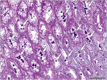
Gastroscopy performed on day 45 of the observation period revealed areas of evident gastric mucosal oedema localized predominantly in the pyloric antrum. Tissue samples were collected from these regions for histological examination, which demonstrated the presence of developmental stages of Cryptosporidium (Figure 2).
Histological section of a gastric biopsy demonstrating developmental stages of C. muris CAT21 in a naturally infected domestic cat (PAS stain, 400 × magnification). Arrows: Developmental stages of C. muris CAT21.

Oocyst morphology
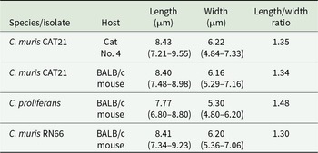
Oocysts of C. muris CAT21 from the naturally infected cat did not differ morphometrically from those obtained from experimentally infected BALB/c mice (T 2 = 4.32, df1 = 2, df2 = 24.62, P = 0.2208; Table 2). Statistical analysis showed that C. muris CAT21 oocysts were not distinguishable in size from C. muris RN66 oocysts (T 2 = 12.32, df1 = 2, df2 = 25.63, P = 0.1907), but they were larger than C. proliferans oocysts (T 2 = 32.56, df1 = 2, df2 = 32.27, P < 0.0001). Oocysts of C. muris isolate CAT21 obtained from a naturally infected domestic cat and from experimentally infected model hosts were detectable in faecal smears using standard staining techniques for the detection of Cryptosporidium (Figure 3).
Oocysts of Cryptosporidium muris CAT21 (A) in differential interference contrast microscopy, (B) stained by aniline–carbol–methyl violet staining, (C) stained by Ziehl–Neelsen staining, (D) stained by auramine-phenol staining and (E) labelled with anti-Cryptosporidium FITC-conjugated. Bars = 10 μm.

Morphometric characteristics of oocysts of C. muris CAT21, C. proliferans and C. muris RN66 isolated from different hosts

Table 2 Long description
Morphometric measurements are listed for oocysts by species or isolate and host, with columns for length in micrometers, width in micrometers, and length to width ratio. Cryptosporidium muris CAT21 from Cat No. 4 has length 8.43 with range 7.21 to 9.55, width 6.22 with range 4.84 to 7.33, and ratio 1.35. Cryptosporidium muris CAT21 from BALB/c mouse has length 8.40 with range 7.48 to 8.98, width 6.16 with range 5.29 to 7.16, and ratio 1.34. Cryptosporidium proliferans from BALB/c mouse has length 7.77 with range 6.80 to 8.80, width 5.30 with range 4.80 to 6.20, and ratio 1.48. Cryptosporidium muris RN66 from BALB/c mouse has length 8.41 with range 7.34 to 9.23, width 6.20 with range 5.36 to 7.06, and ratio 1.30.
Infectivity and oocyst shedding intensity in experimental hosts
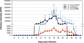
Oocyst shedding differed among the tested isolates (Figure 4). The onset of shedding occurred earliest in C. muris RN66 (day 9 post-infection), followed by C. proliferans (day 10), whereas C. muris CAT21 showed the longest prepatent period, with the first detection on day 12 post-infection. The duration of oocyst excretion also varied among isolates. Shedding persisted until days 26–28 in C. muris RN66, days 25–27 in C. muris CAT21 and days 22–23 in C. proliferans. Remarkable differences were observed in shedding intensity. The highest oocyst output was recorded in C. muris RN66, reaching a peak of 851 200 OPG. Cryptosporidium proliferans showed a lower maximum intensity 493 333 OPG, while C. muris CAT21 produced the lowest oocyst counts with a peak 3156 OPG. The total oocyst output (AUC) differed significantly among isolates (P = 0.00014). Cryptosporidium muris RN66 exhibited the highest total oocyst production (4 080 000 OPG/days), followed by C. proliferans (2 770 000 OPG/days), whereas C. muris CAT21 showed markedly reduced oocyst output (32 300 OPG/days).
Severity of infection, determined by quantification of oocyst excretion expressed as oocysts per gram (OPG) of faeces, in BALB/c mice infected with C. muris CAT21, C. muris RN66, or C. proliferans at a dose of 1 000 000 oocysts per animal. The course represents the mean values derived from 6 infected animals for each isolate.

Figure 4 Long description
The y-axis is labeled, Infection intensity (OPG). The x-axis is labeled, Days post infection. The x-axis runs from 0 to 30 with tick marks every 2. The y-axis is split into two ranges. The lower range runs from 0 to 4000 with tick marks every 1000. The upper range runs from 0 to 1 400 000 with tick marks every 200 000. A legend at the upper right lists three series: C. muris CAT21, C. proliferans and C. muris RN66. Cryptosporidium muris CAT21 is shown with circular markers connected by a line. Values are at 0 from day 0 through day 11. At day 12 the value is about 500. Day 13 about 1 000. Day 14 about 1500. Day 15 about 1 500. Day 16 about 1 700. Day 17 about 1 700. Day 18 about 2 000. Day 19 about 3000. Day 20 about 2 000. Day 21 about 1 500. Day 22 about 1 500. Day 23 about 2 500. Day 24 about 1 500. Day 25 about 2 000. Day 26 about 1 000. Day 27 about 500. Day 28 about 0. Day 29 about 0. Day 30 about 0. Cryptosporidium proliferans is shown with circular markers connected by a line. Values are at 0 from day 0 through day 9. At day 10 the value is about 100 000. Day 11 about 100 000. Day 12 about 150 000. Day 13 about 200 000. Day 14 about 300 000. Day 15 about 200 000. Day 16 about 350 000. Day 17 about 400 000. Day 18 about 450 000. Day 19 about 500 000. Day 20 about 100 000. Day 21 about 100 000. Day 22 about 100 000. Day 23 about 120 000. Day 24 about 0. Day 25 about 0. Day 26 about 0. Day 27 about 0. Day 28 about 0. Day 29 about 0. Day 30 about 0. Cryptosporidium muris RN66 is shown with downward triangle markers connected by a line. Values are at 0 from day 0 through day 11. At day 12 the value is about 100 000. Day 13 about 100 000. Day 14 about 200 000. Day 15 about 300 000. Day 16 about 350 000. Day 17 about 400 000. Day 18 about 850 000. Day 19 about 350 000. Day 20 about 400 000. Day 21 about 350 000. Day 22 about 350 000. Day 23 about 250 000. Day 24 about 200 000. Day 25 about 200 000. Day 26 about 150 000. Day 27 about 150 000. Day 28 about 0. Day 29 about 0. Day 30 about 0.
Degradation of oocysts
A significant difference in the loss of oocyst integrity was observed for C. muris CAT21 compared with C. proliferans and C. muris RN66. No significant difference in oocyst integrity was detected between C. proliferans and C. muris RN66 during the 60-day observation period, with a decrease in oocyst numbers of approximately 35–40%. In contrast, C. muris CAT21 showed a marked decline in oocyst numbers during storage already after 15 days (−20%), followed by a sharp decrease by day 30 (−90%; Figure 5).
Degradation of oocysts of C. muris CAT21, C. muris RN66 and C. proliferans stored in distilled water at 4°C over a 60-day period, expressed relative to the values measured on day 0. Statistical significance was evaluated as follows: P < 0.001 (***).

Figure 5 Long description
The bar graph has a legend with three series, C. proliferans, C. muris RN66 and C. muris CAT21. The x axis label is Storage period left parenthesis days right parenthesis, with tick labels 0, 15, 30, 45, 60. The y axis label is Reduction in oocyst count left parenthesis percent right parenthesis, with tick labels 0, 20, 40, 60, 80, 100. At 0 days, C. proliferans 100 percent, C. muris RN66 100 percent, C. muris CAT21 100 percent. At 15 days, C. proliferans about 95 percent, C. muris RN66 about 90 percent, C. muris CAT21 about 80 percent. A bracket above the 15 day group shows * * asterisk. At 30 days, C. proliferans about 80 percent, C. muris RN66 about 75 percent, C. muris CAT21 about 12 percent. Brackets above the 30 day group show * * asterisk. At 45 days, C. proliferans about 65 percent, C. muris RN66 about 70 percent, C. muris CAT21 about 12 percent. Brackets above the 45 day group show * * asterisk. At 60 days, C. proliferans about 60 percent, C. muris RN66 about 62 percent, C. muris CAT21 about 8 percent. Brackets above the 60 day group show * * asterisk.
Discussion
In this study, we describe a natural infection of a domestic cat caused by C. muris CAT21, manifested by a relatively high intensity of infection for more than 90 days, but with production of oocysts displaying phenotypic properties different from those expected for this Cryptosporidium species. The study therefore does not represent merely another host record, but primarily a biologically unusual interaction between a parasite and a non-specific host, suggesting an altered course of parasite development.
The typical hosts of C. muris are rodents, especially mice and rats, in which the parasite produces stable infections characterized by relatively high oocyst shedding and a patent period usually lasting several weeks, with typical tissue localization in the gastric glands (Iseki et al., Reference Iseki, Maekawa, Moriya, Uni and Takada1989; Kváč et al., Reference Kváč, Kodádková, Sak, Květoňová, Jalovecká, Rost and Salát2011). Although C. muris has also been repeatedly recorded in other mammalian hosts, including humans, non-human primates, other rodents, ruminants, rabbits, seals, dogs or cats, described infections outside rodent hosts are rare and the biological properties of such isolates remain poorly explored (Iseki et al., Reference Iseki, Maekawa, Moriya, Uni and Takada1989; Gatei et al., Reference Gatei, Ashford, Beeching, Kamwati, Greensill and Hart2002; Warren et al., Reference Warren, Swan, Morgan-Ryan, Friend and Elliot2003; Pavlásek and Ryan, Reference Pavlásek and Ryan2007; Lupo et al., Reference Lupo, Langer-curry, Robinson, Okhuysen and Chappell2008; Feng, Reference Feng2010; Kodádková et al., Reference Kodádková, Kváč, Ditrich, Sak and Xiao2010; Kváč et al., Reference Kváč, Havrdová, Hlasková, Daňková, Kanděra, Ježková, Vítovec, Sak, Ortega, Xiao, Modrý, Chelladurai, Prantlová and McEvoy2016).
Iseki et al. (Reference Iseki, Maekawa, Moriya, Uni and Takada1989) experimentally demonstrated that cats are susceptible to C. muris infection. However, only a few cases of natural detection of oocysts or specific C. muris DNA in cats have been described to date (Table 1). Only in 3 of them, an active infection can be stated with certainty, based either on histological findings in a gastric biopsy or on long-term monitoring of parasite shedding in faeces (Iseki et al., Reference Iseki, Maekawa, Moriya, Uni and Takada1989; Pavlásek and Ryan, Reference Pavlásek and Ryan2007; FitzGerald et al., Reference FitzGerald, Bennett, Ng, Nicholls, James, Elliot, Slaven and Ryan2011), which excludes mechanical transport of oocysts from food (infected rodents). Detection of parasite-specific DNA in an atypical host during a single examination, without evidence of developmental stages, is often interpreted in the literature as passive passage of oocysts after ingestion of the parasite’s natural host, serving as a prey (Xiao et al., Reference Xiao, Fayer, Ryan and Upton2004; Ng et al., Reference Ng, Pavlásek and Ryan2006; Kváč et al., Reference Kváč, Kestřánová, Květoňová, Kotková, Ortega, McEvoy and Sak2012). In our case, active infection was demonstrated by a combination of several independent findings. The monitored cat shed oocysts daily for more than 90 days, which is incompatible with simple mechanical passage through the digestive tract after ingestion of infected prey. Active infection was further confirmed by histological detection of developmental stages in the gastric mucosa. Together, these findings reliably document completion of the parasite life cycle in the feline host.
Molecular analysis of 5 selected loci showed 100% identity between the isolate obtained from the cat and isolates originating from mice. Despite identical sequences at all analysed genetic loci, and while acknowledging that genome-wide differences cannot be completely excluded, oocysts obtained from the feline host exhibited distinct biological properties. The morphometry and morphology of C. muris CAT21 oocysts obtained from the cat and from experimentally infected rodents were indistinguishable. This observation is consistent with previous studies showing that oocysts of Cryptosporidium spp. retain their characteristic shape and size after passing through different host species (Holubová et al., Reference Holubová, Sak, Horčičková, Hlásková, Květoňová, Menchaca, McEvoy and Kváč2016; Ježková et al., Reference Ježková, Limpouchová, Prediger, Holubová, Sak, Konečný, Květoňová, Hlásková, Rost, McEvoy, Rajský, Feng and Kváč2021). A marked difference was observed in oocyst integrity during storage, which directly affected survival in the external environment. Most oocysts of the C. muris CAT21 isolate lost integrity/spontaneously excysted already between days 15–30 of storage, which is not typical for mammalian gastric cryptosporidia. Our comparative observations with C. proliferans and C. muris RN66 (reference strain), as well as previously published data on C. andersoni, show that oocysts of these species retain integrity for several months under comparable conditions (Kváč et al., Reference Kváč, Květoňová, Salát and Ditrich2007).
Consistent with the results of Pavlásek and Ryan (Reference Pavlásek and Ryan2007), we demonstrated that C. muris oocysts originating from a naturally infected cat retain their infectivity and are capable of infecting experimental mice. However, the course of infection differed in dynamics compared with the reference isolate C. muris RN66. While neither the prepatent nor patent period differed significantly, the C. muris CAT21 isolate showed several orders of magnitude lower oocyst shedding intensity. These results indicate that parasite development in the feline host did not lead to a loss of viability but to a reduction in the ability to transmit oocysts. The parasite was able to complete its life cycle yet produced a transmissible stage with a limited capacity to infect subsequent hosts.
Reduction of transmission fitness after development in a non-specific/non-optimal host is well documented in other members of the phylum Apicomplexa. In Toxoplasma gondii, adaptation of isolates to cell culture and repeated passage led to marked phenotypic changes, including reduced ability to form tissue cysts and altered virulence in experimental mice (Villard et al., Reference Villard, Candolfi, Ferguson, Marcellin and Kien1997; Colos-arango et al., Reference Colos-arango, Largo-de la Torre, Calero-bernal, Ortega-mora and Regidor-cerrillo2023). Similarly, in Babesia bovis, repeated passage in an immunologically non-optimal host selects populations with reduced virulence, while retaining the capacity for invasion and multiplication (Callow and Stewart, Reference Callow and Stewart1978). In coccidia of the genus Eimeria, selection of so-called precocious (attenuated) lines leads to phenotypic changes. These lines complete their life cycle and produce morphologically normal and viable oocysts, but exhibit reduced reproductive capacity and lower infectivity in subsequent hosts (Richards et al., Reference Richards, Whithear, Scott and Gasser2006; McDonald and Shirley, Reference McDonald and Shirley2009). Our observations in C. muris CAT21 correspond to this biological principle, according to which the parasite remains infectious and able to complete development, but the properties of its transmissive stages and subsequent infectivity may change substantially.
From a clinical perspective, infection in the monitored cat proceeded without apparent symptoms, consistent with the results of experimental infection of a cat with the C. muris RN66 isolate (Iseki et al., Reference Iseki, Maekawa, Moriya, Uni and Takada1989) and the general fact that infection with gastric cryptosporidia of mammals is usually asymptomatic (Rhee et al., Reference Rhee, Seu and Park1991; Ozkul and Aydin, Reference Ozkul and Aydin1994; Hikosaka and Nakai, Reference Hikosaka and Nakai2005; Kváč et al., Reference Kváč, Sak, Květoňová, Ditrich, Hofmannová, Modrý, Vítovec and Xiao2008). In contrast, Pavlásek and Ryan (Reference Pavlásek and Ryan2007) described cats with C. muris infection showing marked clinical signs including diarrhoea and vomiting. The authors attributed these signs to C. muris infection but completely overlooked that the cat was simultaneously infected with the roundworm Toxocara cati. It is therefore likely that the health problems in that cat were caused by T. cati infection, for which the described clinical signs are typical (Overgaauw, Reference Overgaauw1997; Ursache et al., Reference Ursache, Gyorke, Mircean, Dumitrache, Codea and Cozma2021). FitzGerald et al. (Reference FitzGerald, Bennett, Ng, Nicholls, James, Elliot, Slaven and Ryan2011) also described clinical signs associated with mixed infection by C. muris and C. felis, and noted that it is difficult to attribute the clinical presentations to either species.
Conclusions
The results of our study, in agreement with previous observations, confirm that the cat may serve as a susceptible but epidemiologically inefficient host of C. muris. Although long-term oocyst shedding occurs in cats (Iseki et al., Reference Iseki, Maekawa, Moriya, Uni and Takada1989; FitzGerald et al., Reference FitzGerald, Bennett, Ng, Nicholls, James, Elliot, Slaven and Ryan2011), their reduced infectivity to the natural rodent host indicates a limited capacity for further transmission (this study). The cat therefore probably does not represent an important infection reservoir. The mere presence of oocysts in faeces does not necessarily reflect the epidemiological importance of a host, because the quality of the transmissive stage may be strongly influenced by the host environment in which it was produced. The precise reason why only some hosts show susceptibility to infection by atypical parasites remains unclear. Exposure alone is probably insufficient, as other cats from the same environment remained uninfected. Host factors (individual immune response, physiological conditions of the gastrointestinal tract) likely play a role, although an influence of the isolate itself, such as its adaptation state or quality of transmissive stages, cannot be excluded. Occurrence of C. muris in atypical hosts may thus reflect a complex host–parasite interaction, the mechanism of which remains a subject for further research. Elucidating these mechanisms may also help explain why human infections with C. muris remain exceptionally rare, despite the parasite’s ability to infect humans (Chappell et al., Reference Chappell, Okhuysen, Langer-curry, Lupo, Widmer and Tzipori2015), and may enhance our understanding of cross-species transmission within a One Health framework.
Data availability statement
All data are available in the publication. Raw data may be obtained from the Institute of Parasitology, Biology Centre, Czech Academy of Sciences, České Budějovice, Czech Republic.
Acknowledgements
During the preparation of this manuscript, the authors used ChatGPT (OpenAI, GPT-5.5; 2026) solely to improve the English language, grammar and readability of the manuscript. All scientific content, interpretations and conclusions were developed, reviewed and verified by the authors, who take full responsibility for the final version of the manuscript.
Author contributions
J.F.: Conceptualization, data curation, formal analysis, investigation, resources, validation, writing – original draft; N.H.: Data curation, investigation, resources, visualization; L.H.: Investigation, resources, validation; M.S.: Data curation, funding acquisition, software, writing – review and editing; B.S.: Formal analysis, Methodology, Software, Validation, Writing – original draft; M.K.: Conceptualization, formal analysis, funding acquisition, methodology, project administration, supervision, visualization, writing – review and editing.
Financial support
This research was funded by the Grant Agency of the Czech Republic, grant number GACR 21-23773S and by the Polish Ministry of Health subvention according to number SUBZ.A060.26.072 from the IT Simple system of Wroclaw Medical University.
Competing interests
The authors declare that there are no conflicts of interest.
Ethical standards
All samples were collected by animal breeders in accordance with Czech legislation (Act No. 246/1992 Coll. on the Protection of Animals Against Cruelty) and relevant European regulations. The study design was reviewed and approved by the Ethics Committee of the Biology Centre of the Czech Academy of Sciences (protocol No. 73-2024-P).





 Open access

