Introduction
Production of cloned animals through somatic cell nuclear transfer (SCNT), using the genetic material available in any form, can enable bringing dead animals back to life. Although getting appropriate genetic material from wild animals or extinct species is a major hurdle in restoring them, dead farm animals can be restored using genetic material available in the form of cryopreserved semen. Recently, we have successfully isolated and established somatic cells from a progeny-tested bull that had died over 10 years before, the cryopreserved semen of which was available, and used these cells as donor cells for producing a live buffalo calf by SCNT through the hand-made cloning (HMC) technique (Selokar et al., Reference Selokar, Saini, Palta, Chauhan, Manik and Singla2014). This approach, which could enable restoration of valuable high genetic merit progeny-tested bulls, holds considerable economic importance as such bulls could be used for collection of semen at the earliest possible age, without being subjected to a progeny-testing program. In addition to their use in multiplication of superior germplasm, such high genetic merit bulls could serve as a potential source of novel genes that could be reincorporated into an animal population with a limited gene pool.
However, the success of this approach is dependent upon increasing the live offspring rate of cloned embryos which, at <5% for most mammalian species, is much lesser than that of over 40% with embryos produced by in vitro fertilization (IVF) (Campbell et al., Reference Campbell, Fisher, Chen, Choi, Kelly, Lee and Xhu2007). The unacceptably low live offspring rate obtained with SCNT embryos is believed to be due primarily to aberrant epigenetic reprogramming, including abnormal histone modification, which results in production of abnormal embryos. Among many epigenetic modulators that have been used across many species to correct the epigenetic remodelling during reprogramming, trichostatin A (TSA), a potent histone deacetylase inhibitor, has been shown to increase blastocyst rate and quality by inducing hyperacetylation in cattle (Ding et al. Reference Ding, Wang, Zhang, Wang, Guo and Zhang2008) and buffalo cloned embryos (Luo et al., Reference Luo, Lu, Wang, Wang, Li, Gong, Jiang, Liu and Shi2013; Saini et al., Reference Saini, Selokar, Agrawal, Singla, Chauhan, Manik and Palta2014). The aim of the present study was to examine the effects of treatment of reconstructed embryos with TSA on the in vitro and in vivo developmental competence, quality and epigenetic status of cloned buffalo embryos produced using somatic cells isolated in our previous study from the cryopreserved semen of a bull that had died over 10 years before (Selokar et al., Reference Selokar, Saini, Palta, Chauhan, Manik and Singla2014).
Materials and Methods
Chemicals were obtained from Sigma Chemical Company (St. Louis, MO, USA), media were purchased from GIBCO (Grand Island, NY, USA) and the disposable plasticware was from Nunc (Roskilde, Denmark) unless otherwise mentioned. Animal experiments were conducted after following the guidelines laid down by Committee for the Purpose of Control and Supervision on Experiments on Animals and the Institute Animal Ethics Committee.
Production of cloned embryos and TSA treatment
Somatic cells that had been isolated from over a decade old frozen semen and established in culture in our previous report (Selokar et al., Reference Selokar, Saini, Palta, Chauhan, Manik and Singla2014), were used as donor cells in the present study. Synchronization of the cultured cells in G0/G1 stage of cell cycle using contact inhibition, preparation of recipient oocytes (in vitro maturation, cumulus/zona removal and manual enucleation), fusion, activation and culture of cloned embryos were performed as described earlier (Selokar et al., Reference Selokar, Saini, Palta, Chauhan, Manik and Singla2014). In the present study, the fused embryos were treated for 10 h with TSA (0, 50 or 75 nM) before culture. The cleavage and blastocyst rate were recorded for measuring the in vitro developmental competence. The total cell number (TCN) and the level of apoptosis in day 8 blastocysts, used as indicators of quality, were determined by TUNEL staining as described previously (Saini et al., Reference Saini, Selokar, Agrawal, Singla, Chauhan, Manik and Palta2014).
Immunofluorescence staining for epigenetic markers in embryos
Global level of acetylation of lysine 18 at histone 3 (H3K18ac), lysine 5 at histone 4 (H4K5ac) and trimethylation of lysine 27 at histone 3 (H3K27me3) was determined in cloned blastocysts by immunofluorescence staining as described earlier (Saini et al., Reference Saini, Selokar, Agrawal, Singla, Chauhan, Manik and Palta2014). NIS-element basic research image processing software (Nikon) equipped with the microscope was used for image acquisition and quantitative measurements of the mean pixel intensity emitted by each individual nucleus. At least 10 blastocysts (~75 nuclei/blastocyst) were analyzed for each epigenetic marker.
Embryo transfer and detection of pregnancies
Cycling buffaloes possessing a functional corpus luteum were treated with PGF2α analogue (cloprostenol sodium, 500 mg) intramuscularly. Those exhibiting estrus 72 h after the treatment were selected as recipients. Two, day-8 blastocysts produced from control and TSA-treated donor cells were transferred to each recipient, one each to the ipsilateral and contralateral uterine horn. Pregnancies were confirmed at day 60 by transrectal palpation.
Experimental design and statistical analysis
In Experiment 1, the fused embryos were treated with TSA (0, 50 or 75 nM) prior to culture. The developmental competence was measured by recording the cleavage and blastocyst rate, and the embryo quality was assessed by determining the TCN and the apoptotic index by TUNEL assay. In Experiment 2, the control and TSA-treated embryos were transferred to synchronized recipients to determine their in vivo developmental potential. In Experiment 3, the global level of H3K18ac, H4K5ac and H3K27me3 in cloned blastocysts was examined by immunofluorescence staining. Analysis was performed using SYSTAT 12 (Systat Software Inc., Chicago, IL, USA) software. Percentage values were analyzed after arcsine transformation. One-way analysis of variance (ANOVA), together with Fisher's least significant difference (LSD) test was used to compare the means of different groups. The differences were considered to be significant at P < 0.05.
Results and discussion
It was demonstrated in the present study that TSA treatment of cloned buffalo embryos, produced using somatic cells isolated from frozen–thawed semen, improved their epigenetic status but not the in vitro developmental potential and offspring rate.
We had earlier isolated somatic cells from over a decade old cryopreserved semen, established them in culture and successfully used them for producing two live offspring, one of which died after 12 h of birth (Selokar et al., Reference Selokar, Saini, Palta, Chauhan, Manik and Singla2014), and the other which is now 9 months old (Selokar et al., unpublished data). We used these cells as donor cells in the present study. The reconstructed embryos were treated with TSA (0, 50 or 75 nM) prior to culture to find out if it could improve the in vitro and in vivo developmental competence and the epigenetic status of the cloned embryos. The concentrations of TSA and the time of treatment were based on those used for treatment of reconstructed embryos in previous studies in cattle (Ding et al., Reference Ding, Wang, Zhang, Wang, Guo and Zhang2008) and buffalo (Saini M, unpublished data). In Experiment 1, following treatment of fused embryos with TSA (50 or 75 nM), the cleavage and blastocyst rate, TCN and apoptotic index were not found to be significantly different from those of controls, at both the concentrations examined (Table 1) indicating that neither the in vitro developmental competence nor the embryo quality were improved by TSA treatment. This is contrary to the results of earlier reports in cattle (Enright et al., Reference Enright, Kubota, Yang and Tian2003; Ding et al., Reference Ding, Wang, Zhang, Wang, Guo and Zhang2008) and those of previous study in buffalo (Luo et al., Reference Luo, Lu, Wang, Wang, Li, Gong, Jiang, Liu and Shi2013; Srirattana et al., Reference Srirattana, Ketudat-Cairns, Nagai, Kaneda and Parnpai2014; Saini et al., Reference Saini, Selokar, Agrawal, Singla, Chauhan, Manik and Palta2014) in which TSA treatment of donor cells or embryos or both was found to improve the blastocyst rate and/or TCN of blastocysts. As the ability to be reprogrammed varies considerably among different cell types (Oback & Wells Reference Oback and Wells2002), is possible that this disparity in the effects of TSA may be because we have used semen-derived epithelial cells in the present study whereas skin-derived fibroblasts had been used in the earlier studies mentioned above. Skin fibroblasts are much easier to reprogram than epithelial cells as these cells have been reported to offer better cloning efficiency and live birth rate compared to mammary epithelial cells in cattle (Tian et al. Reference Tian, Kubota, Enright and Yang2003) and oviductal epithelial cells in pig (Zhang et al. Reference Zhang, Song, Kim, Cong, Lee, Lee, Yi and Park2009). We have found the blastocyst rate with skin fibroblasts to be over 2.5-fold (P < 0.05) higher than that with skin epithelial cells for the production of inter-species cloned buffalo embryos (Saini et al., Reference Saini, Selokar, Raja, Sahare, Singla, Chauhan, Manik and Palta2015).
Effect of treatment of fused embryos with trichostatin A on the in vitro and in vivo developmental competence and level of apoptosis in cloned embryos produced from somatic cells isolated from frozen–thawed semen

Data from 10 trials. Values are mean ± standard error of the mean (SEM).
The ability to result in pregnancy leading to birth of live offspring is the only true test of the quality of cloned embryos because there are no reliable markers indicative of their developmental competence. In Experiment 2, transfer of embryos treated with 75 nM TSA (n = 10) resulted in two pregnancies (20%), however, one pregnancy was aborted in the second and the other in the third trimester (Table 1). Transfer of control embryos (n = 20) or those treated with 50 nM TSA (n = 12) to recipients did not result in any pregnancy. This indicates that treatment of reconstructed embryos with TSA (75 nM) improves the conception rate but not the live birth rate of cloned embryos.
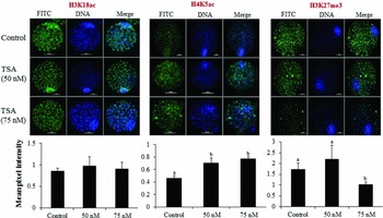
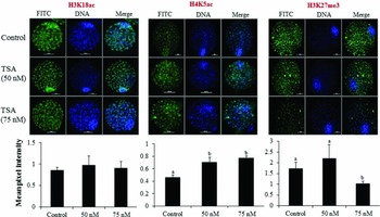
Histone modifications, which are important epigenetic markers associated with reprogramming in the cloned embryos, can occur at several amino acid residues, of which the lysine residues can be either acetylated or methylated. In general, acetylated lysine residues of histones represent gene transcription active status while methylated lysine residues represent gene silencing. In Experiment 3, we examined the effect of TSA treatment on the global level of H4K5ac, H3K18ac and H3K27me3. We found that TSA treatment of reconstructed embryos caused hyperacetylation and hypomethylation of histones as indicated by an increase (P < 0.05) in the global level of H4K5ac in embryos treated with 50 or 75 nM TSA than that in controls and a decrease in the global level of H3K27me3 in embryos treated with 75 nM TSA than that in embryos treated with 50 nM TSA or that in controls (Figure 1). However, the global level of H3K18ac was not significantly different among the three groups. This agrees with earlier reports in which TSA treatment of cloned embryos was found to lower H3K27me3 level in buffalo (Saini et al., Reference Saini, Selokar, Agrawal, Singla, Chauhan, Manik and Palta2014), treatment of donor cells with TSA resulted in an increase in the AcH4K8 level of SCNT buffalo embryos (Luo et al., Reference Luo, Lu, Wang, Wang, Li, Gong, Jiang, Liu and Shi2013) and increase that of H4K5ac in cattle blastocysts (Iager et al. Reference Iager, Ragina, Ross, Beyhan, Cunniff, Rodriguez and Cibelli2008). Besides its direct effects on histones, TSA treatment may also affect DNA methylation which is considered to be a very important epigenetic modification and is generally abnormal and aberrant in SCNT embryos. Specific histone modifications and DNA methylation are believed to influence each other with the relationship working in both directions: histone methylation can help to direct DNA methylation patterns, and DNA methylation might serve as a template for some histone modifications after DNA replication (Cedar & Bergman Reference Cedar and Bergman2009). Because of its influence on regulation of gene expression through alteration in DNA methylation, H3K27me3 is considered an important histone modification. Whereas histone modifications providing labile transcriptional repression, DNA methylation is a highly stable silencing mark that is not easily reversed (Cedar & Bergman Reference Cedar and Bergman2009). Coupled with the results of Experiment 2 in which TSA (75 nM) treatment of embryos was found to improve the conception rate, these results suggest that the beneficial effects of exposing reconstructed cloned embryos to TSA may be primarily due to improvement in the epigenetic status of the blastocysts, which was, however, not reflected in the form of an increased blastocyst rate or TCN or lower apoptotic index.
Effect of treatment of reconstructed embryos with TSA on the global level of H3K18ac, H4K5ac and H3K27me3 in cloned blastocysts produced from somatic cells isolated from frozen–thawed semen. a,bBars with different superscripts differ significantly (P < 0.05).
In conclusion, the results of the present study suggest that TSA treatment of cloned buffalo embryos produced using somatic cells isolated from frozen–thawed semen improved their epigenetic status but not the in vitro developmental potential and offspring rate.
Acknowledgements
The present work was funded by a National Agriculture Innovative Project (NAIP) grant to SKS (C 2–1-(5)/2007) and MSC (C-2067 and 075). MS and NLS are recipients of a CSIR-SRF fellowship.
Conflict of interest statement
None of the authors has any conflict of interest to declare.
Authors’ contributions
MS, NLS and HA carried out the experiments. SKS and PP designed the experiments and prepared and revised the manuscript. SKS, MSC and RSM contributed funds, reagents and infrastructure.