Autophagy derives from the Greek words auto (self) and phagia (eating) and is a term that quite aptly describes the process of self-eating, or consumption and digestion of the cell's own constituents (Ref. Reference De Duve and Wattiaux1). If in anthropomorphic terms apoptosis is used as a term to describe cell suicide, by analogy, autophagy could be envisioned as a form of self-cannibalism. Autophagy is but one of the mechanisms the cell has at its disposal to degrade intracellular proteins. Although there are many protein degradation (proteolytic) systems in cells, the two major ones are the ubiquitin–proteasome system (UPS) and the autophagy–lysosome pathway (ALP). These degradation systems are important for the normal turnover of cell components both under normal housekeeping conditions and under conditions of stress. Such turnover is important for the renewal of the cell and the removal of potentially damaging constituents, but also for fine-tuning the cell phenotype, so that it can respond more readily to changes in its environment and its own metabolic changes.
The ALP differs from the UPS in a number of ways, most notably because degradation through the ALP is relatively more important for long-lived proteins, and because the ALP can also degrade cellular organelles through the process of macroautophagy, not only smaller proteins. Macroautophagy is one of the three major avenues through which the ALP degrades intracellular constituents, the other two being chaperone-mediated autophagy (CMA) and microautophagy. All three pathways result in delivery of intracellular components to the lysosomes; within this acidic environment, and through the effects of lysosomal hydrolases, they are digested into their composite parts, which can then be reused by the cell. In this way, the cell can reorganise its arsenal to best fit its needs.
What distinguishes the different types of autophagy is the manner in which the cellular constituents reach the lysosomes. In microautophagy, invaginations are formed at the level of the lysosomal membrane; such invaginations engulf neighbouring areas of the cytosol, which may include organelles, lipids or proteins (Ref. Reference Muller2). The invaginations eventually round up into vesicles that are digested within the lysosomes, together with their engulfed constituents. The lysosomal membrane itself can be engulfed in this manner, leading to the adjustment of the size of the vacuole. Very little is known about microautophagy in mammalian systems, in particular regarding its possible role in neurodegenerative disease; therefore, it will not be discussed further here. It should be noted, however, that it could conceivably have an important role in protein degradation in neuronal systems, especially if other proteolytic systems are dysfunctional.
Macroautophagy
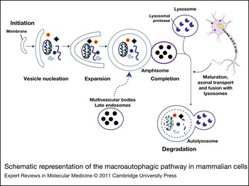
Macroautophagy is sometimes used synonymously with autophagy, although it is clear that it just represents a subset of autophagic pathways in mammalian systems (Refs Reference Shintani and Klionsky3, Reference Mizushima4). In macroautophagy, cup- or rod-shaped isolation double-membrane structures, called phagophores, are first formed within the cell (Fig. 1). The origin of this isolation membrane is still under investigation, but so far the endoplasmic reticulum, Golgi, mitochondria and the plasma membrane are all proposed to be the membrane source (Refs Reference Hayashi-Nishino5, Reference Tooze and Yoshimori6, Reference Yang and Klionsky7). These structures gradually form the so-called autophagosome or autophagic vacuole (AV), which is characterised by the external boundary of the double membrane. Within this structure, there are engulfed cytosolic constituents such as organelles, proteins and lipids. The autophagosome fuses with lysosomes, creating a structure called the autophagolysosome, wherein the vesicular components are eventually degraded. This process, similarly to microautophagy, involves delimitation of cystosolic constituents by a vesicular membrane. A major difference, however, is that such autophagosomes can be formed anywhere within the cell, and not necessarily within close proximity to the lysosomal membrane (Ref. Reference Kochl8). In fact, on many occasions such structures, especially in neurons, can be found at long distances from the perinuclearly located lysosomes, for example along neuronal processes (Ref. Reference Hollenbeck9). It follows that an important aspect of macroautophagy involves the movement of autophagosomes from such remote areas to a perinuclear location, hence the importance of the cellular transport machinery in the efficient functioning of this system. The endosomal system also converges on macroautophagy. Vesicular structures from the late endosomal pathway and multivesicular bodies fuse with autophagosomes to create amphisomes, which enable full maturation of the vesicle and subsequent fusion with lysosomes. Therefore, a dysfunction of either the late endosomal system or the cellular transport machinery may secondarily cause macroautophagy impairment.
Schematic representation of the macroautophagic pathway in mammalian cells. The macroautophagy pathway can be divided into several sequential steps. The first step is the formation of autophagosomes, a process that includes initiation, nucleation, cargo recognition and expansion events that result in completion of the autophagosomes. The completed autophagosomes undergo multiple maturation steps and fusion events with multivesicular bodies or endosomes (amphisomes). An important feature of the process is the movement of autophagosomes (axonal transport) from neuronal axons to a perinuclear location and vice versa (bidirectional transport). Finally, degradation of the cargo takes place in the acidic environment of the autolysosomes by lysosomal proteases, and the constituents of the outer membrane are available for reuse.

Macroautophagy is a highly regulated process and has been studied in great detail in yeast, and more recently, has also been described in mammalian systems. The process of autophagosome formation and maturation in particular includes a series of separate steps: initiation, nucleation, cargo recognition, expansion and completion of the autophagosomes (Fig. 1). Several autophagy-related genes involved in induction and completion of macroautophagy have been well characterised in yeast and in mammals (Refs Reference Ohsumi10, Reference Klionsky11). Macroautophagy occurs constitutively in cells, but is markedly induced under certain circumstances. The classical example is that of starvation or relative nutrient deprivation. In mammalian cells, this can be mimicked by deprivation of amino acid or insulin or growth factor signals, which leads to inactivation of the serine/threonine kinase mTOR (mammalian target of rapamycin), the master regulator of nutrient signalling (Ref. Reference Jung12). Therefore, the mTOR inhibitor rapamycin is the usual agent used to activate macroautophagy. It should be noted, however, that in mammalian cells there are indications that macroautophagy can also be induced by mTOR-independent pathways (Refs Reference Ravikumar13, Reference He and Klionsky14). Regulation of autophagy downstream of mTOR is not yet fully understood, but the protein complex made up of ULK1 (yeast Atg1), Atg13 and FIP200 has a pivotal role (Ref. Reference Jung12). Recently, a novel Atg protein, Atg101, was identified, which interacts with the mammalian ULK1–Atg13–FIP200 complex and localises to the isolation membrane or phagophore (Ref. Reference Hosokawa15). The nucleation phase of initial phagophore formation requires the interaction of ULK1 with complexes of Vps34 and PI3K class III, which are made up of beclin-1 (Atg6), p150/Vps15, AMBRA1, Atg14L and UVRAG (UV irradiation resistance-associated gene) proteins (Refs Reference Klionsky16, Reference Yan17, Reference Levine, Sinha and Kroemer18, Reference Thoresen19, Reference Funderburk, Wang and Yue20). Other beclin-1-binding proteins include Rubicon (RUN domain, a cysteine-rich domain containing beclin-1-interacting protein), Bcl-2, Rab5, AMBRA1 and Bif-1 (Refs Reference Thoresen19, Reference Funderburk, Wang and Yue20).
The classical pharmacological inhibitors of macroautophagy, 3-methyladenine (3-MA) and wortmannin, act at this stage of the pathway as PI3K class III inhibitors. The further expansion of autophagosomes is mediated by two ubiquitin-like conjugation systems. One is the conjugation of Atg5 with Atg12 and the other is the covalent linkage of MAP1LC3 (microtubule-associated proteins 1A/1B light chain 3; Atg8) to phosphatidylethanolamine. This linkage leads to two changes that are useful as specific indices of macroautophagy: the change of LC3 labelling from a diffuse to a punctuate pattern, which can be detected by immunostaining, and a shift of the molecular mass of the protein, which can be detected on western immunoblots as LC3-II. Another marker that is potentially useful (although experience is still rather limited, and there may be issues of specificity) is p62 (sequestosome 1, SQSTM1). This appears to be a substrate for macroautophagy; hence its levels increase when macroautophagy is inhibited, and decrease when macroautophagy is induced (Ref. Reference Bjorkoy21). p62 binds directly to LC3 through a specific sequence motif at the same time as it binds to polyubiquitylated proteins. Thus, p62 provides a link between macroautophagy and the UPS; it is thought to sequester polyubiquitylated proteins away from the UPS and direct them towards macroautophagic-dependent lysosomal degradation (Refs Reference Bjorkoy, Lamark and Johansen22, Reference Bjorkoy23). In combination with LC3-II, it can provide an index of autophagic flux, as explained in detail in Table 1. It should be noted that electron microscopy evidence is still ideally required to conclusively show autophagosome accumulation, although because of the biochemical tools now available, it is not always performed (Ref. Reference Klionsky24).
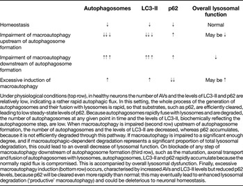
Alterations in the number of autophagosomes and LC3-II and p62 levels in neurons under physiological and pathological conditions

Under physiological conditions (top row), in healthy neurons the number of AVs and the levels of LC3-II and p62 are relatively low, indicating a rather rapid autophagic flux. In this setting, the whole process of the generation of autophagosomes and their fusion with lysosomes is rapid, so that substrates, such as p62, are efficiently cleared, leading to low steady-state levels of p62. Because autophagosomes rapidly fuse with lysosomes and are degraded, the number of autophagosomes at any given point in time and the levels of LC3-II, biochemically reflecting the autophagosome step, are low. When macroautophagy is impaired (second row) upstream of autophagosome formation, the number of autophagosomes and the levels of LC3-II are decreased, whereas p62 accumulates, because it is not efficiently degraded through this pathway. If macroautophagy is impaired to a significant enough degree, and if macroautophagic-dependent degradation represents a significant proportion of total lysosomal degradation, this could lead to an overall decrease of lysosomal function. On blockade of any step of macroautophagy downstream of autophagosome formation (third row), such as the maturation, axonal transport and fusion of autophagosomes with lysosomes, autophagosomes, LC3-II and p62 rapidly accumulate because the normally rapid flux is compromised. This is accompanied by overall lysosomal dysfunction. Finally, excessive macroautophagy induction (bottom row) occurs, characterised by increased AVs and LC3-II levels but reduced p62 levels, because p62 will be cleared even more rapidly than normal; this may eventually lead to enhanced lysosomal degradation (‘productive’ macroautophagy) and could be deleterious to neuronal homeostasis.
Recently, macroautophagy has been shown to mediate selective degradation of various targets such as aggregated proteins and damaged organelles. In this process, p62 and perhaps HDAC6 proteins have a vital role. HDAC6 has the capacity to bind both polyU misfolded proteins and dynein motors, thus targeting polyubiquitylated aggregates and damaged mitochondria to aggresomes for degradation (Ref. Reference Kawaguchi25). Moreover, several other proteins that link the autophagic machinery to its substrates have been identified in mammalian systems. These are designated ‘autophagy receptors’ and are involved in the cytosol-to-vacuole targeting pathway (Atg19) (Ref. Reference Chang and Huang26) and in the degradation of ubiquitylated bacteria (NDP52) (Ref. Reference Thurston27), mitochondria (Nix-1, Atg32) (Refs Reference Novak28, Reference Kanki29) and protein aggregates (NBR1) (Ref. Reference Kirkin30). All proteins contain an LC3-interacting region and a substrate-recognition domain. Recently, it has also been shown that Alfy (autophagy-linked FYVE protein, also known as WDFY) is required for the macroautophagic elimination of aggregated proteins, but not for macroautophagic elimination of bulk cytosol in response to starvation (Ref. Reference Filimonenko31).
Macroautophagy in the nervous system
The fact that constitutive macroautophagy is essential for neuronal homeostasis was highlighted by two landmark studies which showed that genetic ablation of essential macroautophagy genes (Atg5 or Atg7) in the mouse control nervous system (CNS) led to neuronal degeneration within the first few months of life (Refs Reference Hara32, Reference Komatsu33). This was surprisingly accompanied by extensive accumulation of ubiquitylated inclusions, which would have been expected only with UPS dysfunction; in fact, enzymatic proteasomal activity was found to be unaffected in these mice. Two additional pieces of work seem to provide an answer to this conundrum. Mating of Atg7-deficient mice with p62-knockout mice leads to drastic diminution of the inclusions, although neurodegeneration per se is not affected (Ref. Reference Komatsu34). Furthermore, p62 is upregulated following impairment of macroautophagy and leads to enhanced recruitment of ubiquitylated substrates away from the UPS, thus impeding their degradation (Ref. Reference Korolchuk35). It appears therefore that p62 has a key role in this respect, and its regulation may be important for a variety of situations where ubiquitylated inclusions accumulate in neurodegenerative conditions.
From the above-mentioned studies it became clear that post-mitotic neurons rely heavily on macroautophagic machinery to degrade cellular components as part of their normal physiological function. A number of additional studies in a variety of organisms have shown that elimination of components of the macroautophagic pathway leads to neuronal degeneration and lysosomal abnormalities (Refs Reference Zhou36, Reference McIntire37, Reference Pickford38, Reference Fimia39, Reference Lee40). In fact, it seems that healthy neurons normally have an increased autophagic flux compared with other cells, meaning that the process of autophagosome formation, maturation and digestion within lysosomes is especially efficient and rapid. A correlate of this is that LC3-II and autophagosomes are barely detectable in healthy cultured neuronal cells and in nervous system tissue. However, upon blockade of lysosomal function or of any step downstream of autophagosome formation, such as the maturation and fusion of autophagosomes with lysosomes, LC3-II rapidly accumulates (Refs Reference Mizushima41, Reference Mizushima and Kuma42, Reference Mizushima43, Reference Tsvetkov, Mitra and Finkbeiner44, Reference Boland45, Reference Mitra, Tsvetkov and Finkbeiner46). In a sense, neurons are functioning in an ‘overdrive’ of macroautophagy, and that is why they are especially vulnerable to its disruption.
The conditional Atg-knockout mice have also served to reveal specific important roles of basal macroautophagy in the homeostasis of neurites. Conditional mutant mice lacking Atg7 in Purkinje neurons displayed cell-autonomous, progressive dystrophy (manifested by axonal swellings), degeneration of axonal terminals and behavioural deficits, in the absence of autophagic vacuole accumulation and in the absence of dendritic pathology (Refs Reference Komatsu47, Reference Yue48); this contrasts with the situation in pathological conditions, such as Alzheimer disease, where autophagosomes accumulate in such axonal dystrophic swellings (see below). These findings underscore a specialised role of neuronal macroautophagy in the maintenance of homeostasis of axonal terminals and protection against axonal degeneration.
Another factor that should be mentioned here is ageing. It appears that the capacity for macroautophagy-mediated protein degradation declines with age, although this has not been specifically studied in the brain (Refs Reference Ward49, Reference Cuervo50, Reference Cuervo and Dice51, Reference Vellai52). In invertebrate organisms, elimination or reduction of macroautophagy-related genes has consistently led to a reduced lifespan (Refs Reference Simonsen53, Reference Simonsen, Cumming and Finley54, Reference Simonsen55).
Concept of (macro)autophagic stress
Macroautophagic stress applies to situations in which there is perturbation of the normal flow of macroautophagic degradation. This is usually apparent as an accumulation of autophagosomes in the material studied, which can be due either to enhanced activation of the macroautophagic pathway or, apparently more often, especially in neurons, to problems further downstream that affect the late endosomal pathway, the cellular transport machinery or general lysosomal function. In the former case, the term ‘productive’ macroautophagy can be used to imply that the enhanced macroautophagy will eventually lead to enhanced lysosomal protein degradation, whereas in the latter case, the macroautophagy is ‘nonproductive’ because the autophagic flux and the rate of lysosomal degradation actually decrease (Table 1). This is an important distinction that needs to be made in situations in which autophagosomes accumulate. However, it should be stressed that the problem might also be due to a dysfunction of the macroautophagy pathway per se, such as seen with the Atg-knockout mice, where autophagosomes fail to form.
Chaperone-mediated autophagy
CMA is a selective mechanism for the degradation of specific cytosolic proteins within lysosomes, and does not involve vesicle formation. In this process, cytosolic proteins with the particular pentapeptide motif KFERQ or a biochemically related sequence are recognised by a complex of chaperones and cochaperones in the cytosol; they are then translocated one by one to the lysosomal membrane, where they bind to another complex and, through this binding, are finally threaded into the lysosomes and degraded (Refs Reference Chiang56, Reference Cuervo, Dice and Knecht57, Reference Cuervo and Dice58, Reference Agarraberes and Dice59, Reference Bandyopadhyay60). The KFERQ-like motif is present in about 30% of all cytosolic proteins, but even more proteins could become substrates if they are post-translationally modified (Refs Reference Dice61, Reference Chiang and Dice62).
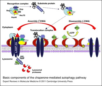
The laboratories of Fred Dice, Erwin Knecht and, more recently, Ana Maria Cuervo have been instrumental in the discovery and elucidation of the mechanism underlying this pathway of lysosomal degradation. The chaperone–cochaperone complex involved in recognition is made up of the heat shock cognate protein of 70 kDa (Hsc70), a cytosolic member of the Hsp70 chaperone family (Ref. Reference Chiang56) and Hsp70 cochaperones (such as Hsp40, Hsp90, Hip, Hop and Bag-1). The main element in the lysosomal membrane complex is the lysosome-associated membrane protein type 2A (LAMP-2A), an alternatively spliced form of the Lamp2 gene, and the only one known to participate in CMA (Ref. Reference Hatem, Gough and Fambrough63). LAMP-2A is an integral lysosomal membrane protein with a single transmembrane region, a short cytosolic tail and a large, highly glycosylated luminal region. LAMP-2A can form oligomers, which are thought to form the protein translocation pore. The substrate protein is subsequently degraded after unfolding and translocation into the lysosomal lumen, with the help of lysosomal Hsc70 (a luminal form of Hsc70). Lysosomal Hsc70 and lysosomal Hsp90 are required for stabilisation, assembly and disassembly of the LAMP-2A-enriched multimeric complexes (Ref. Reference Bandyopadhyay60). A very recent study from the Cuervo group has added a further layer of complexity to this process, because they identified glial fibrillary acidic protein (GFAP) and elongation factor 1 α (EF1α; EEF1A1) as regulators of the disassembly of the transmembrane translocation complex; through these regulatory proteins, guanosine triphosphate (GTP) mediates an inhibitory effect on CMA (Ref. Reference Bandyopadhyay64) (Fig. 2). CMA activity is directly dependent on the levels of LAMP-2A at the lysosomal membrane and on lysosomal Hsc70 levels in the lysosomal lumen (Refs Reference Cuervo and Dice65, Reference Cuervo66, Reference Agarraberes, Terlecky and Dice67). Basal levels of CMA activity are detectable – although variable – in most cells and tissues, but CMA is often upregulated under conditions of stress or nutrient deprivation (Ref. Reference Martinez-Vicente and Cuervo68). Macroautophagy and CMA are interconnected, although the molecular mechanisms that modulate this crosstalk are not fully understood. Experimental blockage of one of them results in compensatory upregulation of the other (Refs Reference Vogiatzi69, Reference Massey70, Reference Wang71, Reference Kaushik72).
Basic components of the CMA pathway. CMA substrates are translocated across the lysosomal membrane by the participation of two major protein complexes: a group of cytosolic chaperones and cochaperones (cargo recognition complex) and a group of lysosomal proteins located on both sides of the lysosomal membrane (cargo translocation complex). Cytosolic Hsc70 recognises the CMA-targeting motif in the substrate protein, and in collaboration with chaperones and cochaperones delivers it to the surface of the lysosomes where it binds to LAMP-2A monomers. The stability of the CMA translocation complex is modulated by GFAP and EF1α. GFAP binds to LAMP-2A at the complex and stabilises it. However, GFAP may also form dimers with its phosphorylated form (GFAP-P), leading thus to disassembly of the translocation complex. Such dimerisation is inhibited by EF1α binding to phosphorylated GFAP. In the presence of GTP, EF1α is removed from the lysosomal membrane, thus leading to GFAP dimerisation and translocation complex disassembly (modified from Ref. Reference Bandyopadhyay64). Lys-Hsc70 and Lys-Hsp90 are required for stabilisation, assembly and disassembly of the LAMP-2A-enriched multimeric complexes. Abbreviations: CMA, chaperone-mediated autophagy; EF1α, elongation factor 1 α; GFAP, glial fibrillary acidic protein; GTP, guanosine triphosphate; Hsc, heat shock cognate protein; Hsp, heat shock protein; LAMP-2A, lysosome-associated membrane protein type 2a.

Importantly, CMA activity declines with age in almost all tissues analysed so far, mainly as a consequence of decreased LAMP-2A levels at the lysosomal membrane (Ref. Reference Cuervo and Dice51). In various cell systems, overexpression of LAMP-2A is able to ameliorate CMA function, indicating that increased LAMP-2A levels are sufficient for this effect (Ref. Reference Cuervo and Dice73). Consistent with this idea is a recent in vivo study which showed that transgenic overexpression of LAMP-2A in the liver not only reversed age-asociated CMA impairment, but also led to improvement of liver function (Ref. Reference Zhang and Cuervo74). This stresses the importance of LAMP-2A as a rate-limiting step in the pathway and underscores the utility of targeting this molecule in order to improve CMA function.
Not much is known about the role of CMA in the nervous system. Recent data suggest that Hsc70 and LAMP-2A are expressed in rodent and human brain (Refs Reference Vogiatzi69, Reference Alvarez-Erviti75). In one study, LAMP-2A levels were developmentally induced in rat brain during the first days after birth. There was no decrease of total LAMP-2A levels with ageing in the rat brain, but lysosomal-bound LAMP-2A was not specifically assessed (Ref. Reference Vogiatzi69). Furthermore, various studies in neuronal cells and cultured primary neurons, largely based on inactivation of CMA components, indicate that CMA indeed operates as a mechanism for lysosomal degradation in these cellular systems (Refs Reference Vogiatzi69, Reference Alvarez-Erviti75, Reference Xilouri76, Reference Cuervo77, Reference Martinez-Vicente78, Reference Yang79). The particular culture conditions appear to influence the degree of participation of the CMA pathway in lysosomal degradation.
Autophagy in neurodegenerative diseases
In various neurodegenerative conditions, a common denominator is the presence of ubiquitylated inclusions, which are indicative of failure of protein degradation systems. Although this was originally construed as evidence for UPS dysfunction, conditional-knockout Atg mice (Refs Reference Hara32, Reference Komatsu33) have shown us that this is not necessarily the case, and that macroautophagy impairment can lead to similar results. There is in fact accumulating evidence of increased accumulation of autophagic vacuoles in various neurodegenerative diseases, which, as mentioned above, could be an indication of excessive induction of macroautophagy or a failure of macroautophagy completion. Autophagy imbalance has been implicated as a pathogenic mechanism in various situations of acute injury, but this will not be discussed further here. There are indications that link macroautophagic dysfunction to Huntington disease and other triple-repeat disorders, a rare form of frontotemporal dementia, prion disease, Alzheimer disease and motor neuron disease. The reader is advised to seek reviews that cover more broadly the link of autophagy to neurodegenerative diseases (Refs Reference Martinez-Vicente and Cuervo68, Reference Jaeger and Wyss-Coray80, Reference Rubinsztein81, Reference Cherra and Chu82, Reference Wong and Cuervo83) because we focus here more on Parkinson disease (PD) and related Lewy body disorders.
A central issue in neurodegenerative diseases is whether the observed accumulation of autophagic vacuoles represents a protective effect, or at least a step in this direction, or, instead, a pathway for cell death, in which case the term ‘autophagic cell death’ or type II cell death, differentiating it from apoptosis or necrosis, can be applied (Refs Reference Shintani and Klionsky3, Reference Rubinsztein81, Reference Cherra and Chu82, Reference Kunchithapautham and Rohrer84, Reference Hornung, Koppel and Clarke85, Reference Lohr86). A corollary to this is whether strategies targeted towards macroautophagy, which would theoretically improve clearance of aberrant protein conformations linked to such proteinopathies, could be applied as therapeutic agents. This is a continuously evolving field, and it is clear that many of the effects observed are context dependent. To give an illustrative example from outside the field of neurodegenerative disease, mice with conditional Atg7 deletion in the CNS that show severe neurodegeneration are protected from hypoxia- or ischaemia-induced neuronal death (Ref. Reference Koike87). An example from the field of Alzheimer disease research comes from flies partially deficient in Atg1: although they demonstrated a reduced lifespan, they are partially protected against neurodegeneration induced by β-amyloid (APP or Aβ) because of aberrant activation of macroautophagy. Aβ1–42 triggered a neurodegenerative cascade, accompanied by excessive accumulation of autophagic vacuoles, which was not only partially rescued by macroautophagy inhibition, but also enhanced by macroautophagy activation with rapamycin. Thus, in this system, the usual prosurvival role of neuronal macroautophagy changes to a prodeath role in the presence of Aβ1–42 (Ref. Reference Ling88).
Parkinson disease
PD represents one of the most common neurodegenerative conditions. It is classically diagnosed when the motor symptoms of tremor, bradykinesia, rigidity and postural imbalance appear (Refs Reference Forno89, Reference Gibb90). However, it is now recognised that the disease is also characterised by a host of nonmotor symptoms, some of which might even precede the motor manifestations. A major problem is the gradual emergence of cognitive decline in most patients, which can lead to dementia. In the closely related condition, dementia with Lewy bodies (DLB), cognitive decline precedes or appears concurrently with the motor defects.
At the neuropathological level, the disease was traditionally viewed as a dopamine deficiency state, with loss of dopaminergic nigral neurons and their nigrostriatal projections. However, it is now well appreciated that the neuropathological process is much more widespread, involving many neuronal populations across the peripheral and central nervous system. The neuropathogical hallmark of the disease is the appearance, in surviving neurons, of cytoplasmic inclusions, termed Lewy bodies (LBs), whereas analogous aggregates, termed Lewy neurites (LNs), accumulate in neuronal processes (Ref. Reference Gibb90). These aggregates stain strongly for the presynaptic protein α-synuclein (encoded by SNCA) and are present in DLB and a host of other neurodegenerative conditions, collectively termed synucleinopathies (Refs Reference Spillantini91, Reference Spillantini92). Management of PD consists of various forms of dopamine replacement therapy, which is effective in the initial disease stages. However, the emergence of motor complications and severe nonmotor symptomatology, especially dementia, complicates management, hence the need for disease-modifying therapies that attack the disease at its source.
The aetiology of PD is not known, although various theories have been proposed, including oxidative stress, mitochondrial toxin exposure and altered protein handling (Refs Reference Ross and Poirier93, Reference Dauer and Przedborski94, Reference Abou-Sleiman, Muqit and Wood95, Reference Giasson96, Reference Levy, Malagelada and Greene97). This last theory was initially confined to the idea of UPS dysfunction, but more recent data suggest that the autophagy–lysosome system is also affected in PD. Evidence for this has accumulated in part through the association of several of the proteins linked genetically to PD with autophagic pathways. Although PD is largely sporadic, about 10–20% of patients exhibit a clear pattern of inheritance. This has led to the identification of a number of genetic defects that lead to PD. The genes involved can be broadly categorised into two groups: one leading to autosomal dominant PD, and the other to autosomal recessive PD. It is generally assumed that in the former group, a toxic gain of function is operating, whereas the latter is due to a loss of function. Although relatively rare, these genetic defects have opened new windows to the investigation of PD pathogenesis (Ref. Reference Gasser98).
PD and autophagic pathways
Neuropathological evidence linking autophagic pathways to the disease
The first mention of autophagy in the context of analysis of pathological specimens of PD brains came from the study of Anglade and co-workers, where ultrastructural examination of the substantia nigra pars compacta of such brains revealed some evidence of accumulation of autophagic vacuoles (Ref. Reference Anglade99). This was confirmed in another study (Ref. Reference Zhu100). However, it should be stated that many years before, Lydia Forno had observed that LBs within the stellate ganglion of PD patients displayed a rather vesicular nature, suggesting the participation of the autophagic–lysosomal pathway in the formation or dissolution of LBs (Ref. Reference Forno and Norville101). A recent study has shown that LC3 accumulates within LBs and other α-synuclein-immunoreactive structures, supporting this possibility. However, whether LC3-II or LC3-I is the form that accumulates within the inclusions could not be determined. Analysis of LC3 by western blotting revealed a tendency for an increase of the LC3-II band in PD patients, but this was not statistically significant, probably because of the small number of brains analysed (Ref. Reference Alvarez-Erviti75). The accumulation of autophagic vacuoles, as well as the existence of macroautophagic components within LBs in postmortem PD brains, was also shown (Ref. Reference Dehay102). In this work, evidence for lysosomal disruption and release of lysosomal hydrolases was found in MPTP-treated mice, which represent a well-characterised neurotoxin model of Parkinsonism induced by mitochondrial dysfunction and oxidative stress. This led to a net loss of lysosomes in the mouse nigra. These changes were attributed to release of reactive oxygen species from the mitochondria, which damaged the lysosomal membrane. An increase of lysosomal biogenesis, which was achieved through an upregulation of the lysosomal transcription factor EB or, remarkably, the macroautophagy enhancer rapamycin, partially prevented neurodegeneration in the MPTP model (Ref. Reference Dehay102). A decrease in general lysosomal markers, such as cathepsin D and LAMP-1, in PD nigra was also reported in another study. This phenomenon was more marked in LB-containing neurons (Ref. Reference Chu103). This exciting new set of data suggests that the consistent accumulation of autophagic vacuoles in the nigra in PD is secondary to lysosomal dysfunction. The physiological neuromelanin granules in human nigra are proposed represent aborted autophagic vacuoles, containing pigments derived from catecholamine oxidation, that have not been digested by lysosomes (Ref. Reference Sulzer104). Macroautophagic markers were also examined in brain material from patients with DLB. Interestingly, in one study, levels of Atg7 were found to be reduced, whereas those of mTOR were increased, which suggests that the net effect could be inhibition of the progression through the macroautophagy pathway. Despite this, abnormal macroautophagic structures were identified, indicating that the process was still operating but was perhaps also dysfunctional further downstream. LC3 labelling was again observed within such LB-like structures (Ref. Reference Crews105). In another study, levels of LC3-II and beclin-1 were found to be increased in DLB brains (Ref. Reference Yu106).
The existence of potential abnormalities in CMA components has also started to receive some attention in neuropathological studies of PD brains. In one of the above-mentioned studies (Ref. Reference Chu103), a decrease in Hsc70 was observed in PD nigral neurons, but this did not appear to be entirely specific, because other lysosomal markers were also affected. mRNA encoding Hsc70 was also found to be downregulated in PD nigra in a gene-profiling study (Ref. Reference Mandel107). In a careful study, which examined a relatively limited number of brains, western immunoblots showed decreased levels of the two main CMA proteins, LAMP-2A and Hsc70, in both the nigra and amygdala of PD brains (Ref. Reference Alvarez-Erviti75). The authors confirmed that at least in the amygdala, the decrease in LAMP-2A was reflected in lysosomal fractions, in which they controlled for LAMP-1 levels, which were unchanged in the disease brains. These important findings suggest that there may be a specific impairment of CMA that is widespread in PD brains, and is not confined to areas of major cell loss, such as the nigra. In fact, based on our findings of the relationship of α-synuclein to CMA and macroautophagy, we have hypothesised that a triggering or at least contributing event in the generation of the PD pathogenetic cascade could be a malfunction of such degradation systems (Ref. Reference Xilouri108). In support of this idea, two genetic defects associated with PD appear to cause direct general lysosomal dysfunction. These include loss-of-function mutations in ATP13A2, a lysosomal ATPase, which leads to a rare familial form of Parkinsonism with dementia and pyramidal tract signs (Refs Reference Di Fonzo109, Reference Ramirez110), and mutations in the lysosomal enzyme glucocerebrosidase, normally associated with the quintessential lysosomal storage disorder Gaucher disease, which leads to an increased risk of PD (Ref. Reference Sidransky111).
α-Synuclein and autophagic pathways
α-Synuclein, which, as mentioned above, is the major constituent of LBs and LNs, is also linked genetically to the disease through point mutations or multiplications of its gene locus, SNCA (Refs Reference Chartier-Harlin112, Reference Polymeropoulos113, Reference Kruger114, Reference Singleton115). Furthermore, polymorphisms within the SNCA locus confer increased risk of sporadic PD (Ref. Reference Maraganore116). α-Synuclein is accordingly thought to be a major pathogenetic factor in both familial and sporadic PD (Refs Reference Gasser98, Reference Simon-Sanchez117). A major issue has been the manner of α-synuclein degradation, because impairment of such degradation could lead to accumulation of α-synuclein, which characterises the disease. Although some studies have suggested that the UPS is the main system for α-synuclein degradation (Refs Reference Bennett118, Reference Imai, Soda and Takahashi119), we and others have suggested that such degradation is mainly carried out by lysosomal pathways, and in particular by macroautophagy and CMA (Refs Reference Cuervo77, Reference Webb120, Reference Lee121). Initially, wild-type α-synuclein was identified as a substrate for CMA in in vitro assays.Two of the α-synuclein mutants, A30P and A53T, bound strongly to LAMP-2A but were not internalised and degraded, and thus acted as inhibitors of CMA degradation of other substrates (Ref. Reference Cuervo77). In subsequent studies, using siRNAs against LAMP-2A and expressing forms of α-synuclein that lack the CMA recognition motif, we were able to confirm that CMA is an important pathway for normal α-synuclein turnover, especially in primary neurons, including those derived from rat ventral midbrain (Ref. Reference Vogiatzi69). Detergent-insoluble or high molecular weight oligomeric conformations of α-synuclein also increased following CMA inhibition, suggesting that CMA dysfunction could eventually lead to α-synuclein-related pathology, although discrete aggregates were not detected within the time frame of our experiments (Ref. Reference Vogiatzi69). We found that macroautophagy inhibition by 3-MA led to accumulation of wild-type (WT) α-synuclein (Ref. Reference Webb120), suggesting that macroautophagy is also important for normal α-synuclein turnover. The results implicating CMA in degradation of WT α-synuclein in neuronal cells have recently been confirmed (Ref. Reference Alvarez-Erviti75). Furthermore, transgenic mice for α-synuclein or mice exposed to the mitochondrial toxin paraquat upregulate lysosomal LAMP-2A and are able to degrade α-synuclein more efficiently, providing indirect evidence that CMA is a mechanism for α-synuclein degradation in vivo (Ref. Reference Mak122).
Based on the link of α-synuclein to PD pathogenesis, many cellular and animal models of ‘synucleinopathy’ have been created. In one of these cellular models, we noted that stable expression of the A53T mutant form of α-synuclein in PC12 cells induced a marked accumulation of autophagic vacuoles (Ref. Reference Stefanis123). Labelling with Lysotracker and a form of dextran that was taken up and translocated to the lysosomes disclosed a significant decrease of lysosomal acidification in these cells, suggesting that lysosomes were dysfunctional. In fact, we found that the ability of lysosomes to degrade long-lived proteins was severely impaired in the A53T-expressing cells (Ref. Reference Cuervo77). As discussed earlier, this could have led to the dramatic macroautophagy phenotype that was observed (Ref. Reference Stefanis123). The increase in autophagosomes in cells and transgenic mice overexpressing mutant, or, in some cases, wild-type α-synuclein, has now been replicated in a number of studies (Refs Reference Crews105, Reference Yu106, Reference Spencer124) and mirrors the neuropathological data mentioned above.
In subsequent work, we have extended these studies to differentiated human neuroblastoma cells inducibly expressing α-synuclein and rat cortical neurons adenovirally transduced with α-synuclein. Although we saw consistent CMA dysfunction, autophagosome accumulation (as defined in this case by an increase of LC3-II immunolabelling) and neuronal toxicity, general lysosomal dysfunction was not always observed. Macroautophagic alterations and neuronal toxicity were secondary to CMA impairment, because forms of α-synuclein that were not targeted to CMA did not induce autophagosome accumulation and induced death to a lesser extent (Ref. Reference Xilouri76).
An earlier study showed that post-translational modifications of α-synuclein differentially affect the ability of the protein to be degraded by CMA and the degradation of other CMA substrates (Ref. Reference Martinez-Vicente78). More particularly, it was shown that only monomers and dimers of α-synuclein, but not oligomers, are degraded by CMA, whereas oxidation and nitration of the protein slightly inhibit its degradation by CMA (Ref. Reference Martinez-Vicente78). By contrast, phosphorylation and dopamine modification of α-synuclein almost completely prevented the degradation of the protein by CMA. Only dopamine modification of α-synuclein was able to significantly inhibit the degradation of other CMA substrates. Consistent with these findings, we found, in dopaminergic cells, that even wild-type α-synuclein caused CMA dysfunction and CMA-targeting-dependent death in a dopamine-dependent fashion (Ref. Reference Xilouri76). This is important, because it extends CMA dysfunction as a more general pathogenetic effect of α-synuclein within dopaminergic neurons beyond the rare familial cases.
Another link of pathogenic effects of α-synuclein with CMA is provided by a study which showed that both WT and A53T α-synuclein caused CMA dysfunction, and that this, in turn, led to mislocalisation of the neuronal survival factor and CMA substrate MEF2D to the cytosol, with resultant loss of its protective function. Adding to the physiological relevance of these findings, mislocalised MEF2D was increased in A53T transgenic mice and in PD patients (Ref. Reference Yang79).
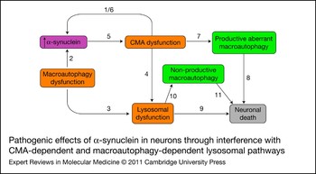
These results highlight the possibility of toxic effects of α-synuclein through direct interference with the CMA pathway, and also the crosstalk between the two major autophagic pathways. It would appear therefore that aberrant α-synuclein can lead to increased autophagosome accumulation either through lysosomal dysfunction or through specific CMA targeting. In the former case macroautophagy is ‘nonproductive’, and in the latter it is ‘productive’. It is unclear at this point what determines each effect, and they could both potentially operate under certain circumstances (Fig. 3). A recent study has raised the additional possibility that α-synuclein could inhibit an early point in autophagosome formation, through interaction with Rab1a (Ref. Reference Winslow125). It is difficult at this point to reconcile these data with our own or those that suggest substantial accumulation of autophagosomes in synucleinopathy models, as mentioned above.
Pathogenic effects of α-synuclein in neurons through interference with CMA-dependent and macroautophagy-dependent lysosomal pathways. The diagram represents a hypothetical scheme of the link between α-synuclein and autophagic pathways, based on currently available data. At early stages of PD, dysfunction of CMA-dependent (arrow 1) or of macroautophagy-dependent (arrow 2) degradation pathways might lead to α-synuclein accumulation, which characterises the disease. Dysfunction of such pathways might in part be a consequence of ageing. Such dysfunction would be expected to lead in parallel to generalised lysosomal impairment (arrows 3 and 4), because the lysosome relies on these systems to efficiently degrade intracellular proteins. As aberrant α-synuclein continues to accumulate over time, it can cause specific CMA impairment (arrow 5), which can feed back into further α-synuclein accumulation (arrow 6). CMA dysfunction at this stage can also lead to aberrant activation of ‘productive’ macroautophagy (arrow 7), which can lead to neuronal degeneration (arrow 8). At the same time, the increasing generalised lysosomal dysfunction can lead to neuronal death either directly (arrow 9) or through aberrant accumulation of autophagosomes that cannot be degraded by the dysfunctional lysosomes (‘nonproductive’ macroautophagy) (arrow 10). Accumulation of such malfunctioning autophagosomes could be further detrimental to neuronal survival (arrow 11). The end result from both productive and nonproductive macroautophagic pathways at late stages of the disease would be neuronal death with evidence of accumulation of autophagosomes in dying neurons (‘autophagic cell death’). Abbreviations: CMA, chaperone-mediated autophagy; PD, Parkinson disease.

An important question is whether macroautophagy in our experimental setting of cultured differentiated neuroblastoma and cortical neurons served a protective or deleterious effect. We found that pharmacological or molecular inhibition of macroautophagy was protective. This occurred even in cortical neurons, where macroautophagy was ‘productive’ (Ref. Reference Xilouri76). There could be various mechanisms through which such death could occur, such as excess consumption of vital cellular constituents, leakage of lysosomal hydrolases into the cytosol or enhanced production of damaging molecules within autophagic vacuoles or autophagolysosomes.
Such results run against an accumulating body of literature suggesting that enhancement of macroautophagy is a treatment strategy against synucleinopathies or, for that matter, other neurodegenerative conditions. Induction of macroautophagy by treatment with rapamycin or overexpression of Atg7 or beclin-1 led to reversal of pathological effects induced by α-synuclein in cellular and in vivo models (Refs Reference Crews105, Reference Yu106, Reference Spencer124). These apparently differential effects could be due to timing, because it is conceivable that early in the pathological process macroautophagy might be beneficial as a clearing mechanism; however, later on, when excessively activated, in cells that are already suffering and close to death, it may be deleterious (Fig. 3). Alternatively, it could be an issue of differences in the particular models used. We are currently examining our cellular systems for the effects of macroautophagy enhancers to address this possibility.
Linkage of UCH-L1 and LRRK2 to autophagic pathways
A very rare genetic cause of PD is a mutant form of ubiquitin C-terminal esterase L1 (UCH-L1), I93M, which has been identified in only a single family and has also been linked to the CMA pathway. This mutant form, but not WT UCH-L1, interacted aberrantly with the CMA components LAMP-2A and Hsc70, inhibited CMA and caused an increase in α-synuclein levels, indicating that the aberrant interaction of mutant UCH-L1 with CMA might underlie its pathogenic role (Ref. Reference Kabuta126).
Another protein linked genetically to PD through an assumed toxic gain of function is LRRK2 (leucine-rich repeat serine/threonine protein kinase 2 or PARK8), which might also be intricately linked to the ALP. In brains and cells, LRRK2 appears to localise mainly to vesicular granular structures of the late endosomal–lysosomal pathway (Refs Reference Alegre-Abarrategui127, Reference Alegre-Abarrategui and Wade-Martins128, Reference Higashi129). Aged Lrrk2-null mice display an autophagy-relevant phenotype in the kidney, where LRRK2 is normally highly expressed (Ref. Reference Tong130). There is induction of LC3-II and p62, and a parallel accumulation of α-synuclein and ubiquitin-positive aggregates, accompanied by extensive cellular degeneration and inflammatory responses. These results indicate a profound impairment of the macroautophagy pathway downstream of autophagosome formation. In apparent contradiction, siRNA-mediated knockdown of LRRK2 in cultured human cells increased autophagic activity and prevented starvation-induced cell death when autophagy was inhibited with bafilomycin (Refs Reference Alegre-Abarrategui127, Reference Alegre-Abarrategui and Wade-Martins128). It will be interesting to decipher the basis for these differences, and to further understand the physiological role of LRRK2 in the regulation of macroautophagy and broader lysosomal function.
However, the main link of LRRK2 to PD is provided through mutant forms identified in patients in families with autosomal dominant inheritance (Ref. Reference Gasser98). Expression of the mutant G2019S form of LRRK2 led to neuritic retraction and accumulation of autophagosomes in differentiated human neuroblastoma SH-SY5Y cells (Ref. Reference Plowey131). Inhibition of macroautophagy through molecular means reversed the detrimental effects of mutant G2019S LRRK2 on the length of neuronal processes (Ref. Reference Plowey131), a prominent feature of the degenerative phenotype associated with PD-associated LRRK2 mutations. In partial agreement with this work, expression of another PD-linked LRRK2 mutant, R1441G, induced autophagic stress characterised by the accumulation of abnormal multivesicular bodies and enlarged autophagosomes with high levels of p62 and by the appearance of skein-like inclusions (Ref. Reference Alegre-Abarrategui127). In the latter study, however, no attempt was made to examine the effect on cellular homeostasis by macroautophagy modulation.
It would appear therefore that, as in our own synucleinopathy cellular models, expression of mutant forms linked to autosomal dominant PD might lead to induction of the presence of autophagosomes, and that in these settings, the macroautophagy pathway may be aberrantly activated. The data obtained with mutant LRRK2 (Ref. Reference Plowey131), together with the previously mentioned data illustrating selective vulnerability of neurites to genetically induced macroautophagy impairment (Ref. Reference Komatsu47), show that macroautophagy balance is especially critical in neurites.
Proteins linked to autosomal recessive Parkinsonism provide a link to mitophagy
Apparent loss of function of Parkin (PARK2) and PINK1 constitutes the bulk of cases with autosomal recessive Parkinsonism (Ref. Reference Gasser98). Given the dramatic mitochondrial deficits present in fly models with deletion of the corresponding genes, it was assumed that the function of both Parkin and PINK1 was to preserve mitochondrial integrity (Refs Reference Gasser98, Reference Yao and Wood132). This tied in well with the traditional notion that PD is related to mitochondrial dysfunction (Ref. Reference Yao and Wood132). In fact, both proteins were found to function in a pathway that controlled fusion–fission events in mitochondria (Ref. Reference Yao and Wood132). An unexpected discovery showed that Parkin is recruited to depolarised mitochondria and enabled their autophagic degradation, in a process termed mitophagy, because damaged mitochondria are selectively targeted (Ref. Reference Narendra133). It has subsequently been shown that PINK1 is necessary for this action of Parkin, thus linking both these proteins to mitophagy (Refs Reference Narendra134, Reference Vives-Bauza135). In apparent contradiction, probably because depolarisation conditions were not used, loss of PINK1 enhanced autophagy and mitophagy in another study (Ref. Reference Dagda136). The weight of evidence suggests that inability to degrade damaged mitochondria through mitophagy might be a factor leading to autosomal recessive PD. PINK1 might have a more general role in macroautophagy because, on overexpression, it interacted with beclin-1 and enhanced basal and starvation-induced macroautophagy, which was reduced by knockdown of beclin-1 expression or by inhibition of the beclin-1 partner Vps34 (Ref. Reference Michiorri137). Adding to the link of recessive forms of Parkinsonism with more general autophagic pathways, the orphan G-protein-coupled receptor 37 (GPR37), a potential Parkin substrate, was also recently demonstrated to induce macroautophagy (Ref. Reference Marazziti138).
Potential therapeutic implications and future directions
Insights gained from the studies mentioned above lead to the following conclusions that: (1) alterations in autophagic pathways occur in PD, DLB and relevant models; (2) genetic defects leading to PD cause alterations of autophagic pathways; and, consequently, (3) targeting such autophagic pathways might represent a valuable therapeutic strategy.
Regarding synucleinopathies, our current view is that because of the double-edged sword action of macroautophagy (which if aberrantly activated in compromised neurons can lead to enhanced neuronal degeneration) targeting bulk macroautophagy might not be the best approach for the treatment of synucleinopathies. We suggest that targeting CMA instead may be a more fruitful approach. CMA dysfunction appears to represent a direct effect of α-synuclein-mediated toxicity and CMA might be a major pathway for α-synuclein degradation. Therefore, improving CMA function may serve to limit α-synuclein-mediated toxic lysosomal effects, while at the same time it would be expected to lead to enhanced α-synuclein clearance. It should be stressed, however, that insights regarding CMA involvement in α-synuclein clearance and neurotoxicity are largely based on cell culture data, and a note of caution is required regarding their applicability to the in vivo situation.
The most obvious strategy of enhancing CMA would be that of inducing expression of LAMP-2A. This strategy, as mentioned, has been successfully implemented in cultured cells and in mouse liver (Refs Reference Zhang and Cuervo74, Reference Alvarez-Erviti75). Given that Hsc70 represents another major component of CMA, and that lysosomes lacking Hsc70 are unable to degrade substrates in a CMA-dependent fashion, Hsc70 may also need to be induced. Apart from the obvious prospect of gene therapy to induce expression of the relevant genes, the possibility exists that pharmacological agents could act as inducers of expression of LAMP-2A and Hsc70. As the mechanism of CMA is further deciphered, more opportunities for therapeutic intervention arise, which might also include stabilisation of LAMP-2A at the level of the lysosomal membrane.
It should be noted that CMA, although selective, is still responsible for the degradation of many other cytosolic proteins, and therefore the consequences of its induction in the nervous system cannot be anticipated with certainty. Further research is clearly needed to identify CMA substrates in the nervous system and to assess the effects of CMA induction on neuronal homeostasis. However, the fact that CMA components appear to be diminished in PD brains (Ref. Reference Xilouri76) provides a rationale for their induction and the therapeutic approach of CMA enhancement in synucleinopathies.
It is also possible that fine-tuning of macroautophagic pathways may be a valid therapeutic option, especially in earlier stages of the disease process, when it can serve to lessen the load of aberrant α-synuclein conformations. Indeed, with the exciting new information regarding selective macroautophagic pathways that are responsible for removal of aggregates, it is conceivable that targeting and enhancement of such selective pathways may prove to be a safer approach, not only for synucleinopathies but also for neurodegenerative ‘proteinopathies’ in general.
Harnessing excessive macroautophagy, which has been proved to be detrimental in some settings, including those of overexpression of α-synuclein and LRRK2 (Refs Reference Cuervo77, Reference Tong130), could also provide a therapeutic strategy. But in this case, it would be critical not to indiscriminately block macroautophagy, because this could have obvious consequences for neuronal homeostasis, especially in the setting of proteinopathies. Rather, research should be directed towards understanding why in certain settings macroautophagy induction can be detrimental, and how such effects could be curtailed.
Further research is clearly needed in the direction of microautophagy and its possible function in PD and related neurodegenerative conditions, because its role in mammalian systems remains largely obscure. Another emerging area is that of the inter-relationship between autophagic pathways and LBs. Exactly how macroautophagic and, more broadly, lysosomal components are involved in the formation and dissolution of LBs will also need to be deciphered. The discovery of the link of mitophagy to autosomal recessive forms of PD has opened up new possibilities for therepautic intervention. Enhancement of this process, as occurs when Parkin is overexpressed, and clearing damaged mitochondria may prove beneficial not only in these rare familial cases, but also in the sporadic disease where mitochondrial function may be impaired and damaged mitochondria that have lost their transmembrane potential could lead to production of oxidative stress.
Finally, autophagic systems do not act in a vacuum. They are intricately related to endosomal and intracellular trafficking systems, and these systems can have reciprocal influences. A case in point is our finding that α-synuclein can be secreted through the exosomal pathway, which is a secretion pathway for vesicles within multivesicular bodies and a late endocytic component. This process is enhanced by lysosomal inhibitors (Ref. Reference Emmanouilidou139). Findings in erythroleukaemic cells indicate that because an alternative pathway for multivesicular bodies is that of fusion with AVs, enhancement of macroautophagy may diminish exosomal release (Ref. Reference Fader140). If indeed α-synuclein secretion by exosomes is proved to be detrimental to neuronal homeostasis, enhancement of macroautophagy might also represent a therapeutic intervention.
Acknowledgements and funding
This work was supported by an EC/People/FP7 ‘NEURASYN’ grant to L.S. and by a PDF post-doctoral fellowship to M.X. and L.S. The authors thank the peer reviewers for their very valuable and thoughtful comments, which have led to substantial improvement of the manuscript.