Introduction
BPA is widely used in the production of polycarbonate plastics and epoxy resins, food packaging, beverage cans, and lacquers for food cans and water pipes. BPA is a xenoestrogen that shares structural features with steroid hormones and modulates the activity of nuclear hormone receptors. Biochemical assays have been used to examine the kinetics of BPA binding to estrogen receptors (ER) and assay results have shown that BPA binds both ERα and ERβ (Gould et al., 1998; Kuiper et al., 1998; Pennie et al., 1998). The affinity of BPA for ERs is 104–105-fold weaker than that for estradiol. Recently, the estrogen-related receptor γ (ERRγ) was identified as a BPA target receptor. BPA strongly binds to ERRγ and has high constitutive basal activity (Liu et al., Reference Liu, Matsushima, Okada, Tokunaga, Isozaki and Shimohigashi2007; Okada et al., Reference Okada, Tokunaga, Liu, Takayanagi, Matsushima and Shimohigashi2008; Takayanagi et al., Reference Takayanagi, Tokunaga, Liu, Okada, Matsushima and Shimohigashi2006). In addition, Bouskine et al. (Reference Bouskine, Nebout, Brucker-Davis, Benahmed and Fenichel2009) reported that, after binding to a membrane G-protein-coupled receptor (GPCR), BPA transmits the extracellular signal through the PKA and PKG pathway to induce a marked increase in JKT-1 cell proliferation (Bouskine et al., Reference Bouskine, Nebout, Brucker-Davis, Benahmed and Fenichel2009).
The exposure of experimental animals to BPA has been shown to have developmental and reproductive effects including increased prostate size, decreased epididymal weight, increased androgen receptor binding activity (Nagel et al., Reference Nagel, Saal, Thayer, Dhar, Boechler and Welshons1997), induction of a uterotrophic response, and reduced estrous cycle (Kim et al., Reference Kim, Han, Yoo, Lee and Park2001; Laws et al., Reference Laws, Carey, Ferrell, Bodman and Cooper2000). Moreover, the treatment of mouse and human oocytes with BPA reportedly has adverse effects on meiotic cell cycle progression, spindle formation, centrosome dynamics, and chromosome alignment and segregation (Can et al., Reference Can, Semiz and Cinar2005; Lenie et al., Reference Lenie, Cortvrindt, Eichenlaub-Ritter and Smitz2008).
BPAF is used as an alternative material to BPA for the production of polycarbonate resin (Akahori et al., Reference Akahori, Nakai, Yamasaki, Takatsuki, Shimohigashi and Ohtaki2008; Bermudez et al., Reference Bermudez, Gray and Wilson2010). BPAF, a homolog of BPA, contains perfluorinated methyl groups and is widely used as a substitute chemical for BPA (Yang et al., Reference Yang, Yin, Yang, Zhou, Zhang, Shao and Wu2012). Analysis of the structure–activity relationship of BPA and its related compounds suggests that BPAF is a more dangerous synthetic estrogenic chemical than BPA due to a hydrophobic group substituting for the BPA propane bridge of BPA. This type of substitution has been shown to increase estrogenic activity (Kitamura et al., Reference Kitamura, Suzuki, Sanoh, Kohta, Jinno, Sugihara, Yoshihara, Fujimoto, Watanabe and Ohta2005; Bermudez et al., Reference Bermudez, Gray and Wilson2010). In vitro studies indicate that BPAF has the ability to bind with both estrogen receptors, ERα and ERβ. The binding affinity of BPAF to ERα and ERβ, respectively, is approximately 20- and 48-fold stronger than that of BPA (Matsushima et al., Reference Matsushima, Kakuta, Teramoto, Koshiba, Liu, Okada, Tokunaga, Kawabata, Kimura and Shimohigashi2007). Moreover, Matsushima et al. (Reference Matsushima, Liu, Okada, Shimohigashi and Shimohigashi2010) demonstrated that BPAF is a full agonist of ERα, but a highly specific antagonist for ERβ. However, our understanding of the effects of BPAF on development and reproduction is very limited.
Pfeiffer et al. (Reference Pfeiffer, Rosenberg, Deuschel and Metzler1997) examined the aneuploidogenic potential of BPA and BPAF, in a cell-free system and in V79 cell culture, and demonstrated that both chemicals inhibit microtubule polymerization and increase depolymerisation. Inhibition of microtubule polymerization causes the mis-segregation of chromosomes and can result in the generation of aneuploid embryos with severe birth defects. To ensure faithful chromosome segregation, cells possess a mechanism called the spindle assembly checkpoint (SAC). The SAC is a highly conserved signalling pathway that controls the onset of anaphase, spindle integrity, and corrects chromosome orientation within the spindle (Maney et al., Reference Maney, Ginkel, Hunter and Wordeman2000; Hoyt, Reference Hoyt2001; Wang & Sun, Reference Wang and Sun2006). Abnormalities in the attachment between kinetochores and kinetochore-associated microtubules activate the SAC, inhibiting the anaphase-promoting complex/cyclosome (APC/C) and arresting cells at the metaphase stage (Maney et al., Reference Maney, Ginkel, Hunter and Wordeman2000).
Recent studies have revealed that several mitotic SAC proteins, such as MAD2 (Kallio et al., Reference Kallio, Eriksson and Gorbsky2000; Shannon et al., Reference Shannon, Canman and Salmon2002; Wassmann et al., Reference Wassmann, Niault and Maro2003; Zhang et al., Reference Zhang, Ma, Li, Hou, Li, Meng, Sun, Sun and Wang2004; Homer et al., Reference Homer, McDougall, Levasseur, Murdoch and Herbert2005; Ma et al., Reference Ma, Zhang, Hou, Li, Sun, Sun and Wang2005) also function in meiosis. As a signal transducer in the mitotic checkpoint complex, MAD2 directly binds to cell-division cycle protein 20 (Cdc20) and blocks the activity of APC/C-Cdc20 in a stoichiometric manner (Yu, Reference Yu2002). MAD2-dependent SAC is functional during the first meiotic division in mouse oocytes (Niault et al., Reference Niault, Hached, Sotillo, Sorger, Maro, Benezra and Wassmann2007).
Our aim in this study was to elucidate the effects of BPA and BPAF on oocyte maturation by monitoring maturation and MAD2 localization in oocytes cultured using the hanging drop (HD) method.
Materials and Methods
Oocyte collection
Follicle development was stimulated by intraperitoneal injection of 3- to 4-week-old female ICR strain mice (SLC, Shizuoka, Japan) with 5 IU pregnant mare serum gonadotropin (PMSG; Serotropin, Asuka, Tokyo, Japan). Cumulus–oocyte complexes (COCs) were harvested 46–48 h after injection by puncturing large antral follicles with a 26-gauge needle. Retrieved COCs were pooled in Leibovitz's L-15 medium (Invitrogen, Grand Island, NY, USA) containing 0.1% polyvinyl alcohol (PVA; Sigma, St. Louis, MO, USA) and 4 mM hypoxanthine (Sigma) to prevent spontaneous meiotic resumption.
Culture conditions
Obtained COCs were cultured by the HD method of in vitro maturation as described previously (Nishio et al., Reference Nishio, Hoshino, Tanemura and Sato2014). In this method, a 10-μl droplet of culture medium was deposited in each well of a 96-well plate (Thermo Fisher Scientific) and was overlaid with liquid paraffin (Nacalai Tesque) to prevent contamination and droplet evaporation. A single COC was placed in each droplet, and the plate was inverted and incubated in a humidified atmosphere of 5% CO2 in air at 37°C. Waymouth's MB 752/1 medium (Invitrogen) was supplemented with 5% fetal calf serum (Sankyo Kagaku, Tokyo, Japan), 0.23 mM pyruvic acid (Nacalai Tesque, Kyoto, Japan), 75 mg/l penicillin G (Meiji Seika, Tokyo, Japan), 50 mg/l streptomycin sulphate (Meiji Seika), 4 mM hypoxanthine (Sigma), 0.1% PVA, and 100 IU/l follicle-stimulating hormone (Sigma). 2, 20, 50 and 100μg/ml BPA or BPAF were added (Wako, Oosaka, Japan) the medium, respectively. BPA or BPAF was dissolved in dimethyl sulphoxide (DMSO), such that the final concentration in the medium was 0.1% (v/v). Four concentrations of BPA or BPAF (2, 20, 50, and 100 μg/ml) were tested. As a control, the medium containing 0.1% DMSO was used.
To assess the concentration-dependent effects on the nucleus maturation rate, COCs were cultured for 18 h. To examine the effects of the cell cycle delay during the maturation, COCs were collected at 6, 9, 12, 15, and 18 h and the nucleus status was examined. To test the effect of 50 μg/ml of BPA and BPAF on the progression of meiosis, COCs were cultured for 18 or 21 h. To clarify whether oocytes progress to metaphase II (MII) after BPA or BPAF exposure, COCs were cultured for 12 h in the medium containing 50 μg/ml of BPA or BPAF, followed by 9 h in control medium. Oocyte maturity was evaluated by monitoring first polar body extrusion.
Immunostaining of oocytes
Oocyte immunostaining was performed as described previously (Hoshino and Sato, Reference Hoshino and Sato2008). Briefly, oocytes exposed to the effective concentration of BPA or BPAF (50 μg/ml) were immunostained with α-tubulin and MAD2 antibodies. Oocytes were denuded from the cumulus cells using 0.1% hyaluronidase (Sigma) and pipetting, fixed at room temperature for 60 min in 2% paraformaldehyde (Sigma) in Dulbecco's phosphate-buffered saline (PBS) without magnesium or calcium PBS (Nissui Pharmaceutical, Ueno, Tokyo, Japan) containing 0.1% PVA and 0.2% Triton X-100 (Wako).
The antibodies used were as follows: rabbit monoclonal anti-MAD2 antibody (Santa Cruz Biotechnology Inc., Santa Cruz, CA, USA; 1:100 dilution); Alexa Fluor 594 donkey anti-rabbit IgG (Molecular Probes, Eugene, Oregano, USA; 1:100 dilution); Alexa Fluor 488 goat anti-mouse IgG (Molecular Probes; 1:100 dilution); and mouse monoclonal anti-α-tubulin (Sigma; 1:200 dilution). Nuclei were labelled using 10 mg/ml Hoechst 33342 (Sigma; 1:100 dilution). Images were acquired using a Zeiss LSM700 confocal microscope.
Statistical analysis
The data were tested statistically using one–way analysis of variance (ANOVA) and Dunnett's test subsequently confirmed homoscedasticity and normality. The analysis software used was SAS 1996 (SAS Institute, USA). Each experiment was repeated three times with at least 30 oocytes. A P-value < 0.05 was considered to be statistically significant.
Results
Effect of BPAF and BPA on oocyte maturation
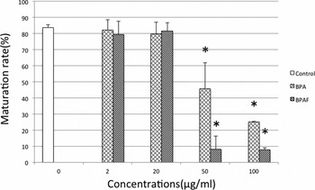
To analyse the effects of BPA and BPAF on the oocyte maturation, COCs were exposed to 2, 20, 50 or 100 μg/ml BPA and BPAF. Oocytes were cultured for 18 h, the time required to complete oocyte maturation in mice. Compared with controls, cells treated with BPA or BPAF (2 or 20 μg/ml) showed no significant difference in maturity (Fig. 1). However, at concentrations of 50 μg/ml and higher, BPA caused a dose-dependent inhibition of meiotic progress, while BPAF had more potent effects. In the 50 μg/ml BPA-treated group, 45.8% of oocytes reached MII. In comparison, 8.27% of the 50 μg/ml BPAF-treated reached MII. In the groups treated with 100 μg/ml BPA or BPAF, there was also significant decrease in maturation(25.2%, 7.83%).
Maturation rate of oocytes exposed to BPA or BPAF at different concentrations. Oocytes were cultured for 18 h. Values are the means ± standard error of the mean (SEM) of three replicates. Bars with different letters are significantly different (*P < 0.05).

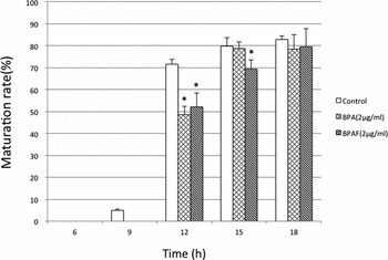
Because BPA and BPAF at a dose of 2 μg/ml did not suppress meiotic progression within the 18 h culture period, we analysed the effect of briefer BPA and BPAF exposures on the maturation of oocytes (Fig. 2). COCs were exposed to 2 μg/ml BPA or BPAF and cultured for 6, 9, 12, 15 or 18 h. In the 12 h culture group, BPA- or BPAF-exposed COCs had matured to a lesser extent than controls (48.8 and 51.1%, respectively). However, BPAF-exposed oocytes after 15 h of culture had matured less than BPA-treated oocytes (67.9 and 79.2%, no significant difference).
Maturation rate of 2 μg/ml BPA or BPAF-exposed oocytes under various culture conditions. Oocytes were cultured for 6, 9, 12, 15 or 18 h. Values are the means ± standard error of the mean (SEM) of three replicates. Bars with different letters are significantly different (*P < 0.05).

Effect of high concentrations of BPAF on meiotic progression
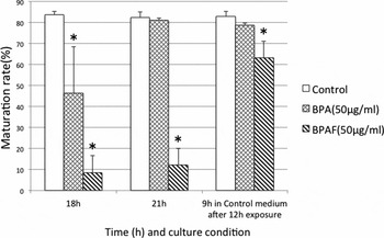
To determine whether the meiotic inhibition reported in Fig. 1 was the result of a delay or arrest, COCs were exposed to 50 μg/ml BPA or BPAF for 21 h (Fig. 3). In the BPA-treated group, 46.8 and 81.1% of oocytes reached MII after 18 and 21 h of culture, respectively (Fig. 3). In contrast, 8.5 and 12.3% of BPAF-treated oocytes reached MII after 18 and 21 h of culture.
Maturation rate of 50 μg/ml BPA or BPAF-exposed oocytes for 18 h or 21 h, and for 9 h in the control medium after 12 h exposure. Values are the means ± standard error of the mean (SEM) of three replicates. Bars with different letters are significantly different (*P < 0.05).

To determine whether the effect on oocyte maturation was reversible, oocytes were cultured under BPA or BPAF conditions for 12 h and transferred to control medium for 9 h. Under these conditions, 63.3% of BPAF-treated oocytes matured to MII, a percentage that was still significantly lower than the control.
Effect of high concentrations of BPA and BPAF on MAD2 localization
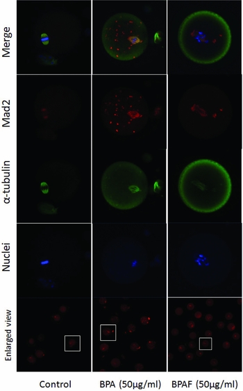
To identify the effect of BPA and BPAF exposure on MAD2 localization in arrested oocytes, treated COCs (50 μg/ml BPA or BPAF, 21 h) were denuded and co-stained using α-tubulin and MAD2 antibodies (Fig. 4). In oocytes exposed to 50 μg/ml BPA, MAD2 was detected not only in the spindle, but also in the cytoplasm. Conversely, in 95% of the oocytes exposed to 50 μg/ml BPAF, MAD2 was detected only in the spindle.
Localization of α-tubulin and Mad2 in oocytes with and without 50 μg/ml of BPA or BPAF treatment.

Discussion
BPA is a chemical widely used in the manufacturing of polycarbonate plastics (Ehrlich et al., Reference Ehrlich, Williams, Missmer, Flaws, Ye, Calafat, Petrozza, Wright and Hauser2012), epoxy resins, and lacquer lining of food and beverage cans (Bae et al., 2002; Carwile & Michels, 2011). BPA is also used in the fabrication of some dental sealants and composites (Sasaki et al., 2005; Joskow et al., 2006) and has been found in thermal receipt paper (Myers et al., 2009; Biedermann et al., 2010). Notably, from several studies, scientists have reported that these products leach BPA. It has been detected in human serum and poses danger to human health (Hunt et al., Reference Hunt, Koehler, Susiarjo, Hodges, Ilagan, Voigt, Thomas, Thomas and Hassold2003; Ikezuki et al., Reference Ikezuki, Tsutsumi, Takai, Kamei and Taketani2002). As alternative to BPA, BPAF is often used. An estimated 10,000–5,000,000 lbs of BPAF are produced annually in the USA (Stout, 2008). Recently, BPAF was nominated for comprehensive toxicological characterization by the US National Institute of Environmental Health Sciences (National Toxicology Program, 2008). However, the information regarding BPAF remains limited. In this study, we analysed the effects of BPA and BPAF on oocyte maturation by altering the concentrations of BPA or BPAF and culture time. The concentration range of BPA and BPAF was selected based on two observations. First, the migration rate of BPA has beem observed to be 2.5 μg/ml. In addition, 45.5 μg/ml of BPA or BPAF fully inhibits microtubule polymerization in cultured V79 cells 140 (Pfeiffer et al., Reference Pfeiffer, Rosenberg, Deuschel and Metzler1997).
Our data indicated that both BPA and BPAF inhibit oocyte maturation (Fig. 1). However, BPAF showed sharp reduction in oocytes reaching maturation, whereas BPA led to a dose-dependent reduction. Compared with recent reports, overall oocyte maturation was better in our study. In recent studies, 30 μM (approximately 6.82 μg/ml) and 10 μg/ml doses of BPA led to decreased oocyte maturation (Eichenlaub-Ritter et al., Reference Eichenlaub-Ritter, Vogt, Cukurcam, Sun, Pacchierotti and Parry2008; Lenie et al., Reference Lenie, Cortvrindt, Eichenlaub-Ritter and Smitz2008). This differed from the culture methods used here. We carried out IVM using a single culture system, a method in which oocytes progressed faster than in mass culture (unpublished data in our laboratory).
BPA reportedly induces meiotic cell cycle delay in mouse oocytes (Can et al., Reference Can, Semiz and Cinar2005). As show in Fig. 2, we monitored the maturation of oocytes treated with 2 μg/ml BPA or BPAF. As shown in Fig. 1, this concentration had no effect on oocyte maturation. When exposed to 2 μg/ml BPA or BPAF, the majority of oocytes progressed to MII after 15 and 18 h of IVM, respectively. After 12 h of culture, the proportion of BPA- and BPAF-treated oocytes reaching MII (48.5 and 52.2%, respectively) was significantly lower than for the controls (71.6%). Therefore, our data indicate that BPA and BPAF inhibit oocyte maturation; however, at a dose of 2 μg/ml, BPAF is more effective than BPA.
As shown in Fig. 1, oocytes exposed to 50 μg/ml BPA or BPAF for 18 h showed inhibited maturation. Therefore, we examined the extent of maturation in oocytes cultured for 21 h (Fig. 3). The cell cycle was delayed in response 50 μg/ml BPA, whereas oocytes treated with the same concentration of BPAF did not progress to MII, indicating that they had experienced a meiotic arrest.
To analyse the condition of the spindle and SAC in treated oocytes, immunofluorescence staining was performed using antibodies against α-tubulin and MAD2. The exposure to 2 μg/ml BPA and BPAF caused spindle abnormalities but showed no difference in the MAD2 localization as controls. Consistent with previous reports, 2 μg/ml BPA and BPAF treatments caused spindle abnormalities (Can et al., Reference Can, Semiz and Cinar2005; Eichenlaub-Ritter et al., Reference Eichenlaub-Ritter, Vogt, Cukurcam, Sun, Pacchierotti and Parry2008; Lenie et al., Reference Lenie, Cortvrindt, Eichenlaub-Ritter and Smitz2008; Machtinger et al., Reference Machtinger, Combelles, Missmer, Correia, Williams, Hauser and Racowsky2013). Figure 4 shows images of α-tubulin and MAD2 in 50 μg/ml BPA- and BPAF-treated oocytes. In BPAF-treated oocytes (50 μg/ml), which arrest after germinal vesicle breakdown, MAD2 was localized to the spindle. This staining pattern is reminiscent of oocytes over-expressing MAD2 (Wassmann et al., Reference Wassmann, Niault and Maro2003). The MAD2-dependent SAC is functional during the first meiotic division in mouse oocytes (Niault et al., Reference Niault, Hached, Sotillo, Sorger, Maro, Benezra and Wassmann2007). Therefore, we speculate that the BPAF-induced meiotic delay and arrest result from SAC activity. Figure 4 showed the localization of MAD2 in BPA-treated oocytes. Multiple small MAD2-positive foci were observed throughout the ooplasm as oocytes progressed to MII. The foci are indicative of SAC dysfunction, which may lead to meiosis delay rather than full arrest. The localization of MAD2 changed with the deformation of the spindle in BPA-treated oocytes.
Here, we report the different effects of BPA and BPAF on oocyte maturation. Our data are consistent with that of previous studies. Because of the CH3–CF3 substitution on the bisphenol backbone, differences in receptor selectivity between BPAF and BPA exist. BPA binds strongly to ERRγ, but very weakly to ERα and ERβ (Nagel et al., Reference Nagel, Saal, Thayer, Dhar, Boechler and Welshons1997; Blair et al., Reference Blair, Fang, Branham, Hass, Dial, Moland, Tong, Shi, Perkins and Sheehan2000; Sheeler et al., Reference Sheeler, Dudley and Khan2000; Matsushima et al., Reference Matsushima, Kakuta, Teramoto, Koshiba, Liu, Okada, Tokunaga, Kawabata, Kimura and Shimohigashi2007). BPAF is a full agonist for ERα and acts as a specific and strong antagonist of the endogenous ERβ agonist 17β-estradiol (Matsushima et al., Reference Matsushima, Liu, Okada, Shimohigashi and Shimohigashi2010). These reports suggest that BPAF and BPA have adverse effects on human health.
In conclusion, BPA and BPAF have been shown to act as meiosis-disrupting agents that interfere with microtubule organization and SAC activity. BPA-treated oocytes apparently underwent polar body extrusion normally, but had some abnormalities. Conversely, high concentrations of BPAF arrested oocyte maturation. Thus, we conclude that both BPA and BPAF have toxic effects on reproduction. Also, our laboratory showed that the HD monoculture system is an efficient method to screen substances that affect oocyte culture (Ishikawa et al., Reference Ishikawa, Machida, Hiraga, Hiradate, Suda and Tanemura2014; Nishio et al., Reference Nishio, Hoshino, Tanemura and Sato2014). The monoculture system can circumvent several problems associated with group culture, a system that is frequently used in artificial reproductive technologies and animal reproduction. The monoculture system requires smaller numbers of oocytes and animals for each experiment thereby making it amenable to high throughput testing. In addition, it is easy to modify and control the microenvironment in monocultures. In this study, we also established that the HD monoculture system, a method in which oocytes have no paracrine effects, is more useful to screen chemicals that effect oocyte maturation.