Brief reviews of advanced imaging techniques in adverse pregnancy settings in the setting of DOHaD
Imaging fetal growth and health with ultrasound
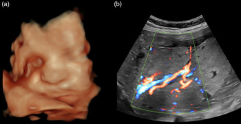
The fetus grows inside the mother, which creates a black box of wonder for moms, dads, grandparents, and healthcare providers. However, a window into this black box is important for diagnosing problems with fetal growth and development that can result in significant early life or subsequent health problems (Fig. 1). Ultrasound is an important part of the clinical management of pregnancy, where it is used to determine fetal viability, gestational age, singleton versus multiple pregnancies, morphology, growth, fetal weight, and fetal cardiac function. Reference Abinader and Warsof1 Progress assessment of most pregnancies in developed countries involves a morphology ultrasound at approximately 12 weeks of gestation for accurate dating, and from which to infer healthy fetal growth later in gestation. This is followed by a mid-gestation ultrasound scan to measure the trajectory of fetal growth, and to identify any developmental abnormalities that may require a more detailed follow-up. Later, gestation serial assessment of fetal size by ultrasound is usually recommended in women with risk factors for poor fetal growth and placentation, such as hypertension and diabetes, so as to identify those fetuses that are small or are failing to achieve their growth potential. 2 For the low-risk pregnant woman, it has been difficult to demonstrate the benefit of serial fetal size assessment, although a recent prospective study found that screening of nulliparous women with universal third-trimester fetal biometry improved detection of the small fetus and identified a subset that was at increased risk of neonatal morbidity. Reference Sovio, White, Dacey, Pasupathy and Smith3
Four-dimensional ultrasound image showing the face of a fetus at 34 weeks’ gestational age (a). Example of Doppler ultrasound of placental chorionic vessels (red = positive flow, blue = negative flow) in a 34-week gestational age fetus (b).

The addition of Doppler ultrasound permits assessment of blood velocity dynamics and waveform patterns in multiple vessels inclusive of the uterine arteries, umbilical arteries, middle cerebral artery, and ductus venosus, with the calculation of resistance and pulsatility of blood flow.
This allows obstetricians to infer fetal health based on expected blood flow responses to the intrauterine environment. Uterine artery Doppler studies can be used to predict as early as in the first trimester those women at increased risk of pregnancy complications such as pre-eclampsia. The second trimester raised pulsatility index and “notched” abnormal utero-placental flow velocity waveforms also provide evidence of poor placentation, characterized histologically as reduced or absent trophoblast invasion of the spiral arteries. Reference Lyall, Robson and Bulmer4 The first-trimester detection of pre-eclampsia is improved when uterine artery assessment is paired with maternal serum screening and mean arterial blood pressure. Reference Drouin, Boutin and Paquette5,Reference Poon, Shennan and Hyett6 Women found to be at high risk can be prescribed low-dose aspirin as a means to reduce pre-eclampsia and fetal growth restriction (FGR). Reference Groom and David7 However, ultrasound screening does not ensure that all complicated pregnancies are detected. Reference Cnossen, Morris and ter Riet8,Reference English, Kenny and McCarthy9
Ultrasound visualization of the placenta is important, as most placentas examined following extremely preterm births (<28 weeks of gestation) with pre-eclampsia or FGR are small and grossly abnormal. Reference Mifsud and Sebire10 Two-dimensional ultrasound can be useful to assess the maximum placental length, thickness, overall placental shape, and cord insertion, as gross abnormalities such as a narrow thick placenta or a velamentous cord insertion confer an increased risk of perinatal complications. Reference Kingdom, Audette, Hobson, Windrim and Morgen11 Assessment of feto-placental, cerebral and cardiovascular hemodynamics, fetal movements, and liquor volume are key to the management of pregnancies with early and late-onset FGR. Reference Figueras and Gratacos12–Reference Morrison15 Despite its limitations, ultrasound screening remains the current standard for clinical care because of its safety profile, affordability, and ease of use.
In a research environment, the impact of various pregnancy complications, such as FGR, prenatal iron deficiency, and an altered immune profile on uterine and umbilical blood flow velocity and resistance to blood flow can be controlled and evaluated. Reference Woodman, Care and Mansour16 For example, given reports of increasing gestational cannabis consumption and risk of associated impaired placental blood flow, increased placental vascular resistance, and FGR, ultrasound may have a role in guiding the antenatal care of pregnant women and fetuses at risk. Reference Corsi, Walsh and Weiss17–Reference Benevenuto, Domenico and Martins19 Antenatal ultrasound scans can provide information of value to both mother and fetus, including maternal adiposity (e.g. subcutaneous fat thickness and fatty liver), fetal growth and adiposity distribution evidenced by fetal abdominal circumference, and possibly fetal fatty liver. These features provide a window to the possible insulin-resistant maternal–fetal metabolic environment. Furthermore, the utility of ultrasound is enhanced by laser photoacoustic capability, enabling the assessment of oxygenation and hemoglobin saturation in the fetus and placenta, and how these change in animal models of pregnancy complications. Reference Yamaleyeva, Sun, Bledsoe, Hoke, Gurley and Brosnihan20–Reference Maneas, Aughwane and Huynh22
Imaging real-time blood flow and oxygenation in the fetus with ultrasound and magnetic resonance imaging
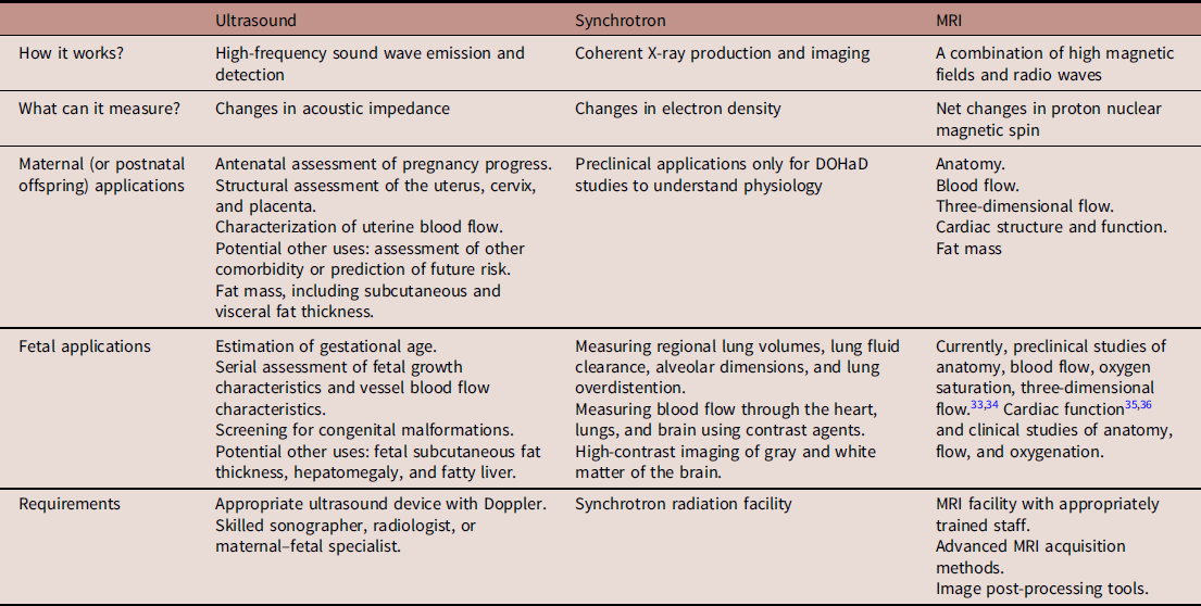
Magnetic resonance imaging (MRI) is increasingly used in fetal imaging applications (Table 1). This technology uses strong magnetic fields (up to 3.0 T) and radiofrequency pulses to generate signals from 1H nuclei. Time-varying magnetic fields are used to encode spatial information into this signal so that images can be created. MRI can generate exquisite high-contrast images of soft tissues in the body because they contain high concentrations of protons (1H) in water and lipid molecules. High-spatial resolution structural information about the fetus and placenta can be obtained, and this is supplemented by the menagerie of unique imaging contrasts that can be obtained using different pulse sequences. MRI is nonionizing and considered safe in pregnancy, Reference Gowland and Prayer23,Reference Ray, Vermeulen, Bharatha, Montanera and Park24 but is limited by its high cost, the significant time needed to acquire images, and maternal comfort because of the small-bore size and claustrophobia. Despite this, recent advances in MRI have allowed not just morphological 3D images of the fetus to be obtained, but methods to measure blood flow in major vessels (volume flow in ml/min) throughout the body, so that the absolute distribution of cardiac output can be determined in preclinical models of human pregnancies Reference Duan, Darby and Soo25 and clinically. Reference Prsa, Sun and van Amerom26 This information is useful clinically because it is known that the fetus responds to acute hypoxia by redistributing cardiac output to maintain perfusion of the brain, heart, and adrenal glands. Reference Giussani27,Reference Bennet28 Thus, detection of a redistribution of cardiac output in the fetus is a sign of fetal hypoxemia, and coupled with other measures, may signal the need for preterm delivery to reduce the risk of stillbirth. Interestingly, when this method was used to measure the distribution of cardiac output in the FGR human fetus, Reference Zhu, Milligan and Keating29 there was no change in the distribution of cardiac output, a finding that is consistent with sheep studies of FGR with chronic hypoxemia. Reference Poudel, McMillen, Dunn, Zhang and Morrison30,Reference Miller, Supramaniam, Jenkin, Walker and Wallace31 However, hyperoxygenation does change fetal hemodynamics as measured using MRI. Reference Porayette, Madathil and Sun32
Comparative applications of different imaging approaches

Even with a direct measure of blood flow, a finding of no redistribution may not mean that the fetus is doing well, since chronically hypoxemic fetuses may not redistribute their cardiac output in late gestation. Reference Zhu, Milligan and Keating29–Reference Miller, Supramaniam, Jenkin, Walker and Wallace31 A more direct measure would be oxygen saturation, which can be sampled invasively (cordocentesis) in animal models, but presents risks of miscarriage or preterm birth in humans. Reference Soothill, Nicolaides and Campbell37 Thus, recent advances in MRI that allow accurate and precise measurement of fetal oxygen saturation will have a significant impact on clinical practice. Reference Portnoy, Seed, Sled and Macgowan38–Reference Luo, Abaci Turk and Bibbo41 This methodology has recently been validated in a sheep model where it was possible to use a blood gas machine to measure blood oxygen saturation to create a relationship with T2 measures. Reference Saini, Darby and Portnoy42,Reference Morrison, Berry and Botting43 Researchers have also investigated measuring oxygen levels in the placenta, which is less susceptible to fetal movement. Recent models have attempted to separate the fetal and maternal contributions of the MRI signal to analyze oxygen saturation and exchange between mother and fetus in controls and FGR. Reference Melbourne, Aughwane and Sokolska39,Reference Aughwane, Ingram, Johnstone, Salomon, David and Melbourne44,Reference Aughwane, Mufti and Flouri45 Placental MRI thus promises to be a powerful tool both for monitoring pregnancy and measuring the efficacy of new therapy. Knowing that a fetus is small but has a normal oxygen saturation may give more confidence that delaying delivery to gain time in utero will not compromise the fetus significantly, whereas a low oxygen saturation may trigger a decision to deliver to prevent stillbirth. This could have a significant impact on neonatal outcomes and health throughout the life course.
It is also possible to use MRI to image the fetal heart. Human studies have employed accelerated and motion-corrected MRI approaches to achieve multi-slice and volumetric dynamic anatomical imaging of the fetal heart, relying on retrospective data analysis to achieve cardiac gating. Reference Roy, Marini and Lloyd46–Reference Chaptinel, Yerly and Mivelaz48 Conversely, preclinical sheep models of pregnancy provide reliable prospective fetal cardiac gating signals using invasive probes. Reference Yamamura, Kopp and Frisch49,Reference Yamamura, Schnackenburg and Kooijmann50 This latter approach has been applied to identify a cardiac infarct in the fetus and determine cardiac function. Reference Duan, Lock and Perumal36 2D phase-contrast MRI encodes the velocity of moving tissue such as blood flow directly into the MRI acquisition and can be used to determine cardiac output, while ventricular volumetric can be used to determine additional factors such as ejection fraction and stroke volume. Reference Cho, Darby and Saini35
Understanding brain maturation with MRI
Advances in MRI have allowed us to better understand the causes of fetal and neonatal brain injury, and provide early detection to improve management to limit the long term consequences of brain injury in compromised newborns. Reference Melbourne, Eaton-Rosen and Orasanu51–Reference Ball, Srinivasan and Aljabar56 MRI provides greater soft tissue contrast than transcranial ultrasound, although it is technically more challenging. The neonatal brain has a higher tissue water content and less myelinated white matter than the mature brain. MRI is particularly useful for assessing processes associated with myelination, which occurs at different rates in different brain regions Reference Dai, Muller, Wang and Deoni57 and species Reference Morrison, Berry and Botting58 ; however, this is more challenging in the fetus. Investigators have found white matter abnormalities to be predictive for severe cognitive delay and other neurodevelopmental impairments in preterm infants. Reference Woodward, Anderson, Austin, Howard and Inder59
Clinically, structural T1- and T2-weighted MRI can be used to differentiate white matter from gray matter, calculate regional brain volumes, and perform cortical surface reconstruction and gyrification analysis, Reference Dai, Muller, Wang and Deoni57 as well as assess the development of myelinated white matter. Reference Schmidbauer, Geisl and Diogo60 Both structural and diffusion-weighted MRI can also be used to detect overt brain injury associated with insults such as hypoxia–ischemia, encephalopathy, hemorrhage, infection, and trauma, Reference Woodward, Anderson, Austin, Howard and Inder59 as well as visualize fiber tracts in the brain, a process known as MR tractography. Reference van de Looij, Dean, Gunn, Huppi and Sizonenko61,Reference van de Looij, Lodygensky and Dean62 Hypoxia–ischemia is a relatively common insult to the developing brain, and can impair normal brain development or in extreme cases, lead to acute brain damage. Reference Davidson, Dean and Fraser63 After the insult, a rapid depletion of brain cell metabolites occurs. This is followed by partial recovery of oxidative metabolism, but without the treatment, another decline in energy metabolism will follow leading to cell death hours to days later. Severe trauma may substantially reduce this critical window. Magnetic resonance spectroscopy (MRS) is also used to provide additional information on changes in metabolism occurring while the brain adapts to hypoxic challenges. Reference van de Looij, Dean, Gunn, Huppi and Sizonenko61,Reference Azzopardi, Wyatt and Cady64–Reference Malhotra, Sepehrizadeh and Dhollander66 Thus, the ability to noninvasively evaluate the structural and metabolic status of the injured neonatal brain allows opportunities for improved understanding of the underlying physiology, while also functioning as a tool to help evaluate the efficacy of potential therapies.
Advanced diffusion imaging techniques can also provide sensitive biomarkers for assessing the microstructural development of the brain, and the efficacy of potential therapies in the neonate. In the third trimester and in early postnatal life, there are marked changes in the growth and maturation of the white matter and gray matter regions of the human brain, including the onset and progression of axonal myelination, Reference Back, Luo, Borenstein, Levine, Volpe and Kinney67–Reference de Graaf-Peters and Hadders-Algra69 and the differentiation, branching, and connectivity of neuronal dendrites. Reference Kriegstein70–Reference Mrzljak, Uylings, Kostovic and van Eden72 Critically, in the absence of overt brain lesions, MRI studies combined with pathological data suggest that these key developmental events are particularly vulnerable to disruption following premature birth or other prenatal/postnatal insults. Reference Dean, Bennet, Back, McClendon, Riddle and Gunn73–Reference Back and Miller75 For example, extensive diffusion tensor (DTI) studies using the index fractional anisotropy have shown persisting alterations in white matter and gray matter microstructural development in infants born prematurely, Reference Ball, Srinivasan and Aljabar56,Reference Thompson, Kelly and Chen76,Reference Ment, Hirtz and Hüppi77 with histological correlates of reduced white matter myelination and reduced dendritic arborization of cortical neurons. Reference Dean, McClendon and Hansen78–Reference Favrais, van de Looij and Fleiss81 The recent development of neurite-orientation dispersion and density imaging (NODDI) also provides more specific markers of cellular microstructure than for DTI, including separate measures of neurite density and neurite dispersion/fanning, Reference Zhang, Schneider, Wheeler-Kingshott and Alexander82 allowing more detailed assessment of underlying cellular changes during brain development Reference Lynch, Cabeen, Toga and Clark83,Reference Jelescu, Veraart, Adisetiyo, Milla, Novikov and Fieremans84 and following neonatal brain injury. Reference Batalle, O’Muircheartaigh and Makropoulos85 Changes in these NODDI indices in the white matter and cerebral cortex, and their associations with functional outcomes, have been reported in very preterm infants, Reference Batalle, O’Muircheartaigh and Makropoulos85–Reference Kelly, Thompson and Chen87 as well as in experimental studies of neonatal hypoxia–ischemia. Reference Ohki, Saito, Hata, Okano, Higuchi and Fukuchi88 Finally, investigators are also using blood-oxygen-level-dependent (BOLD) functional MRI to evaluate the relationships between structural and functional connectivity in the neonatal brain. Reference Omidvarnia, Fransson, Metsaranta and Vanhatalo89
There are significant technical challenges associated with MRI of infants. Special equipment, especially specialized radiofrequency coils are required. Mitigating image degradation because of involuntary motion is a constant challenge. Reference Denisova90 Since important aspects of the brain’s adaptation to an injury can occur in utero, investigators are extending the methods previously used on infants to image the fetal brain. Reference van de Looij, Lodygensky and Dean62,Reference Gholipour, Estroff and Warfield91,Reference Ebner, Wang and Li92 Growth restriction in the human fetus is one such pathology in which the brain has been extensively studied by investigators using MRI in utero. Smaller brain volume, insular cortical thickness, and corpus callosum length, thickness, and area have been observed in growth-restricted cohorts at term in comparison to appropriate for gestational age fetuses. Reference Egana-Ugrinovic, Sanz-Cortes, Figueras, Bargallo and Gratacos93 These findings correlate significantly with a poorer neurobehavioral performance at birth. Similarly, there is evidence for cerebellar and brainstem differences in term small for gestational age fetuses, which are associated with altered neurobehavioral during the neonatal period. Reference Egana-Ugrinovic, Sanz-Cortes, Couve-Perez, Figueras and Gratacos94 MRS of the fetal brain has also been studied in term fetuses with growth restriction for brain metabolism assessment. A significantly decreased N-acetylaspartate (NAA)/choline (Cho) ratio has been observed, indicating subtle reorganization of the brain in response to hypoxia. Reference Sanz-Cortes, Simoes, Bargallo, Masoller, Figueras and Gratacos95 In summary, methods for evaluating reproducibility and standardizing interpretation and analysis must be generally adopted, which requires the active participation of multi-institutional teams of investigators. Reference Makropoulos, Robinson and Schuh96
Synchrotron imaging of cardiorespiratory function: improving the transition at birth
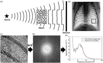
Synchrotron-based phase-contrast X-ray (PCX) imaging has provided invaluable insights into previously unrealized mechanisms regulating development and the transition at birth. Synchrotron-based PCX imaging technology uses the refractive index difference between air and water to image the airways as they fill with air at birth. The phase shift of the X-rays as they pass across the air/water boundary produces strong contrast that is orders of magnitude greater than the absorption contrast. When multiple air-filled structures (e.g. alveoli), overlie each other in projection, a complex speckle pattern is formed that can be analyzed to determine total and regional lung gas volumes as well as the size and number of alveolar units in projection (<100 µm; Fig. 2). The resulting information has been used to provide a fundamental understanding of the process of lung aeration and how this process can be assisted to improve interventions in the delivery room for subpopulations of newborns. Implementing the most supportive interventions that promote respiratory function in the vital minutes after birth while not causing lung injury are critical for preserving life without significant lifelong consequences. The strength and clinical relevance of this work lies in the multidisciplinary collaborations whereby innovative imaging techniques are used to investigate the most pressing newborn issues, yielding solutions that can be translated back into the clinic to improve outcomes.
Overview of synchrotron-based PCX imaging of lung aeration in a newborn rabbit. (a) Illumination of a phase object by partially coherent radiation source creates phase-contrast fringes upon propagation through space to the detector as a result of interference effects. The airways in lung tissue can serve as a random phase screen for X-rays, resulting in a speckled pattern providing details about lung aeration in the entire lung (b). Zooming in on the black box from (b) reveals the speckle detail in the terminal airways in (c). The two-dimensional (2D) power spectrum of (c) is shown in (d) (log scale). (e) Azimuthal average of (d) showing the raw data fit with a Pearson type VII function for measuring the position of the peak in the power spectrum caused by the speckle. The peak centroid at 6.2 ± 0.4 mm−1 reveals that the dominant length scale of this region of lung speckle is 159 ± 10 μm. (From99, Copyright Institute of Physics and Engineering in Medicine. Reproduced by permission of IOP Publishing. All rights reserved).

This improvement in our fundamental knowledge of newborn physiology has revolutionized the support of premature newborns with worldwide implications. Ongoing work has focussed on understanding the early origins of lung disease caused by a congenital condition leading to a diaphragmatic hernia, which results in severe lung hypoplasia (smaller lungs). This can have a devastating consequence for these newborns, leading to poor outcomes including air leaks, pulmonary hypertension, and death. Reference Flemmer, Thio and Wallace97 More recently, this technology has begun to tease apart mechanisms underlying the pathophysiology of respiratory distress in near-term newborns caused by elevated airway liquid volumes at birth, which represents a substantial and growing burden on newborn intensive care resources in clinical practice. Reference McGillick, Lee and Yamaoka98 Further advances in this field are in providing greater insights into the mechanisms and interventions to improve newborn cardiorespiratory outcomes.
Using synchrotron imaging, Kitchen et al. showed that speckle patterns created by the lungs in PCX images enable quantitative measurement of the mean airway dimensions in projection through the lungs. Reference Kitchen, Buckley and Leong99 Using an animal model, they showed that the airways, which is determined by the speckles, change size during respiration in a manner that could readily distinguish between immature lungs and those that had begun alveolar development near-term. The use of this technique highlighted the tendency of the premature newborn lung to collapse (de-recruit) or refill with lung liquid between breaths at low airway pressures, while the terminal airways in a more mature lung remained open between breaths but with smaller dominant airway size, highlighting more uniform distribution of airway size within the lung. More recently, this technology has provided an understanding of the effects of elevated airway liquid volume at birth as a cause of poor lung function in near-term newborns who experience respiratory distress. Reference McGillick, Lee and Yamaoka98 The advances using synchrotron imaging technology combined with our growing understanding of factors affecting abnormal cardiorespiratory development and the transition at birth help ensure that some of the biggest questions in neonatology are answered, and that the safest and most effective interventions are used in early life to reduce the lifelong burden of disease in newborn subpopulations.
While other imaging advances in DOHaD are focussed on developing techniques to take the imaging from preclinical to validated clinical methods, the availability of synchrotron imaging for routine diagnostics is not feasible for newborns. Instead, synchrotron PCX imaging provides a firm foundation using preclinical models that mimic morbidity in clinical practice to understand the physiology, which can then inform clinical trials and ultimately change clinical practice to improve newborn outcomes. Synchrotron imaging has a growing interest in clinical radiography diagnostics/treatments, and recent advances have increased the sensitivity of detection and resulted in significant reductions in radiation dose. Reference Kitchen, Buckley and Gureyev100–Reference Pacile, Baran and Dullin102 In the future, this will have significant implications in both clinical and veterinary medicine, using either non-synchrotron sources or compact X-ray sources.
Using imaging techniques to visualize early distribution and accumulation of fat and its associations early in life in the clinical setting
Nonalcoholic fatty liver disease (NAFLD) is a complex liver disorder associated with excessive adiposity, trajectories of weight gain, and insulin resistance unrelated to alcohol at any stage during the life course. NAFLD is the most common cause of chronic liver disease worldwide. Though commonly recognized as an adult-onset disease, NAFLD or risk factors for NAFLD may have their onset from as early as prenatal life, early–late childhood, adolescence, or adulthood. As NAFLD prevalence in the general population increases, the importance of maternal prepregnancy and pregnancy factors, including antenatal ultrasound examination, becomes important in predicting maternal and offspring cardiometabolic outcomes, including NAFLD. Currently, maternal obesity, gestational diabetes, excessive maternal weight gain, and excessive high-fat intake during pregnancy are associated with NAFLD in offspring.
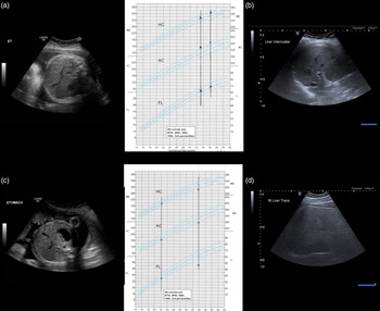
The earliest risk factors for NAFLD are attributable to maternal prepregnancy and gestational health and lifestyle. Reference Ayonrinde, Adams and Mori103–Reference Abdelmalek, Liu, Shuster, Nelson and Asal112 This is evidenced by the observation of NAFLD in stillborn autopsy studies, in neonates using MRI, and in infants using ultrasound. Reference Patel, White and Deutsch113–Reference Bedogni, De Matteis and Fabrizi116 Small for gestational age or microsomy suggestive of intrauterine growth restriction, with accelerated catch-up growth, as well as large for gestational age or macrosomy, is associated with subsequent NAFLD during early life. Reference Patel, White and Deutsch113,Reference Nobili, Marcellini and Marchesini117 Both ultrasound and MRI scanning display the abdominal girth, liver size, and thickness of the fetal abdominal subcutaneous fat compartment (Fig. 3a, 3b), as well as identifying gestational fatty liver in the mother. Additionally, maternal fatty liver during the first trimester of pregnancy is associated with fetal macrosomy, independent of maternal gestational diabetes mellitus. Reference Lee, Kim and Koo118 Thus, antenatal ultrasound has the potential to identify not only maternal metabolic risk, but also extrapolate the quality of the metabolic milieu in which the fetus resides. There is controversy related to whether the perinatal or postnatal environment contributes most to a future diagnosis of NAFLD; however, it is known that NAFLD prevalence increases with age from childhood through adolescence and adulthood, and is associated with increased risk of diabetes and cardiovascular disease. Reference Ayonrinde, Olynyk and Beilin119 Recent MRI evidence from nonhuman primates suggests that prenatal corticoid administration also may also influence liver steatosis in adult life. Reference Kuo, Li and Li120 Excessive weight gain during early childhood also increases the risk of NAFLD during childhood and adolescence. Reference Ayonrinde, Olynyk and Marsh121 Fatty liver with corresponding increased subcutaneous fat thickness can be detected in children using ultrasound (Fig. 3c, 3d).
Antenatal fetal morphometry ultrasound scans with corresponding growth charts in (a) a large for gestational age (macrosomic) fetus with increased abdominal subcutaneous fat, abdominal circumference, and estimated fetal weight >97% centile, hepatomegaly, and presumed fatty liver, (b) a small for gestational age (microsomic) fetus with thin abdominal subcutaneous fat, abdominal circumference <5th centile, and estimated fetal weight <3rd centile (i.e. future risk of developing fatty liver associated with catch-up growth), (c) a fatty liver with a 27-mm abdominal subcutaneous fat thickness in an 8-year-old child, and (d) a severe fatty liver with a 43-mm abdominal subcutaneous fat thickness in a 15-year-old adolescent. (Scan images courtesy of Marilyn Zelesco, Lead Sonographer, Fiona Stanley Hospital).

Liver ultrasound and other imaging modalities such as controlled attenuation parameter (CAP), MRI, and computed tomography (CT) scans are useful for noninvasive diagnosis of NAFLD in children; however availability of CAP and MRI are limited, and CT scan radiation dose limits its utility in children. Reference Li, Dhyani, Grajo, Sirlin and Samir122–Reference Chartampilas124 In the future, there will be a need for more detailed fetal anatomy scans incorporating adipose distribution in the metabolically active sites of the liver, subcutaneous, and visceral fat compartments. This will be particularly important in understanding associations between intrauterine exposures, adipose distribution, and longer-term cardiometabolic risk.
Brown adipose tissue (BAT) differs from white adipose tissue (WAT) as it can utilize energy and become exoergic, so that it plays a role in thermoregulation. In humans, BAT is metabolically active at birth with depots in the shoulders, suprarenal areas, pericardial regions, around the aorta, and within the abdominal cavity. Reference Symonds125 During development, brown fat recedes or is replaced by white fat in all of those locations, so that there typically exist only small, metabolically-inactive substernal and supraclavicular depots by adulthood. For humans living in colder climates, there may be greater retention of BAT during growth. BAT cells have an embryonic origin that is more akin to that of muscle cells and connective tissues than with white fat. Reference Darby, Sorvina and Bader126,Reference Ghazanfar, Vuocolo and Morrison127 The fact that BAT is prominent at birth and rapidly diminishes has led to speculation that proper characterization of BAT volume and metabolism over time may inform our understanding of the development of obesity and metabolic disorders in adult life. Reference Symonds, Pearce, Bispham, Gardner and Stephenson128
Multiple imaging technologies have been employed to image BAT. Reference Sun, Yan, Sun, Velan and Leow129 In adults, positron emission tomography (PET)/CT has proved the most successful; however, PET is inherently limited by only detecting metabolically active BAT, and its low spatial resolution limits the ability to detect small and/or diluted amounts. Infrared thermography (IRT) has been used to image BAT in children, but is limited to evaluations of only superficial tissues. Reference Symonds, Henderson and Elvidge130 MRI also has the potential for assessing the amount and metabolic activity of BAT. Multi-echo water-fat MRI studies of neonates reported that WAT fat fraction was significantly greater than BAT fat fraction in spinal, supraclavicular, and axillary regions. Reference Rasmussen, Entringer and Nguyen131 Using this MRI fat-fraction difference to identify BAT versus WAT, an inverse relationship between BAT loss and muscle mass increase between birth and 6 months of age was reported in healthy infants. Reference Ponrartana, Aggabao, Chavez, Dharmavaram and Gilsanz132 An MRI study in healthy infants supports the hypothesis that higher BAT concentration at birth may be protective against excess adiposity gain during the first 6 months of life. Reference Entringer, Rasmussen and Cooper133 Also, since the color of BAT is primarily because of its high concentration of iron-rich mitochondria, T2 and T2* parametric imaging can be used along with fat-fraction MRI for segmentation of BAT from WAT. Reference Bhanu Prakash, Verma and Yaligar134 This approach has been used in human infants and children to confirm that T2* and fat fraction are complimentary MRI biomarkers for evaluating BAT. Reference Hu, Yin, Aggabao, Perkins, Chia and Gilsanz135
Limitations of BMI: how imaging can get us to the next level?
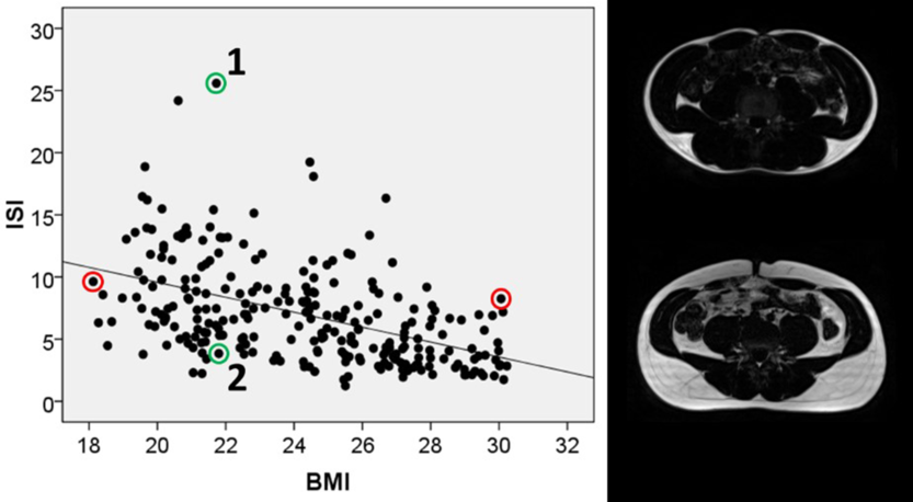
BMI provides information about whole-body adiposity, but masks a great deal of information regarding the body composition and fat compartmentation. BMI does not differentiate between fat mass and lean mass, hence a person with high muscle mass can be placed into the overweight/obese categories based on his/her BMI. The body’s fat/fat-free mass composition is associated with metabolic health, and individuals with comparable levels of obesity can have different fat partitioning profiles. This is illustrated by the relationship between insulin sensitivity index and BMI of the subjects in the Singapore Adults Metabolism Study, which included normal and overweight Singaporean adult men aged 20−40 years (Fig. 4). Reference Khoo, Leow and Sadananthan136 Subjects 1 and 2 have a similar normal range of BMI, but have varied insulin sensitivity values and different body fat distribution. Subject 1 has a lower percentage of body fat compared with subject 2 (9.7% vs. 25.3%, respectively). Subject 2 has increased abdominal subcutaneous (SAT) and visceral adipose tissue (VAT) depot volumes and a lower insulin sensitivity index than subject 1. Thus, a person can be obese based on BMI, yet metabolically healthy, or lean and yet metabolically unhealthy. Ethnic and sex differences exist in body fat partitioning and several studies have investigated the strong links with insulin resistance and cardiovascular health. Reference Khoo, Leow and Sadananthan136–Reference Revenga-Frauca, González-Gil and Bueno-Lozano140
Individuals with similar body mass index (BMI) and different insulin sensitivity levels, and their body fat partitioning profile, from the Singapore Adults Metabolism Study. Reference Khoo, Leow and Sadananthan136

MRI and MRS provide a noninvasive and comprehensive way to accurately assess and estimate body fat distribution. Different MRI and MRS techniques can be employed to determine fat in the abdomen, liver, muscle, pancreas, and heart, etc. MRI protocols have been optimized for imaging abdominal fat, liver fat, and muscle fat in adult men. Reference Sadananthan, Prakash and Leow141 In the abdomen, the adipose tissue compartment is divided into visceral (VAT) and subcutaneous adipose tissues (SAT). The SAT is separated into deep (DSAT) and superficial subcutaneous adipose tissue (SSAT) compartments by a fascial plane known as the fascia superficialis. These adipose tissue depots are structurally and metabolically different, hence accurate quantification of these fat depots will provide a better marker of metabolic risk than the whole abdominal fat volume. Reference Lee, Wu and Fried142,Reference Walker, Verti and Marzullo143 With the aim of determining the individual fat depot volumes, a fully automated algorithm based on graph cuts and level sets to segment and quantify the volume of SAT, SSAT, and VAT depots has been developed. Reference Sadananthan, Prakash and Leow141
Pediatric MRI protocols and data processing pipeline had been established to assess body fat partitioning in infancy and early childhood Reference Tint, Fortier and Godfrey144–Reference Michael, Gupta and Sadananthan146 through the Growing Up in Singapore Towards Healthy Outcomes (GUSTO) study, which investigated maternal and early-life determinants of metabolic health and disease later in life. Reference Soh, Tint and Gluckman147 MRI provides a feasible, reliable, and safe approach to longitudinally track the changes in fat accumulation as the children age. Indian and Malay neonates had higher levels of DSAT than Chinese neonates,. Reference Tint, Fortier and Godfrey144 recapitulating a phenotype of elevated deep and SSAT reported in adult men in the SAMS study. Reference Khoo, Leow and Sadananthan136 This ethnic patterning of abdominal fat persisted at age 4.5 years, with the highest SSAT and DSAT levels in Indian children and the lowest levels in Chinese children. Reference Sadananthan, Tint and Michael145 No significant sex or ethnic differences were observed in VAT. Skeletal muscle insulin resistance plays a role in the pathophysiology of type 2 diabetes, and cytosolic accumulation of lipid droplets within myocytes, referred to as intramyocellular lipids (IMCLs), has been implicated in the early pathogenesis of insulin resistance. Indian adults have a higher predisposition to accumulating IMCLs compared with Chinese and Malays. Reference Khoo, Leow and Sadananthan136 We found this ethnic variation to emerge in early childhood, with Indian children having the highest IMCL levels at age 4.5 years. Reference Michael, Gupta and Sadananthan146 Thus, MRI provides an effective way to investigate body fat partitioning in children and adults because accurate estimates of the different fat depots in the context of understanding ethnic diversity can help in predicting the risk of adverse metabolic health.
New innovative studies with imaging modalities in DOHaD research
Application of MRI to visualize the foramen ovale in the human fetus to understand the hemodynamics of streaming
X-ray imaging during infusion of iodine contrast can be used to visualize complex patterns of fetal blood flow, but this approach is limited in clinical practice by the risk of exposure to ionizing radiation and the complications of infection from intravenous injection and allergic reaction to iodine. Reference Wang, Chong and Kielar148 Imaging the fetal heart with MRI is difficult because of the small size structures with the average vessel diameter <10 mm. Additionally, there will be motion during the acquisition, including cyclic maternal respiration and sporadic gross fetal movement. The beating heart also moves rapidly, and accurate measurements of blood flow and cardiac output require imaging that is synchronized to the fetal heart rate. As opposed to postnatal imaging using electrocardiogram (ECG), monitoring of the fetal heart rate is not trivial. As a result, MRI research in the human fetal heart has focused on acquisition and reconstruction methods to generate images with high resolution, cardiac gating, and that can compensate for various sources of motion.
Two approaches to noninvasively detect the fetal heart rate in humans exist. First, a non-imaging Doppler ultrasound probe that is MRI compatible and enables detection of signals related to the fetal cardiac cycle has been developed. Reference Haris, Hedstrom and Kording149,Reference Kording, Schoennagel and de Sousa150 These are then relayed to the MRI scanner to provide prospective cardiac gating. Second, retrospective cardiac gating has been performed by multiple MR research groups using the raw MR signal. Reference Roy, Seed and Macgowan151
Meanwhile, dramatic reductions in fetal MRI scan time have been achieved using advanced acquisition strategies together with modern iterative reconstruction methods. Reference Wang, Chong and Kielar148,Reference Holm, Bastani and Holme152 Such acquisitions enable reconstruction of real-time images to discard data acquired during active fetal movement, with subsequent spatial registration of the remaining data enabling reconstruction of the final high-quality dynamic images. Reference Roy, Seed, Kingdom and Macgowan153 Using such approaches, single-slice phase-contrast MRI flow data may be acquired in <10 s. Extensions to this approach include the addition of flow sensitivity along all spatial dimensions, enabling multi-slice acquisitions of flow. Reference Goolaub, Roy and Schrauben154 Such data may be combined to create volumetric flow maps (4D flow) that may improve our understanding of the intricate blood flow patterns in the human fetus, including visualizing and quantifying the streaming of oxygen and nutrient-rich blood through the foramen ovale.
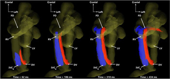
These human studies could then be contrasted with MRI flow acquisitions in sheep, which may be performed using conventional 4D flow acquisitions in the anesthetized and invasively-gated fetus to visualize preferential streaming of blood from the placenta toward the fetal brain Reference Schrauben, Darby and Saini33,Reference Schrauben, Saini and Darby34 (Fig. 5).
4D flow magnetic resonance imaging particle trace visualization in a 128-day gestation fetal sheep (term = 150 days). This ventral view shows preferential delivery of DV blood to the left side of the heart. DV (red) and IVCd (blue) particles are shown at four time points over one cardiac cycle. The two streams remain well-separated, with blood from the DV primarily entering the left heart, while IVCd blood passes into the right ventricle and main pulmonary artery. DV: ductus venosus; IVCd: distal inferior vena cava; FO: foramen ovale; RV: right ventricle; LV: left ventricle (used with permission Reference Schrauben, Saini and Darby34 under the terms of the Creative Commons Attribution 4.0 International License (http://creativecommons.org/licenses/by/4.0/)).

Application of hyperpolarized MRI to unravel the impact of maternal diet upon placental function
Globally, an increasing number of people consume a Western diet, one that is high in fats and processed sugars. Reference Cena and Calder155,Reference Huth, Fulgoni, Keast, Park and Auestad156 The consumption of such a diet is strongly associated with the development of obesity and other metabolic diseases, in conjunction with their preclinical markers such as NAFLD and type 2 diabetes. Reference Scorletti and Byrne157,Reference Mikolasevic, Milic and Turk Wensveen158 Historically, studies have reported the prevalence of NAFLD in combination with obesity to be as high as 80%. Indeed, a recent meta-analysis reports a dose-dependent relationship between NAFLD and BMI, whereby the risk of developing NAFLD increases by ~1.2 per unit increment in BMI. Reference Li, Liu, Yan, Wang, Zhao and Wang159 New and emerging data identify that NAFLD is not an outcome solely associated with an altered body composition. Recent reports are highlighting that there is an increasing prevalence of NAFLD in lean populations, Reference Wattacheril and Sanyal160–Reference Younossi, Anstee and Marietti162 highlighting that body composition alone may not be an accurate predictor of metabolic health. Lean NAFLD has become a major clinical challenge because in the absence of obesity – the primary risk factor perceived by patients, the diagnosis of the liver disease, is often delayed or overlooked, resulting in compromised effectiveness or complete absence of the required treatment. Reference Li, Liu, Yan, Wang, Zhao and Wang159
In reproduction, the Western diet is associated with altered body composition and detrimental impacts on gametes, Reference Hohos and Skaznik-Wikiel163,Reference Salas-Huetos, Bulló and Salas-Salvadó164 and together these effects have significant impacts on placental and fetal development and the long-term metabolic health of offspring. Moreover, in a maternal obesogenic environment, fetal cells are understood to inherit malfunctioning mitochondria and/or deregulated epigenetic signatures in a germline fashion, impacting the immediate generation and possible future generations. Reference Oestreich and Moley165 While altered maternal and paternal body composition is recognized as a contributing factor to an altered offspring metabolic health profile, maternal diet (and fetal diet), independent of parental body composition, may also, independent of body composition, have significant impacts upon placental and fetal development as well as their metabolic outcomes. Indeed, two-cell stage embryo transfer studies have highlighted that only pups exposed to maternal high-fat diet during gestation displayed obesity into adulthood. Reference Sasson, Vitins, Mainigi, Moley and Simmons166 Furthermore, subsequent pregnancy diet reversal studies, where an obese mothers’ diet has been switched to control during pregnancy, have highlighted improved placental function in addition to improved liver metabolic health and function in offspring.
Many animal model systems have been developed, in conjunction with human observation and intervention studies, to understand the impact of Western, or so-called “cafeteria” diets, and obesity on reproduction and offspring outcomes. Reference Oestreich and Moley165,Reference Lomas-Soria, Reyes-Castro and Rodríguez-González167 However, few studies have explored the development of NAFLD in a phenotypically lean population living in an “obesogenic” environment, and fewer have examined how these specific conditions may interact with aspects of reproduction over the life course. Maternal Western diet consumption prior to and during pregnancy in a guinea pig model induces differences in maternal liver fat content, fetal liver volume, and liver fat storage, as well as changes in fetal adipose tissue deposition that can be measured in utero using MRI. Reference Sinclair, Friesen-Waldner and McCurdy168,Reference Morrison, Botting and Darby169 These studies highlighted the propensity for the fetus to store fat in the liver and adipose tissue, as increased lipid deposition, especially in organs and intra-abdominally near term (~ 60 days, term 69 days); changes that are outcomes associated with later-life metabolic and cardiovascular disease. Importantly, these outcomes occurred in association with a lifelong exposure to the consumption of a Western diet, where maternal NAFLD developed in the absence of maternal obesity. Reference Sinclair, Friesen-Waldner and McCurdy168
While using changes in near-term organ size as a predictive marker for future sub-optimal metabolic function is useful for understanding the risk of later-life metabolic disease, identifying, earlier in pregnancy, changes in the metabolic processes that set the stage for metabolic disease would be more useful. Cellular metabolic flexibility is the ability of a cell to adaptively utilize metabolic pathways to maintain energy status and physiological functions when challenged. In a number of disease states, metabolic flexibility is made evident by physiological and pathological alterations affecting cellular function and survival through the induction of a “glycolytic shift”, whereby affected cells favor ATP production by glycolysis over oxidative phosphorylation (OXPHOS), even when oxygen availability is high. Reference Zheng170 Increases in key metabolites such as lactate and alanine are often reported as part of this glycolytic shift. Reference Wang, Ohliger and Larson171 Hyperpolarized carbon-13 MRI (HP13C MRI) is a tool that has been widely used to investigate changes in mitochondrial metabolism, being able to measure changes in lactate and alanine from the metabolism of pyruvate, as a marker of changes in mitochondrial metabolism and disease presence and severity. Reference Kurhanewicz, Vigneron and Ardenkjaer-Larsen172 Recent work has demonstrated the utility of these technologies in identifying, in real time, placental pyruvate metabolism as a measure of mitochondrial function. Reference Friesen-Waldner, Sinclair and Wade173 While these studies were conducted at term in a guinea pig, they highlighted that HP13C MRI could provide a novel method for longitudinal studies of feto-placental abnormalities.
Current preliminary studies conducted in the lifelong Western diet-fed guinea pig pregnancy model show evidence of this glycolytic switch occurring in the placentae of these NAFLD lean pregnancies at mid-gestation (~35 days). Changes in the placental alanine to pyruvate ratio (APR), an indicator of alanine production from pyruvate, and the lactate to pyruvate ratio (LPR), are increased in Western diet pregnancies when compared with control diet pregnancies. These early results provide evidence that lifelong consumption of a Western diet in a nonobese phenotype is associated with altered placental metabolism at mid-gestation. Moreover, the observed increases in placental APR and LPR may be useful as biomarkers of feto-placental metabolic health.
Looking to the future, methods for imaging placental metabolism early in pregnancy, such as HP13C MRI, in conjunction with established metabolomic signature platforms, namely untargeted metabolome analysis, would provide an improved understanding of the mechanisms and development of placenta dysfunction over the course of gestation. HP13C MRI, together with other functional imaging modalities such as PET/CT, will offer functional metabolic information, which may be integrated with corresponding maternal, placental, and fetal metabolomic data in preclinical animal models to validate specific metabolic signatures. These signatures will then serve as a reference to be used for investigating interventions in preclinical animal models and for early detection of potential complications in human pregnancy.
Application of MRI to differentiate between brown and white preadipocytes
MRI is ideally suited to the measurement of the amount, the growth, and the location of fetal fat to help better understand the fetal metabolic state, as altered in utero metabolism can lead to altered fetal fat development. It is uniquely sensitive for imaging lipids, is capable of precise and accurate quantification of tissue lipid content, and its lack of ionizing radiation makes it safe for use in pregnancy. Fetal development of WAT and BAT is important for future metabolic health, because WAT is a storage depot for energy that can be drawn on in critical times in early postnatal life, while BAT is a critical organ for thermoregulation in early life.
MRI has been used to differentiate mature BAT from mature WAT in adults, children, and even neonates. However, it has not been used for investigation of fetal adipose until the recent development of MRI acquisitions fast enough to freeze fetal motion. Reference Giza, Olmstead and McCooeye174 These first studies showed that fetal WAT is significantly different than WAT in children or adults, with substantially lower lipid content – that content is increasing rapidly during the third trimester. These and other differences between fetal BAT and mature WAT complicate the differentiation of WAT and BAT in utero, since MRI methods that can successfully differentiate mature WAT and BAT rely on assumptions that are not valid in fetal life, Reference Hu, Tovar, Pavlova, Smith and Gilsanz175 or use techniques that are poorly suited to fetal imaging such as BOLD, Reference Izzi-Engbeaya, Salem, Atkar and Dhillo176 xenon MRI, Reference Antonacci, McHugh, Kelley, McCallister, Degan and Branca177 and zero-quantum coherence. Reference Branca, Zhang and Warren178
Techniques that show promise for fetal WAT/BAT differentiation include quantification of proton-density fat fraction (PDFF), Reference Giza, Olmstead and McCooeye174,Reference Stojanovska, Lumeng and Griffin179,Reference Hernando, Sharma and Aliyari Ghasabeh180 quantification of fatty acid composition (FAC), Reference Peterson and Månsson181 and adipose tissue relaxometry, particularly T2*; however, all of these methods need further development before they can be considered valid tools for BAT detection. PDFF quantification can differentiate WAT and BAT, but only after 30 weeks of gestational age when the PDFF of WAT and BAT are sufficiently different. Adipose tissue development begins considerably earlier at around 20 weeks, so this leaves a large window of development. Quantification of T2* with MRI relaxometry has similar issues, but perhaps both measures together can probe earlier gestational ages. BAT and WAT are thought to have different fatty acids in their lipid vesicles, Reference Hu, Smith, Nayak, Goran and Nagy182 so MRI FAC quantification is another promising avenue to pursue. Adult adipose tissue FAC has been quantified with MRI, so these methods need to be adapted to the unique in utero environment.
Using synchrotron-based PCX to expand our understanding factors regulating the transition from fetal to newborn life
In addition to the importance of fetal imaging for diagnostic and prognostic purposes, it is critically important to understand (i) the complex physiology underpinning the transition from fetal to newborn life, (ii) how different factors including pregnancy complications influence the success of the transition at birth, and (iii) how complications at the time of birth influence the risk of disease in the newborn period that has consequences extending throughout the life course. Synchrotron PCX imaging using preclinical models has provided invaluable insights into the fundamental factors regulating the transition from fetal to newborn life, by comprehensively unraveling the intricate sequence of cardiorespiratory factors necessary to ensure the successful transition to independent respiration and circulation when the umbilical cord is clamped at birth. Reference Hooper, Kitchen and Siew183–Reference Leong, Buckley, Paganin, Hooper, Wallace and Kitchen187
The pioneering use of PCX imaging technology and development of advanced techniques to image lung aeration, total and regional pulmonary blood flow, as well as airway and vascular structure has redefined our understanding of fetal and newborn physiology. Reference Flemmer, Thio and Wallace97–Reference Kitchen, Buckley and Leong99,Reference Leong, Buckley, Paganin, Hooper, Wallace and Kitchen187–Reference Lang, Pearson and te Pas191 These advances have overturned previously held theories underlying how fetal lung liquid was cleared at birth and how best to support aeration of the newborn lung. Reference Siew, Te Pas and Wallace188–Reference Siew, Wallace and Kitchen190,Reference Dekker, Hooper and Croughan192,Reference Te Pas, Kitchen and Lee193 This new knowledge has also provided critical information about why some babies do not benefit from noninvasive respiratory support at birth, and identified key factors including oxygenation, pharmacological targets, and noninvasive interventions in the delivery room that can be targeted to aid in a more rapid and successful transition to breathing in newborns to reduce the risk of adverse outcomes. Reference Dekker, Hooper and Croughan192,Reference Dekker, Martherus and Lopriore194,Reference Crawshaw, Kitchen and Binder-Heschl195 Together, this use of imaging has provided the vitally important physiological evidence required to drive safe and effective interventions in newborn clinical trials, which have been incorporated into resuscitation guidelines to make resuscitation safer and more effective for newborns worldwide. The use of synchrotron-based PCX imaging technology has enabled the highest level of physiological evidence to be generated by understanding the intricate details of complex processes at birth, rather than evidence solely guided by expert opinion, to aid in the transition at birth to prevent lifelong disease in vulnerable newborns.
An advantage of synchrotron PCX imaging is the ability to resolve features down to the smallest alveoli, to match up lung aeration and perfusion and investigate effects of different ventilation strategies. Taken together, these factors have provided vital pieces of evidence that have improved our understanding of the cardiorespiratory processes in the fetus and newborn. Reference Hooper, Kitchen and Siew183,Reference Hooper, Te Pas and Kitchen186 The expansion of these techniques and sensitivity of imaging has enabled 2D and 3D characterizations of newborn lung structure and function. Reference Flemmer, Thio and Wallace97,Reference Kitchen, Buckley and Gureyev100,Reference Lang, Pearson and te Pas191 Current work is also focused on using this unique technology to image gray and white matter in the brain with high contrast, toward better understanding mechanisms of newborn brain injury. Reference Croton, Morgan and Paganin196 Further advances in this technology combined with the development of more cutting-edge preclinical models will continue to provide practical solutions that can be translated into clinical trials to address the most critical issues facing newborns.
In addition to the development of novel techniques for investigating the transition at birth, the use of placental vascular casting provides a unique perspective of understanding early origins of placental dysfunction. Reference Langheinrich, Vorman and Seidenstucker197–Reference Aughwane, Schaaf and Hutchinson199 The use of routine ultrasound with a focus on the newborn lung has confirmed that lung liquid clearance at birth in human infants is similar to that observed in animal models, and provided a new tool for diagnosing respiratory dysfunction in the newborn. Reference Blank, Rogerson and Kamlin200
Is there a role for artificial intelligence together with advanced imaging techniques in DOHaD?
The power of automated segmentation and plane localization of medical images, particularly those made possible with artificial intelligence (AI) powered by deep learning, is slowly making its way into human fetal imaging. Studies have used both ultrasound and MRI. Reference Gholipour, Estroff and Warfield91,Reference Ebner, Wang and Li92,Reference Chen, Ni and Qin201–Reference Kuklisova-Murgasova, Quaghebeur, Rutherford, Hajnal and Schnabel203 In the coming years, AI approaches to “smart” MRI, including detecting when the fetus moves and automated deep learning reconstructions that take a fraction of the time compared with conventional techniques, will make their way into clinical practice. These approaches will ensure consistency in scan plane placement for longitudinal and cross-sectional comparison studies and simplify data analysis for example by automating vessel segmentation for flow analysis and summarizing hemodynamic features from these complex datasets.
Conclusions
In the context of DOHaD, these advances in imaging have given us the opportunity to understand fundamental processes regulating fetal cardiovascular, lung, metabolic, and brain development as well as the transition at birth. Together, these provide us with the opportunity to investigate how an adverse intrauterine environment or pregnancy exposures and complications affect these processes, leading to consequences that extend past the immediate newborn period into adulthood. The ultimate goal will be to provide technique-specific reference ranges to facilitate the classification of “normal” versus “abnormal”. This will require a range of paired preclinical and clinical studies with the likelihood of those techniques showing immediate short-term clinical significance to be the first to “take off” and leap the fence toward DOHaD research. Indeed, it is our hope that measures taken in utero by the techniques discussed in this review may become part of postnatal health assessments with general practitioners taking an individual’s in utero characteristics into consideration.
The work discussed herein is targeted at identifying the most effective interventions to improve outcomes both in the newborn and in later life. As a group, we are collectively excited to participate in the advancement of imaging methodologies and their application to DOHaD research to improve health across the lifespan.
Acknowledgments
We thank Marilyn Zelesco (Lead Sonographer, Fiona Stanley Hospital) for providing the ultrasound scan images.
Financial Support
JLM was funded by an ARC Future Fellowship (Level 3; FT170100431).
MJK was funded by an ARC Future Fellowship (FT160100454).
CAM and TRHR are supported by the Children’s Health Research Institute and the Eunice Kennedy Shriver National Institute of Child Health and Human Development, Grant/Award Number: U01HD087181.
EVM was supported by a NHMRC Peter Doherty Biomedical Early Career Fellowship (APP1138049).
AM was supported by the Wellcome Trust (210182/Z/18/Z, 101957/Z/13/Z) and EPSRC (NS/A000027/1).
ALD is supported by the National Institute for Health Research Biomedical Research Centre at the University College London Hospitals NHS Foundation Trust and the University College London.
JMD is supported by the Health Research Council, New Zealand (17/076).
ASC was supported by a Future Leader Fellowship (101998) from the National Heart Foundation of Australia.