INTRODUCTION
Mild cognitive impairment (MCI) represents an intermediate condition between normal aging and dementia (Petersen & Morris, Reference Petersen and Morris2005; Winblad et al., Reference Winblad, Palmer, Kivipelto, Jelic, Fratiglioni and Wahlund2004). MCI patients bear a high risk for developing dementia (Flicker, Ferris, & Reisberg, Reference Flicker, Ferris and Reisberg1991; Lopez et al., Reference Lopez, Kuller, Becker, Dulberg, Sweet and Gach2007). Therefore, identifying individuals with MCI is of great importance for early intervention (Rivas-Vazquez, Mendez, Rey, & Carrazana, Reference Rivas-Vazquez, Mendez, Rey and Carrazana2004).
There is considerable heterogeneity regarding the definition of MCI (Petersen & Morris, Reference Petersen and Morris2005; Winblad et al., Reference Winblad, Palmer, Kivipelto, Jelic, Fratiglioni and Wahlund2004). Based on its etiology, MCI is a prophase for many diseases (Petersen & Morris, Reference Petersen and Morris2005; Winblad et al., Reference Winblad, Palmer, Kivipelto, Jelic, Fratiglioni and Wahlund2004). Among them, the prodromal phase of Alzheimer’s disease (MCI-AD) has been an important target for research. A 3-year follow-up of 769 MCI-AD patients yielded a 16% conversion rate to AD (Petersen et al., Reference Petersen, Thomas, Grundman, Bennett, Doody and Ferris2005) per year, approximately 10 times that of healthy elders (Petersen, Smith, Waring, Ivnik, Tangalos, & Kokmen, Reference Petersen, Smith, Waring, Ivnik, Tangalos and Kokmen1999). As new treatments for AD are developed, early identification of MCI-AD becomes more critical.
Cerebral small vessel disease (SVD) is another major cause for cognitive impairment (Chui, Reference Chui2001; Román, Erkinjuntti, Wallin, Pantoni, & Chui, Reference Román, Erkinjuntti, Wallin, Pantoni and Chui2002; van der Flier et al., Reference van der Flier, van Straaten, Barkhof, Verdelho, Madureira and Pantoni2005). Studies have indicated that patients with MCI caused by SVD (MCI-SVD) had a high frequency of adverse outcomes. A 40-month follow-up revealed that there was only a 50% survival rate, and 35% of the survivors who were moved into nursing homes. Even those patients who were living at home showed cognitive deterioration (Frisoni, Galluzzi, Bresciani, Zanetti, & Geroldi, Reference Frisoni, Galluzzi, Bresciani, Zanetti and Geroldi2002). Early detection of MCI-SVD may provide an opportunity for interventions to delay cognitive deterioration.
Unlike typical vascular dementia (VD), the majority of MCI-SVD exhibit a relatively insidious onset, with gradual cognitive deterioration that mimics MCI-AD. This similar pattern of decline makes early differentiation of MCI-SVD and MCI-AD difficult (Meyer, Xu, Thornby, Chowdhury, & Quach, Reference Meyer, Xu, Thornby, Chowdhury and Quach2002). Neuropsychological measures may reveal distinctive cognitive profiles between them and establish a discriminative tool. Many studies indicated that MCI-SVD patients display frontal-subcortical cognitive features with relatively preserved verbal memory and poor performance on executive functions and mental processing speed (Kramer, Reed, Mungas, Weiner, & Chui, Reference Kramer, Reed, Mungas, Weiner and Chui2002; Prins et al., Reference Prins, van Dijk, den Heijer, Vermeer, Jolles and Koudstaal2005). However, the majority of patients with MCI-SVD may show memory deficits (Loewenstein et al., Reference Loewenstein, Acevedo, Agron, Issacson, Strauman and Crocco2006). While memory impairment is the hallmark of MCI-AD, a recent meta-analysis concluded that deficits in multiple cognitive domains were characteristic of AD several years before clinical diagnosis (Bäckman, Jones, Berger, Laukka, & Small, Reference Bäckman, Jones, Berger, Laukka and Small2005). Many studies failed to find differences between MCI-SVD and MCI-AD on memory or nonmemory measures (Loewenstein et al., Reference Loewenstein, Acevedo, Agron, Issacson, Strauman and Crocco2006; Meyer et al., Reference Meyer, Xu, Thornby, Chowdhury and Quach2002). Thus, constitution of the distinctive cognitive profiles to differentiate between MCI-SVD and MCI-AD needs further research.
The purposes of current study were (1) to explore the cognitive profiles of MCI-SVD and MCI-AD, (2) to compare the cognitive features between them, and (3) to determine specific measures that best differentiate the two patient groups.
MATERIALS AND METHODS
Subjects and Diagnostic Procedure
Fifty-six MCI-SVD patients (36 males and 20 females) and 30 MCI-AD subjects (12 males and 18 females) were recruited from our cognitive impairment clinic and stroke clinic from January 2006 to January 2007. A semi-structured interview was performed on all patients and their close informants. Basic demographic data, vascular risk factors, past and present history, general and neurological examinations, neuroimaging data, global cognitive level (Clinical Dementia Rating Scale, CDR), and functional ability (Activities of Daily Living, ADL) were collected and analyzed. The diagnosis was made by consensus of two neurologists. The Ethics Committees of Xuan Wu Hospital, Capital Medical University, gave ethical approval for the study.
MCI-SVD patients were recruited according to the following criteria (Garrett et al., Reference Garrett, Browndyke, Whelihan, Paul, DiCarlo and Moser2004; Ingles, Wentzel, Fisk, & Rockwood, Reference Ingles, Wentzel, Fisk and Rockwood2002; Román et al., Reference Román, Erkinjuntti, Wallin, Pantoni and Chui2002; Winblad et al., Reference Winblad, Palmer, Kivipelto, Jelic, Fratiglioni and Wahlund2004): (1) Subjects had cognitive impairment that did not meet the Diagnostic and Statistical Manual of Mental Disorders, fourth edition (DSM-IV) criteria for dementia (American Psychiatric Association, 1994), and they obtained a score of 0.5 on CDR with preserved ADL skills. (2) The cognitive impairment was attributed to SVD as suggested by the following criteria: (a) one or more of the following neurological signs commonly seen in SVD, such as hemiparesis, lower facial weakness, Babinski sign, sensory deficit, dysarthria, gait disorder, urine urgency, or motor slowness; and (b) moderate to severe white matter changes (at least one region scored ≥2 according to the Wahlund rating scale) (Wahlund et al., Reference Wahlund, Barkhof, Fazekas, Bronge, Augustin and Sjogren2001) and/or multiple lacunar infarcts (≥2) on brain imaging, in the absence of large subcortical infarcts (greater than 15 mm diameter) or infarcts involving in the cerebral cortex. Exclusion criteria were (Román et al., Reference Román, Erkinjuntti, Wallin, Pantoni and Chui2002) as follows: (1) Early onset of memory deficits and progressive worsening of memory and other cognitive functions in the absence of corresponding focal lesions on brain imaging; (2) Cognitive impairments caused by other reasons, such as Parkinson’s disease, Huntington’s disease, multiple sclerosis, tumor, epilepsy, psychiatric disease, systemic disease, alcohol or drug abuse, and any other neurological disease that may result in cognitive impairment; and (3) Subjects with visual abnormalities, severe aphasia or palsy as a significant limiting factor for assessment.
Petersen et al. (Reference Petersen, Thomas, Grundman, Bennett, Doody and Ferris2005) criteria were used to identify participants with MCI-AD, requiring amnestic mild cognitive impairment of a degenerative nature. We made minor modifications according to our clinical work: (1) Memory complaint corroborated by an informant. To ensure identification of patients with a degenerative condition, participants were required to have a history of an insidious onset with at least 3 months’ duration. (2) Objective abnormal memory function. Patients scored 1.0 SD below the age and education corrected MQ on a Chinese memory test known as “the Clinical Memory Test” (The Cooperation Group For The Construction of “the Clinical Memory Test”, 1986) and scored 0.5 or 1 on the CDR memory box. (3) All subjects scored 0.5 on CDR with preserved ADL skills. (4) Patients had no stroke history or other disease that could be responsible for the memory deficit. (5) No focal signs suggestive of cerebral infarction were present. (6) No significant infarct lesions or other pathology noted on brain imaging (e.g., tumor, hydrocephalus). Mild white matter changes or a single nonstrategic lacunar infarction did not preclude the diagnosis.
The control group consisted of 80 normal community volunteers free of past or present neurologic and psychiatric diseases or cognitive symptoms. All controls had a CDR of 0 and were independent in their activities of daily living. Subjects with kidney disease, thyroid gland disorders, drug abuse, and other diseases that may result in cognitive impairment were excluded.
Neuropsychological Assessments
To obtain as complete a cognitive profile as possible, extended neuropsychological assessments were performed and scored on all subjects by an experienced neuropsychologist blinded to the diagnosis. In addition to the Mini-Mental State Examination (MMSE) (Folstein, Folstein, & McHugh, Reference Folstein, Folstein and McHugh1975), the tests can be grouped into 5 categories: (1) Attention: Digit Span Forward subtest of the Chinese version of the Wechsler Adult Intelligence Scale (WAIS-RC) (Dai, Ryan, Paolo, & Harrington, Reference Dai, Ryan, Paolo and Harrington1990). (2) Memory: Verbal memory was assessed with the World Health Organization-University of California-Los Angeles Auditory Verbal Learning test (WHO-UCLA AVLT), including immediate recall, short delay free recall (3 min later), long delay free recall (30 min later), and long delay recognition (Maj et al., Reference Maj, Satz, Janssen, Zaudig, Starace and D’Elia1994). Visual memory was measured with a modified Rey-Osterrieth Complex Figure test (ROCFT, maximum score 16), including the immediate recall and delay recall (30 min later) (Osterrieth, Reference Osterrieth1944). (3) Executive function: Response inhibition was assessed with part C (interference section) of a modified short form of the Stroop test (Jokinen et al., Reference Jokinen, Kalska, Mantyla, Ylikoski, Hietanen and Pohjasvaara2005). Errors and performance time were analyzed. Verbal initiation and flexibility was evaluated using Semantic Category Verbal Fluency test (animal) (Lezak, Reference Lezak1995). Planning, sequencing, and logical reasoning ability were assessed using WAIS-RC Picture Arrangement subtest (the 1st, 3rd, 5th, and 7th items) (Dai et al., Reference Dai, Ryan, Paolo and Harrington1990). Concept formation and shifting were assessed with the California Card Sorting test (CCST) (Delis, Kaplan, & Kramer, Reference Delis, Kaplan and Kramer2000). Abstract reasoning was evaluated with WAIS-RC Similarities subtest (the 1st, 2nd, 3rd, 4th items) (Dai et al., Reference Dai, Ryan, Paolo and Harrington1990). Working memory was measured with WAIS-RC Digit Span Backwards subtest (Dai et al., Reference Dai, Ryan, Paolo and Harrington1990). (4) Mental processing speed: The time to complete Part A (color naming condition) of the modified Stroop test (Jokinen et al., Reference Jokinen, Kalska, Mantyla, Ylikoski, Hietanen and Pohjasvaara2005) and the WAIS-RC Digit Symbol subtest (Dai et al., Reference Dai, Ryan, Paolo and Harrington1990). (5) Visuoconstructive skill: The copy of the modified Rey-Osterrieth Complex Figure (Osterrieth, Reference Osterrieth1944), WAIS-RC Block Design subtest (the 3rd, 4th, 6th, 7th items) (Dai et al., Reference Dai, Ryan, Paolo and Harrington1990), and Clock Drawing test (CDT) (Alzheimer’s Disease Cooperative Study Group, 1999). The diagnosis of MCI-SVD and MCI-AD were made blinded to and independently of these neuropsychological tests.
Statistical Analysis
One way analyses of variance (ANOVAs) were used to assess group differences in age and education, and χ 2 test for gender (p < .05 was considered as significant). Analyses of covariance (ANCOVA) with age as covariate (to correct for group differences in age) were used to compare neuropsychological scores, followed by post hoc tests with Bonferroni correction for multiple comparisons (a significance level of p < .0025 was considered as significant). The effect size (Cohen’s d) of each test between groups was analyzed. Binary logistic regression analyses using the forward conditional method were performed to identify major predictors from tests that were statistically different between MCI-SVD group and MCI-AD group. The sensitivity and specificity of each predictor for differentiating MCI-SVD and MCI-AD were explored using receiver operating characteristic curves (ROC). The validity of these tests in combination was also determined with discriminant analyses.
RESULTS
Demographic Characteristics
As shown in Table 1, there were group differences in age (p = .001). The mean age of MCI-AD group was older than those of the other two groups. There was no difference between the control group and the MCI-SVD group according to post hoc analysis (p = 1.000). No group differences in gender proportions were found with χ 2 analysis (p = .096). Three groups were matched regarding the education (p = .281).
Means (mean ± SD) and differences in demographic characteristics

Note
MCI-SVD = mild cognitive impairment caused by cerebral small vessel disease; MCI-AD = mild cognitive impairment of Alzheimer’s disease origin.
* Difference is significant at the .05 level.
Neuropsychological Assessments
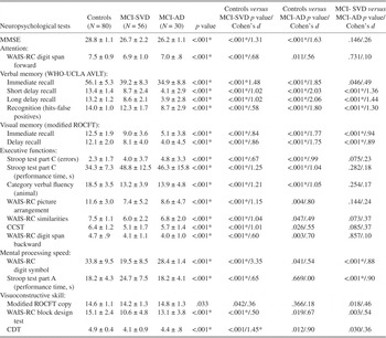
As presented in Table 2, MCI-SVD and MCI-AD groups showed mild cognitive impairment as indicated by lower MMSE performance. Both groups scored slightly but significantly lower than the normal controls (p < .001). The two MCI groups were not significantly different on the MMSE (p = .146).
Means (mean ± SD) and differences in neuropsychological data

Note
MCI-SVD = Mild cognitive impairment caused by cerebral small vessel disease; MCI-AD = Mild cognitive impairment of Alzheimer’s disease origin; * = difference is significant on 0.0025 level; MMSE = Mini-Mental State Examination; WAIS-RC = Chinese version of the Wechsler Adult Intelligence Test; WHO-UCLA AVLT = World Health Organization-University of California-Los Angeles Auditory Verbal Learning Test; ROCFT = Rey-Osterrieth Complex Figure Test; CCST = California Card Sorting Test; CDT = Clock Drawing Test.
Except for the ROCFT copy (p = .033), significant group differences were observed on all other measures (p < .001). Post hoc analyses indicated that the MCI-SVD group performed significantly worse than controls on all tests (p < .001) except for the ROCFT copy (p = .042).
Compared with the controls, the MCI-AD group was mainly impaired on measures of memory and executive function. They performed worse on all memory measures and on several tests tapping executive functions, including the Stroop Test part C (errors), Stroop Test Part C (performance time, s), and Semantic Category Verbal Fluency test (animals) (all p < .001). The MCI-AD group showed no significant differences relative to the control group (Bonferroni-corrected p > .0025) in other cognitive domains (p = .003 to .669).
Post hoc analysis revealed that the two patient groups differed mainly in memory and mental processing speed. Compared with MCI-SVD, MCI-AD patients showed poorer performance on the WHO-UCLA AVLT short delay free recall, long delay free recall, long delay recognition, and ROCFT immediate recall and delay recall. However, they performed better than the MCI-SVD group on the WAIS-RC Digit Symbol subtest and Stroop Test Part A (performance time, s) (all the p < .001). There were no statistical differences between the two groups on other tests tapping attention, executive functions, and visuoconstructive skills (p = .003 to .857).
The effect size (Cohen’s d) of each test between groups was detailed in Table 2. Generally, the Cohen’s d values were consistent with p values. However, there existed medium to large effect size (Cohen’s d > .50) on some tests that did not differ significantly between groups with Bonferroni correction for multiple analyses.
Major Predicting Tests for Differentiating MCI-SVD and MCI-AD
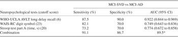
Using the cognitive measures that showed significant differences between MCI-SVD and MCI-AD as the independent variables, and diagnosis as the dependent variable, binary logistic regression analyses using the forward conditional method was performed. To control for confounding effects of age, we simultaneously entered the age variable. Three tests entered the equation. The WHO-UCLA AVLT long delay free recall entered first, and the proportion of correctly classified patients using this equation was 77.9%. The WAIS-RC Digit Symbol subtest also contributed to diagnostic prediction and improved the classification accuracy to 90.7%. The Stroop Test Part A (performance time, s) entered on the 3rd step, and the correctly classified percentage reached 93.0%. Age entered last, adding a minor contribution to prediction accuracy (correctly classified percentage 95.3%).
The cutoff point and diagnostic accuracy of the three major predictors were explored with ROC analysis. The optimal cutoff score was established to maximize the sum of sensitivity and specificity. As presented in Table 3, the long delay free recall indices provided the single best classification accuracy among the individual predictors. The three tests in combination correctly identified 91.1% MCI-SVD patients and 86.7% MCI-AD patients. ROC classification accuracy did not change when age was included in the analysis.
Discriminant validity of major predictors of MCI-SVD and MCI-AD

MCI-SVD = Mild cognitive impairment caused by cerebral small vessel disease; MCI-AD = Mild cognitive impairment of Alzheimer’s disease origin; WHO-UCLA AVLT = World Health Organization-University of California-Los Angeles Auditory Verbal Learning Test; WAIS-RC = Chinese version of the Wechsler Adult Intelligence Test; AUC = area under the ROC curve; * = overall percentage correctly classified (%).
DISCUSSION
The identification and differentiation of MCI-SVD and MCI-AD are important for treatment and prognosis. We administered comprehensive neuropsychological tests to explore the cognitive profiles of each MCI subtype and to establish discriminative measures for these two patient groups.
The current study revealed extensive cognitive deficits involving in five domains among patients with MCI-SVD. These results are inconsistent with previous studies in which significant executive dysfunction contrasting with relatively intact memory were reported (Kramer et al., Reference Kramer, Reed, Mungas, Weiner and Chui2002; Prins et al., Reference Prins, van Dijk, den Heijer, Vermeer, Jolles and Koudstaal2005). However, in agreement with our results, some studies have indicated that the majority of MCI-SVD patients showed memory impairment as well as poor performance in other cognitive domains. Among 21 MCI subjects with vascular disease (most of them were MCI-SVD), Loewenstein and coworkers noted that 52.4% of patients were impaired in multiple domains and 71.4% of patients were impaired on memory measures (Loewenstein et al., Reference Loewenstein, Acevedo, Agron, Issacson, Strauman and Crocco2006). Reed et al. (Reference Reed, Mungas, Kramer, Ellis, Vinters and Zarow2007) found that patients with autopsy-defined small vessel cerebrovascular disease were equally impaired on measures of executive function and memory. Recently, the presence of executive deficits or psychomotor slowing was considered as much weaker diagnostic markers for small vessel subcortical cerebrovascular disease (Gainotti, Ferraccioli, Vita, & Marra, Reference Gainotti, Ferraccioli, Vita and Marra2008). The mechanism underlying the multiple-domain impairment in MCI-SVD was suggested to be a destruction of memory-related loops (e.g., hippocampal-anterior thalamic axis loop) as well as executive-related frontal-subcortical circuits (Mori, Reference Mori2002). In addition, frontal hypometabolism associated with SVD was proposed to be another likely causative factor for impaired memory and global cognitive lesions (Reed, Eberling, Mungas, Weiner, Kramer, & Jagust, Reference Reed, Eberling, Mungas, Weiner, Kramer and Jagust2004).
Our study revealed a relatively specific cognitive profile in MCI-AD patients. Memory impairment is a strong marker of MCI-AD, and our patients were severely impaired in both verbal and visual memory. However, they also showed a relatively slight deficit in executive function. This results were similar to those in other recent studies. One study found that 58.3% of 36 MCI-AD patients were impaired in multiple domains and 22.2% purely in the domain of memory (Loewenstein et al., Reference Loewenstein, Acevedo, Agron, Issacson, Strauman and Crocco2006). In 116 strictly recruited amnestic MCI patients who were possibly at the early stages of AD (Morris et al., Reference Morris, Storandt, Miller, McKeel, Price and Rubin2001), Ribeiro, de Mendonca, and Guerreiro indicated that 52.6% of their patients had at least one other cognitive domain that was affected as severely as memory (Ribeiro et al., Reference Ribeiro, de Mendonca and Guerreiro2006). As a degenerative process, MCI-AD begins insidiously and progresses gradually. Isolated memory impairment may exist at the earliest stages. As the pathology progresses, patients may present with more deficits, but not severe enough to be classified as a frank dementia for several years.
Despite their similar degree of overall cognitive deterioration, the MCI-AD subjects performed significantly worse on verbal and visual delay recall measures and delay recognition tasks relative to the MCI-SVD group, which is consistent with research reported by Gainotti et al. Gainotti and colleagues also found that MCI patients with hippocampal atrophy scored much lower than MCI patients with multiple subcortical infarcts on measures of verbal delay recall and delayed visual-spatial reproduction, but they performed similarly on immediate recall measures (Gainotti et al., Reference Gainotti, Ferraccioli, Vita and Marra2008). The distinctive cognitive differences between MCI-AD and MCI-SVD appear to be consistent with the cognitive patterns seen in the more advanced forms of each condition: AD and subcortical vascular dementia (Graham, Emery, & Hodges, Reference Graham, Emery and Hodges2004).
Some studies found no significant difference on memory measures between MCI-AD and MCI-SVD groups. However, verbal learning tended to be more impaired in MCI-AD in one study (Frisoni et al., Reference Frisoni, Galluzzi, Bresciani, Zanetti and Geroldi2002). Another study also indicated a lower median score for MCI-AD patients on memory measures (Loewenstein et al., Reference Loewenstein, Acevedo, Agron, Issacson, Strauman and Crocco2006). Taken together, we may draw a conclusion that memory performance, especially delayed recall, was more impaired in MCI-AD than in MCI-SVD.
A methodological limitation should be mentioned. We recruited MCI-AD patients demanding memory impairment beforehand and the results showed severe memory impairment in these patients. There may be a circular nature of the memory test. We acknowledge this methodological flaw but find no good solution to avoid it at the moment. As we know, episodic memory impairment is the hallmark and the earliest cognitive manifestation of AD. To recruit MCI-AD subjects as possible, many studies used the same criteria requiring patients score abnormally on memory tests (Loewenstein et al., Reference Loewenstein, Acevedo, Agron, Issacson, Strauman and Crocco2006; Petersen et al., Reference Petersen, Thomas, Grundman, Bennett, Doody and Ferris2005).
In our study, another profound difference between MCI-SVD and MCI-AD groups was mental processing speed. This finding is contrary to the report of Frisoni and co-workers, which suggested greater executive dysfunction in MCI-SVD patients (Frisoni et al., Reference Frisoni, Galluzzi, Bresciani, Zanetti and Geroldi2002). Interestingly, consistent with our results, Gainotti et al. also revealed no difference in the Stroop test interference time between the MCI patients with hippocampal atrophy and multiple subcortical infarct MCI patients (Gainotti et al., Reference Gainotti, Ferraccioli, Vita and Marra2008). Our result was corroborated further by Graham and coworkers, who did not find any difference between patients with subcortical vascular dementia and AD on executive function-related tests, such as the Category Fluency test, Stroop Interference task, and Wisconsin Card Sorting test (Graham et al., Reference Graham, Emery and Hodges2004). A research study on patients with cerebral autosomal dominant arteriopathy with subcortical infarcts and leukoencephalopathy (CADASIL), a monogenic variant of small vessel disease, revealed the greatest impairment in processing speed with slighter deficits in executive functioning (Peters, Opherk, Danek, Ballard, Herzog, & Dichgans, Reference Peters, Opherk, Danek, Ballard, Herzog and Dichgans2005). This finding may partly explain the marked deficit in processing speed in patients with MCI-SVD. MCI-AD patients in our current study showed no deficits on the Stoop Test Part A and the WAIS-RC Digit Symbol subtest, indicating a relatively preserved ability in mental processing speed. Collectively, it appeared that MCI-AD patients were impaired to the same degree on sophisticated executive functioning measures as MCI-SVD patients, but they showed milder deficits on mental processing speed measures.
Visuospatial functions assessed using modified ROCFT copy, WAIS-RC Block Design subtest, and CDT marginally differed between MCI-SVD and MCI-AD groups. Although these tests are traditionally viewed as visuospatial tasks, a wide range of intellectual and perceptual skills are necessary for completion of the tests. They are considered as a mix of visuospatial abilities as well as executive control function (Elderkin-Thompson, Kumar, Mintz, Boone, Baahng, & Lavretsky, Reference Elderkin-Thompson, Kumar, Mintz, Boone, Bahng and Lavretsky2004; Qiu et al., Reference Qiu, Price, Hibberd, Buell, Collins and Leins2006; Shulman, Reference Shulman2000; Watanabea et al., 2005). The more severely impaired visuospatial abilities in MCI-SVD patients may partly reflect the greater executive dysfunction reported by other study (Frisoni et al., Reference Frisoni, Galluzzi, Bresciani, Zanetti and Geroldi2002).
Because of the different treatments and prognosis in MCI-SVD and MCI-AD, it is of great importance to discriminate the two conditions early and accurately. Our study suggested that the long delay free recall measure of the AVLT had the single best predictive accuracy in differentiating the two patient groups. The result was corroborated by Graham and coworkers. They also revealed that delayed recall was the most useful test to discriminate between subcortical vascular dementia and AD (Graham et al., Reference Graham, Emery and Hodges2004). When evaluating the usefulness of a cognitive screening measure, a good sensitivity (>80%) and an acceptably low false positive rate (specificity >60%) are required (Blake, McKinney, Treece, Lee, & Lincoln, Reference Blake, McKinney, Treece, Lee and Lincoln2002). According to this criterion, the WAIS-RC Digit Symbol subtest is also useful in differentiating the two conditions. The combination of the three tests tapping memory and processing speed is valid for differentiating MCI-SVD and MCI-AD.
Our study revealed a multiple-domain cognitive deficit in MCI-SVD patients and a relatively specific cognitive pattern in MCI-AD. The two patient groups differed mainly on measures of memory and mental processing speed. The AVLT long delay recall, the WAIS-RC Digit Symbol subtest, and the Stroop Test Part A for evaluating memory and processing speed may provide a good combination of instruments for discriminating between the two MCI subgroups. Further work is needed to verify the validity of these tests in larger samples in the future.
ACKNOWLEDGMENTS
We thank Dr. John L. Woodard and Dr. Tom Scovel for their invaluable work in language editing of the manuscript. This work was supported by the National Natural Science Key Foundation of China (30830045), National Key Technology R&D Program in the Eleventh Five-year Plan Period (2006BAI02B01), the National Basic Research 973 Program (2006CB500700), Beijing Natural Science Foundation (7071004), and Funding Project for Academic Human Resources Development in Institutions of Higher Learning Under the Jurisdiction of Beijing Municipality.