Congenital anomalies of the mitral valve are rare, and may result in both mitral stenosis and regurgitation. Congenital mitral stenosis occurs in ∼1.2% of patients with CHD, and 0.42% of all cardiac patients.Reference Collins-Nakai, Rosenthal, Castaneda, Bernhard and Nadas 1 Malformations resulting in mitral regurgitation are even more unusual.Reference Séguéla, Houyel and Acar 2 One rare but fascinating anomaly resulting in mitral regurgitation is Ebstein’s malformation of the mitral valve, first described by Ruschhaupt in 1976.Reference Ruschhaupt, Bharati and Lev 3 , Reference Jacob, da Silveira and Braile 4 This anomaly has a highly variable morphology, with an unguarded mitral valve orifice – in which the mitral valve fails to form altogether – speculated to be its most severe form.Reference Anderson, Zuberbuhler, Anderson, Becker and Lie 5 Only four cases of unguarded mitral orifice have been described to date, all of which have been associated with atrioventricular discordance.Reference Yasukochi, Satomi, Park, Ando and Momma 6 – Reference Hwang, Chang, Chu, Lin and Su 8 We report the first known case of unguarded mitral valve orifice, in the setting of atrioventricular concordance, in a newborn with hypoplastic left heart syndrome.
Case report
A 37-week-newborn boy was transferred to our hospital for the management of hypoplastic left heart syndrome and small bowel obstruction. His mother had received comprehensive prenatal care, and the congenital heart defect had been diagnosed prenatally. At birth, the patient was appropriate for gestational age, weighing 2550 g. He was vigorous and maintained oxygen saturations between 80 and 95% on room air. A radiograph of the chest and abdomen revealed a left-sided cardiac apex, mild cardiomegaly, moderate pulmonary vascular congestion, abdominal situs solitus, and severely dilated loops of bowel. A prostaglandin E1 infusion was started, and his postnatal echocardiogram confirmed the diagnosis of hypoplastic left heart syndrome.
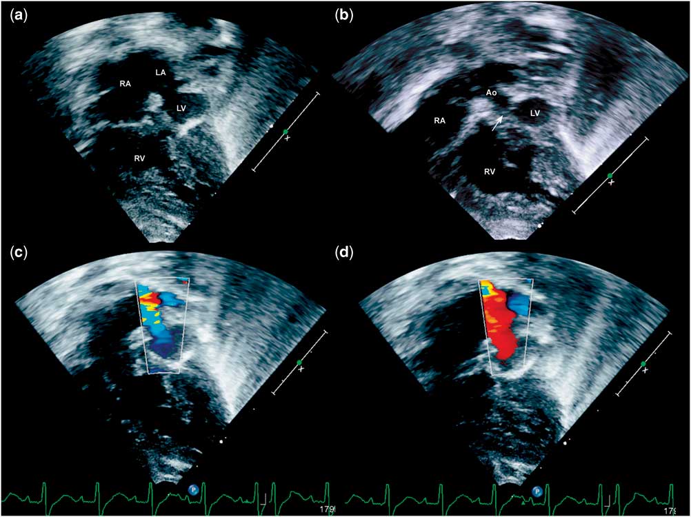
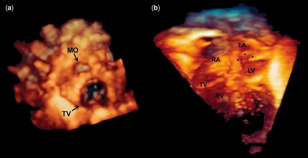
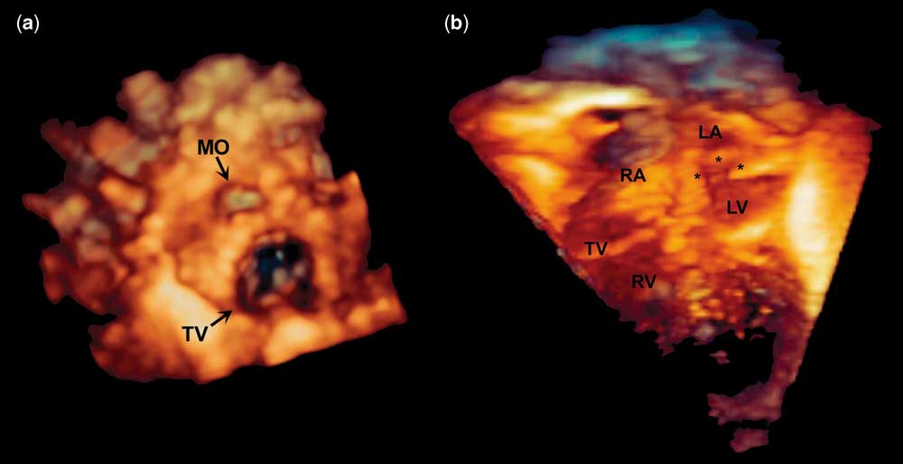
His cardiac segments were {S, D, S}, with a normal right atrium and a left atrium with restrictive left-to-right atrial shunting through a patent foramen ovale. The tricuspid valve was normal and the right ventricle had qualitatively normal systolic function. The pulmonary valve was large with no insufficiency or stenosis. There was no evidence of pulmonary venous stenosis. There was an unguarded mitral valve orifice with virtually absent mitral valve leaflets (Figs 1a and 2). The left ventricular chamber was hypoplastic, but with a bulging cavity and thin, hyperechoic ventricular walls, suggesting possible endocardial fibroelastosis. No identifiable valve chordae or papillary muscles were visualised. The aortic valve, which clearly arose from the left ventricle, was formed but imperforate (Fig 1b). By Doppler interrogation, low-velocity to-and-fro flow was observed between the left atrium and left ventricle. Interestingly, left ventricular filling occurred during systole, suggesting complete absence of left-sided myocardial contraction (Fig 1c), and retrograde flow from the left ventricle to the left atrium occurred during late diastole (Fig 1d).
Transthoracic apical four-chamber views. ( a ) Hypoplastic left heart syndrome status post emergent atrial septostomy, with an unguarded mitral orifice (seen as muscular ridges) between the left atrium (LA) and left ventricle (LV). No visible mitral valve leaflet or chordal tissue is seen. The left ventricle is thin walled, hypoplastic, and hyperechoic. ( b ) Slight anterior probe angulation displays the ascending aorta (Ao) arising from the LV. The formed but imperforate aortic valve (arrow) can be seen between the LV and Ao. ( c ) Diastolic frame demonstrating free retrograde flow through the mitral valve orifice into the LA. ( d ) Late systolic frame demonstrating antegrade blood flow into the LV. RA= right atrium; RV= right ventricle.

Three-dimensional echocardiographic images. ( a ) Cropped view from the superior aspect of the atria, showing en face views of the mitral orifice (MO) and tricuspid valve (TV). No visible mitral leaflets are seen. ( b ) Apical four-chamber view showing the cardiac chambers and the unguarded mitral orifice (asterisks). LA=left atrium; LV=left ventricle; RA=right atrium; RV=right ventricle.
On physical examination, the patient was alert, mildly cyanotic, with no obvious dysmorphic features. His precordium was active with a non-displaced ventricular impulse. Upon auscultation, there was a single S1 and S2, and a grade 2/6 systolic ejection murmur heard along the left upper sternal border with radiation to bilateral axillae. Lung fields were clear and he had a normal respiratory effort. Abdominal examination revealed a non-distended and soft abdomen with decreased bowel sounds, and a liver edge at 2 cm below the right costal margin. Peripheral pulses were 2+ in all extremities.
The patient underwent an emergent atrial septectomy and bilateral pulmonary artery banding to control pulmonary blood flow on day 1 of life. The next day, he underwent an exploratory laparotomy and was found to have jejunal atresia, necessitating small bowel resection and jejunostomy. On day 5 of life, the patient returned to the operating room and underwent a Norwood procedure with placement of a 5 mm right ventricle to pulmonary artery (Sano) shunt. A transoesophageal echocardiogram performed during surgery confirmed the complete absence of identifiable mitral valve tissue.
Cardiac surgery was performed without complications, and the patient returned to the cardiothoracic ICU for continued medical management. His subsequent course was uneventful.
Discussion
Unguarded mitral orifice is an exceedingly rare malformation in which the mitral valve is completely absent, leading to unobstructed to-and-fro flow between the corresponding atrial and ventricular chambers. Previous cases of unguarded mitral orifice have been associated with heterotaxy syndrome including mirror-image atrial arrangements, atrioventricular discordance, and double-outlet right ventricle.Reference Yasukochi, Satomi, Park, Ando and Momma 6 – Reference Hwang, Chang, Chu, Lin and Su 8 This case is unique in that our patient has normal segmental anatomy, indicating that this anomaly does not necessarily involve disturbance of the left–right axis during embryologic development. Moreover, our case is distinct in that the patient carried the diagnosis of hypoplastic left heart syndrome.
Mitral valve development occurs during the 4th and 5th weeks of gestation as a result of leaflet delamination from the left ventricular endomyocardial surface. Interference of this process, either by maldevelopment of the mesenchymal tissue or cardiomyocyte apoptosis, has been speculated to result in an unguarded mitral orifice.Reference Yasukochi, Satomi, Park, Ando and Momma 6 Such disturbances may also affect myocardial development, resulting in an abnormally thinned ventricular wall. This appears to have been the case with our patient – in addition to unguarded mitral orifice leading to unobstructed to-and-fro flow between the left atrium and ventricle, the patient’s left ventricular cavity was thin-walled and essentially akinetic.
It has previously been suggested that unguarded mitral orifice may represent an extreme form of Ebstein’s anomaly of the mitral valve, although the latter is characterised by failure of delamination of the mural leaflet of mitral valve, with the septal leaflet generally formed and attached normally.Reference Anderson, Zuberbuhler, Anderson, Becker and Lie 5 , Reference Cuadra, Muñoz-Castellanos and Espinola-Zavaleta 9 The two anomalies appear to have common features, and may indeed represent a particular spectrum of mitral valve disease. Notably, Ebstein’s malformation of the mitral valve has been associated with aortic obstruction, and unguarded mitral orifice appears to be associated with complete atresia of the corresponding outflow tract.Reference Yasukochi, Satomi, Park, Ando and Momma 6 – Reference Hwang, Chang, Chu, Lin and Su 8 , Reference Erickson and Cocalis 10 In our case, one can cautiously speculate that unguarded mitral orifice, in conjunction with the underlying left ventricular myocardial abnormality, might have led to aortic atresia and evolution of hypoplastic left heart syndrome.
In conclusion, unguarded mitral orifice is a rare and intriguing anomaly that has been previously described in association with heterotaxy and atrioventricular discordance. In this report, we describe a case of a patient with unguarded mitral orifice and normal segmental anatomy. Furthermore, our case represents the first documented occurrence of this very rare anomaly in association with hypoplastic left heart syndrome.
Acknowledgement
None.
Financial Support
This research received no specific grant from any funding agency, commercial, or not-for-profit sectors.
Conflicts of Interest
None.
Supplementary material
To view supplementary material for this article, please visit http://dx.doi.org/10.1017/S1047951114001334