Naturally occurring plant compounds from traditional herbal medicines and the diet represent a largely untapped source of potential therapeutic molecules to control human cancers with reduced side effects. The expanding list of such anticancer phytochemicals includes artemisinin, indole-3-carbinol, 3,3′-diindolylmethane, resveratrol, genistein, kaempferol, curcumin, and epigallocatechin gallate. These phytochemicals have been shown to inhibit growth and induce apoptosis in a wide range of human cancer cell lines and their respective tumour xenografts in athymic mice, which provide a preclinical experimental basis for the eventual development of these compounds in anticancer therapeutic strategies. Considerable recent attention has been focused on the anticancer properties of artemisinin, which has approval by the US Food and Drug Administration (FDA) for use in humans as an antimalaria drug.
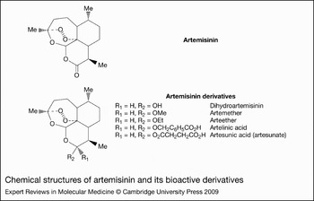
Artemisinin, a sesquiterpene lactone isolated from the Chinese plant Artemisia annua (more commonly known as qinghaosu or sweet wormwood), has been used by Chinese traditional herbal medicine practitioners for at least 2000 years to treat fever and malaria (Refs Reference Duffy and Mutabingwa1, Reference Meshnick2). Its earliest recorded use dates as far back as 340 BC when Ge Hong of the East Jin dynasty described this herbal medicine in The Handbook of Prescriptions for Emergencies. In 1596, the Chinese herbalist Li Shizhen recommended that patients with fever take a handful of qinghaosu, soak it in a sheng (~1 litre) of water, squeeze out the juice and drink it all (Ref. Reference Efferth and Ramawat3). Artemisinin and its bioactive derivatives, such as artesunate, arteether, artemether and dihydroartemisinin (DHA) (Fig. 1), were shown to effectively treat different forms of malarial parasites including multidrug-resistant strains (Refs Reference Meshnick2, Reference Meshnick4). The key structural feature in artemisinin-related molecules that mediates their antimalarial activity, and likely some of their anticancer activities, is an endoperoxide bridge, which is cleaved in the presence of iron leading to generation of free-radical reactive oxygen species (ROS) (Ref. Reference Rosenthal and Meshnick5). In addition to its well-established antimalarial properties, compelling evidence has emerged in the field showing that artemisinin-related compounds have potent anticancer activities in a variety of human cancer cell types.
Chemical structures of artemisinin and its bioactive derivatives. The chemical structures of artemisinin and derivatives show the endoperoxide bridge which is the reactive moiety for the ferrous ion, leading to generation of reactive oxygen species and eventual killing of the malarial parasite. These derivatives differ in their relative stability owing to differences in chemical structure, and have been shown to display different potency as anticancer agents.

Overview of anticancer properties of artemisinin
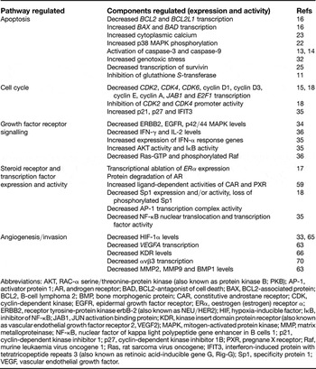
Molecular, cellular and physiological studies have demonstrated that, depending on the tissue type and experimental system, artemisinin and its derivatives arrest the growth, induce an apoptotic response, alter hormone responsive properties and/or inhibit angiogenesis of human cancer cells (summarised in Table 1). The Developmental Therapeutics Program of the National Cancer Institute (NCI), USA, which analysed 55 human cancer cell lines, showed that artesunate, the semisynthetic derivative of artemisinin, has strong anticancer activity against leukaemia and colon cancer cell lines, and has intermediate effects on melanomas, breast, ovarian, prostate, central nervous system, and renal cancer cell lines (Ref. Reference Efferth6). Moreover, the highly stable artemisinin-derived trioxane dimers were shown to inhibit the growth of and selectively kill several human cancer cell lines without inducing cytotoxic effects on normal neighbouring cells (Ref. Reference Paik7). One proposed mechanism by which artemisinin targets cancer cells is cleavage of the endoperoxide bridge by the relatively high concentrations of iron in cancer cells, resulting in free radicals such as ROS and subsequent oxidative damage as well as iron depletion in the cells (Ref. Reference Bustos, Gay and Diquet8). This mechanism resembles the action of artemisinin in malarial parasites. In addition to possessing higher iron influx via transferrin receptors, cancer cells are also sensitive to oxygen radicals because of a relative deficiency in antioxidant enzymes. A significant positive correlation can be made between artesunate sensitivity and transferrin receptor levels as well as between artesunate sensitivity and expression of ATP-binding cassette transporter 6 (ABC6; also involved in iron homeostasis) (IC50 between 0.512 and 124.295 µm, depending on the cancer cell line) (Ref. Reference Kelter9).
Summary of artemisinin-regulated cellular processes and pathways

Abbreviations: AKT, RAC-α serine/threonine-protein kinase (also known as protein kinase B; PKB); AP-1, activator protein 1; AR, androgen receptor; BAD, BCL2-antagonist of cell death; BAX, BCL2-associated protein; BCL2, B-cell lymphoma 2; BMP, bone morphogenic protein; CAR, constitutive androstane receptor; CDK, cyclin-dependent kinase; EGFR, epidermal growth factor receptor; ERα, oestrogen (estrogen) receptor α; ERBB2, receptor tyrosine-protein kinase erbB-2 (also known as NEU/HER2); HIF, hypoxia-inducible factor; IκB, inhibitor of NF-κB; JAB1, JUN activation binding protein; KDR, kinase insert domain protein receptor (also known as vascular endothelial growth factor receptor 2, VEGF2); MAPK, mitogen-activated protein kinase; MMP, matrix metalloproteinase; NF-κB, nuclear factor of kappa light polypeptide gene enhancer in B cells 1; p21, cyclin-dependent kinase inhibitor 1; p27, cyclin-dependent kinase inhibitor 1B; PXR, pregnane X receptor; Raf, murine leukaemia virus oncogene 1; Ras, rat sarcoma virus oncogene; IFIT3, interferon-induced protein with tetratricopeptide repeats 3 (also known as retinoic acid-inducible gene G, Rig-G); Sp1, specificity protein 1; VEGF, vascular endothelial growth factor.
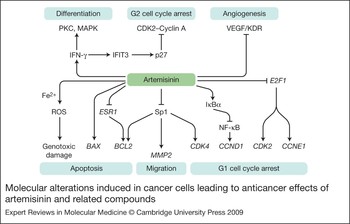
Expression profiling of several classes of tumour cells revealed that artemisinin treatment caused selective expression changes of many more oncogenes and tumour suppressor genes than genes responsible for iron metabolism, which suggests that the anticancer properties of artemisinin cannot be explained simply by the global toxic effects of oxidative damage (Ref. Reference Efferth, Olbrich and Bauer10). Artemisinin has also been observed to attenuate multidrug resistance in cancer patients, an effect due in part to the inhibition of glutathione S-transferase activity (Ref. Reference Mukanganyama11). Artemisinin and its bioactive derivatives elicit their anticancer effects by concurrently activating, inhibiting and/or attenuating multiple complementary cell signalling pathways, which have been described in a variety of human cancer cell systems as well as in athymic mouse xenograft models. The artemisinin compounds exert common as well as distinct cellular effects depending on the phenotype and tissue origin of the examined human cancer cells. This review discusses current evidence for artemisinin-activated antiproliferative cascades and disruption of tumour cell angiogenesis and metastasis and also addresses the role of ROS production in triggering some of these anticancer properties. The key cellular pathways by which artemisinin has been shown to mediate its antiproliferative properties, and which are discussed in this review, are summarised in Table 1 and Figure 2.
Molecular alterations induced in cancer cells leading to anticancer effects of artemisinin and related compounds. The diagram summarises the cellular pathways and molecular components under control of artemisinin and its derivatives that mediate their anticancer properties. Specific subsets of these pathways are regulated depending on the phenotype and tissue origin of the cancer cell. Artemisinin has been shown to induce the generation of reactive oxygen species (ROS) that can lead to genotoxic damage and subsequent apoptosis. In most of the test cancer cell sytems, artemisinin regulates the transcription of critical cell cycle, angiogenic, migration, differentiation and apoptotic regulators by selectively disrupting or enhancing the activity of specific sets of transcription factors in human cancer cells. Abreviations: BAX, BCL2-associated protein; BCL2, B cell lymphoma 2; CCND1, cyclin D1 gene; CCNE1, cyclin E1 gene; CDK, cyclin-dependent kinase; E2F1, early-2 transcription factor 1; MAPK, mitogen-activated protein kinase (also known as extracellular-signal-regulated kinase, ERK); ESR1, oestrogen (estrogen) receptor α (gene); IFIT3, interferon-induced protein with tetratricopeptide repeats 3 (also known as retinoic acid-inducible gene G, Rig-G); IFN, interferon; IκBα, inhibitor of NF-κB; MMP, matrix metalloproteinase; NF-κB, nuclear factor of kappa light polypeptide gene enhancer in B cells; PKC, protein kinase C; Sp1, transcription factor Sp1; VEGF, vascular endothelial growth factor; KDR, vascular endothelial growth factor receptor.

Regulation of apoptotic signalling by artemisinin-related compounds in human cancer cells
Artemisinin and apoptotic regulators
In a variety of human cancer cell systems, artemisinin and its derivatives have been shown to induce an apoptotic response as part of their anticancer effects. In many of the tested systems, lower doses of artemisinin-related compounds induce a cell cycle arrest (see later sections of this review), whereas, at high enough doses, these bioactive molecules cause apoptosis and in some cases necrosis of the tumours. The most potent artemisinin derivative is DHA, which can induce apoptosis after just 12 h of exposure at doses that are a tenth of an efficacious dose of artemisinin in some systems; depending on the cell line, DHA is effective in the 20–50 µm concentration range, compared with the 200–300 µm concentration range required for maximal effect of artemisinin (Ref. Reference Nam12). The effects on expression and activity of critical components of apoptotic cascades were investigated in artemisinin-sensitive cancer cells. In many studies, the apoptotic responses of artemisinin and derivatives occurred with a concomitant activation of caspase-3 and/or caspase-9 (Refs Reference Chen13, Reference Lu14). The levels of the proapoptotic protein BAX were increased and levels of the antiapoptotic regulator BCL2 ablated by DHA treatment, consistent with the strong apoptotic response induced by DHA in HepG2 and Hep3B human hepatoma cells (Ref. Reference Hou15). Interestingly, the IC50 was not different between artemisinin and DHA (10.8 µm artemisinin vs 10.6 µm DHA for 48 h). This effect has also been reported in ovarian cancer cells where DHA exposure (IC50 of 5.5 µm for 48 h) led to ablation of mRNA and protein levels of BCL2 and BCL-XL (BCL2L1) and modest increase in levels of BAX and BAD (Ref. Reference Jiao16). Consistent with these observations, DHA induced apoptosis by reducing the BCL2 to BAX ratio and by increasing capase-9 activation in human pancreatic cancer BxPC-3 and AsPC-1 cells (Ref. Reference Chen13). The exact mechanism of these DHA-mediated transcriptional effects is unknown, although artemisinin treatment inhibits expression of oestrogen (estrogen) receptor α (ERα; ESR1) in human breast cancer cells (Ref. Reference Sundar17) and attenuates Sp1 (SP1) transcription factor activity in human prostate cancer cells (IC50 of 150 µm) (Ref. Reference Willoughby18), and both of these transcription factors are have been shown to play a critical role in the transcription of BCL2 (Ref. Reference Dong19). However, the precise connection between the artemisinin control of ERα and Sp1 expression and DHA inhibition of BCL2 gene expression has not been directly evaluated in the same cancer cell system and represents an intriguing transcriptional control mechanism for induction of the apoptotic response.
Role of p38 MAPK and calcium signalling in the apoptotic response to artemisinin compounds
The p38 mitogen-activated protein kinase (MAPK) family members, which are involved with cell stress signalling and survival, have been paradoxically implicated in the regulation of both apoptosis and cell survival cascades in different cancer cell systems (Refs Reference Lee20, Reference Petersen21). In PC14 lung cancer cells, apoptosis induced by DHA (17.8 µm for 48 h) was accompanied by an increase in phosphorylation of p38 MAPK, which is its activated form, and increased intracellular calcium levels, an event that precedes p38 MAPK phosphorylation (Ref. Reference Mu22). Specific inhibitors of p38 MAPK and chelators of intracellular calcium reversed the DHA-mediated apoptosis in PC14 lung cancer cells, demonstrating the importance of both signalling compounds in this response. Interestingly, the source of calcium was primarily intracellular, because DHA was equally effective in causing apoptosis when PC14 cells were grown in calcium-free or calcium-containing media. Artemisinin had a similar effect on calcium levels in intestinal cancer cells, where artemisinin (IC50 of 10 µm for 5 min) rapidly decreased the activity of smooth endoplasmic reticular calcium ATPase (SERCA), leading to increased cytosolic calcium levels (Ref. Reference Riganti23). Although the specific effects of DHA on SERCA need to be evaluated, an increased level of calcium in the cytosol can modulate many signalling networks, and could possibly explain neurotoxicity of artemisinin at pharmacological doses (1 g/kg body weight, three times a day) (Ref. Reference Genovese and Newman24).
In the PC14 lung cancer cells and the intestinal cancer cells, the precise cell signalling pathways that link the DHA regulation of p38 MAPK and calcium with its apoptotic response have not been characterised. A critical role for increasing intracellular calcium levels and transcriptional downregulation of survivin, a cell-survival-promoting factor, was observed with DHA-induced apoptosis in SPC-A-1 lung cancer cells (Ref. Reference Mu25). Survivin belongs to the class of apoptotic inhibitor proteins, and ectopic expression of survivin confers resistance to apoptosis in these cells (Ref. Reference Mita26). In the ATSC-a-1 pulmonary adenocarcinoma cell line, the DHA-induced apoptotic response occurred with an enhanced nucleosomal DNA fragmentation that was preceded by an activation of caspase-3 with a large increase in mitochondrial width as well as a time-dependent loss of mitochondrial membrane potential (Ref. Reference Lu14). Although DHA is known to cause apoptosis in three lung cancer cell lines, it is not known whether accumulation of calcium and increased p38 MAPK activity is a common mechanism in each cell system.
p53 expression and regulation of the artemisinin apoptotic response
One of the important downstream targets of p38 MAPK is the tumour suppressor p53 (TP53) (Ref. Reference Maruyama27). Although the specific effects of artemisinin or DHA on p53 expression and activity have not been characterised, the mutation status of p53 has been correlated with the sensitivity of human cancer cells to DHA-induced apoptosis in certain cell line models. This effect was demonstrated using knockout HCT116 colon cancer cell lines. Treatment with 10 µm artemisinin was effective at blocking 5-bromo-2′-deoxyuridine (BrDU) incorporation in
TP 53 −/− as well as TP53 +/+ HCT116 cells. Additionally, TP53 was not among the artemisinin-regulated genes uncovered from mRNA profiling of artemisinin-treated versus untreated cells (Ref. Reference Efferth28). DHA was much more effective in causing an apoptotic response in ovarian cancer cells that possess wild-type p53 than in cells expressing mutant p53. The IC50 of DHA in ovarian cancer cell lines (OVCA 420 cells, OVCA 439 cells, OVCA 433 cells, HEY cells and OVCAR-10 cells) expressing wild-type p53 was 3.8–5.7 µm, whereas the IC50 was 13–15 µm in cells that either express mutant p53 (OVCA-432 cells, OVCAR-3 cells, OCC1 cells and ALST cells) or are p53-null (SKOV-3 cells) (Ref. Reference Jiao16). The regulation of DHA sensitivity by p53 warrants investigation and it is tempting to consider that p53 regulates gene expression of specific drug transporter or metabolising enzymes in response to this artemisinin derivative. In contrast, the dependence of cellular sensitivity to DHA-induced apoptosis on p53 status was not observed in human hepatoma cell lines. Even though HepG2 cells express wild-type TP53 and Hep3B cells are TP53-null, IC50 values for these two cell lines were similar, ranging between 10 µm and 14 µm for DHA-induced apoptosis. However, DHA induced expression of the p21 cyclin-dependent kinase (CDK) inhibitor (CDKN1A) in both hepatoma cell lines, suggesting that the DHA induction of p21 can occur by a p53-independent mechanism.
Transferrin and role of iron in the artemisinin apoptotic response
The antimalarial effects of artemisinin have been attributed to generation of ROS and subsequent genotoxic damage in Plasmodium falciparum (Ref. Reference Gautam29), and the role of iron homeostasis in artemisinin-mediated apoptotic effects has been shown to be critical in some but not all cancer cell lines (Ref. Reference Kelter9). It was proposed that the iron released from transferrin could activate artemisinin to generate toxic radical species that kill cells. Additionally, another study reported that artemisinin and derivatives were more effective in causing apoptosis with increased intracellular iron, and failed to cause cell cycle perturbations (Ref. Reference Efferth30). This study also found only 12 iron-responsive genes whose expression was altered in 60 cell lines with eight derivatives of artemisinin, which indicates that cellular iron homeostasis alone may not control cellular sensitivity to the artemisinin apoptotic response. The chemical tagging of artemisinin to transferrin (different numbers of artemisinin units are attached to the N-glycoside chains of transferrin) in androgen-insensitive DU145 human prostate cancer cells increased the apoptotic efficacy of artemisinin (Ref. Reference Nakase31). This study also found that small interfering RNA (siRNA) disruption of transferrin receptor expression inhibited the ability of artemisinin to cause apoptosis, which demonstrates a functional role for the transferrin receptor in this response. However, these same investigators also observed that the DHA-induced apoptosis is somewhat distinct from the effect of artemisinin in that the DHA effect was not affected by disruption of transferrin receptor levels, suggesting that artemisinin and its derivatives may exert effects independent of transferrin signalling.
Role of genotoxic damage in artemisinin-induced apoptosis
Genotoxic damage is a well-established cellular stimulus for apoptosis, and the cellular pathways and regulators have been well characterised (Ref. Reference Mauriz, Tunon and Gonzalez-Gallego32). Although microarray studies have suggested that the anticancer effects of artemisinin may not be attributed to genotoxic damage (Ref. Reference Efferth, Olbrich and Bauer10), artesunate induces apoptosis and necrosis, and causes genotoxic damage (specifically double-strand breaks), in a hamster lung fibroblast cell line as well as in wild-type and DNA-repair-defective Chinese hamster ovarian (CHO) cell lines. Wild-type and transgenic repair-defective VC79-2 hamster lung fibroblast cells and CHO cells that lack base excision repair (POLB), homologous recombination (inactivation of XRCC2 and BRCA2) or nonhomologous end-joining activity (inactivation of XRCC5) were significantly more sensitive to the artesunate apoptotic response than their parental cells. This observation strongly suggests that artesunate-induced DNA damage might be the possible mechanism for its toxicity (Ref. Reference Li33). Studies using rat glioma cells employing ROS scavengers such as edaravone have shown that DHA acts by generating ROS and possible genotoxic damage (Ref. Reference Huang34). Most of these studies were done in rodent model systems and it will be important to directly assess the effects of genotoxic damage in human cancer cells.
Artemisinin-induced cell cycle arrest of human cancer cells
Artemisinin regulation of cell cycle gene expression and function
Several recent studies have uncovered evidence that artemisinin and its derivatives induce a growth arrest that involves selective changes in the expression and activity of cell cycle components. The regulated changes in the cell cycle machinery differ depending on the tissue origin and phenotype of the cancer cells. In androgen-responsive LNCaP prostate cancer cells, artemisinin treatment led to a robust G1 cell cycle arrest marked by reduced levels of phosphorylated retinoblastoma (Rb; RB1) and downregulation of G1 cell cycle regulators in a time- and dose-dependent fashion (Ref. Reference Willoughby18). Artemisinin-treated LNCaP cells showed marked inhibition of expression of two G1-acting CDKs (CDK2 and CDK4) and increased association of the p27 CDK inhibitor (CDKN1B) with the CDK6–Cyclin-D1 complex. The ablation of CDK2 and CDK4 expression is attributable to repression of CDK2 and CDK4 gene promoter activity. Fine mapping of the CDK4 promoter in combination with chromatin immunoprecipitations revealed that artemisinin inhibited binding of endogenous Sp1 transcription factor to the CDK4 promoter in the artemisinin-responsive region, which resulted from a decreased serine phosphorylation of Sp1 (Ref. Reference Willoughby18). Phosphorylation of several serine residues have been reported to be critical to the function of Sp1 and, consistent with this concept, treatment with the phosphatase inhibitor okadaic acid reversed the artemisinin disruption of CDK4 promoter activity (Ref. Reference Willoughby18). These results imply that artemisinin targets specific phosphorylation events that control transcriptional regulation of cell cycle gene expression to block cell cycle progression.
Artemisinin was equally effective in inducing a G1 cell cycle arrest in androgen-unresponsive PC3 and DU145 prostate cancer cells compared with androgen-responsive LNCaP cells (IC50 of 150 µm), although the precise molecular changes were not investigated. It is also very interesting to note that, at this dose of artemisinin, no apoptosis was observed and the expression of p53 was unaltered (Ref. Reference Willoughby18). Similar to the effects in human prostate cancer cells, artemisinin and DHA induced a robust G1 cell cycle arrest of HepG2 and Hep3B human hepatoma cells that precedes the apoptotic response. This growth arrest was accompanied with reduced cellular levels of cyclin D1, E2F1 transcription factor, cyclin E, CDK2 and CDK4, and with elevated levels of the p21 and p27 CDK inhibitors (Ref. Reference Hou15). Interestingly, both artemisinin and DHA displayed a significantly lower cytotoxicity, with no cell cycle effects in normal liver cells.
Although at different efficacies, artemisinin and its derivatives have been shown to inhibit the growth of several types of breast cancer cell lines representing a range of tumourigenic and hormone-responsive phenotypes. The tested cell lines include oestrogen-responsive MCF-7 cells and T47D cells, which represent early-stage breast cancers, as well as oestrogen-unresponsive MDA-MB-231 cells, which represent later-stage breast cancer (Ref. Reference Efferth, Olbrich and Bauer10). Oestrogen-responsive breast cancer cells tend to be more sensitive to the artemisinin-mediated cell cycle arrest than oestrogen-insensitive cells, which is likely due to the ability of artemisinin to disrupt ER responsiveness and oestrogen-dependent cell growth (see later section). In oestrogen-responsive MCF-7 human breast cancer cells, the artemisinin cell cycle arrest was accompanied with markedly reduced expression of RB1, CDK2, CDK4, cyclin E1 (CCNE1) and CCND1. Also, artemisinin treatment strongly repressed CDK2 promoter activity without altering CDK4 promoter function (S.N. Sundar, unpublished). The artemisinin inhibition of CCND1 expression is due in part to the transcriptional ablation of ESR1 expression (Ref. Reference Sundar17), because in MCF-7 human breast cancer cells cyclin D1 is an oestrogen-responsive target gene.
Control of T cell proliferation by artemisinin and its derivatives
Reported effects of artemisinin and its derivatives on T cell function have revealed distinct alterations in cell signalling pathways that control cell proliferation. Artemether induces a G1 cell cycle arrest of primary mouse T cells by downregulation of CDK6 and cyclin D3 levels, as well as an upregulation of p27 levels. Artemether-growth-arrested T cells also displayed a marked depletion of phospho-MAPK, phospho-Raf and Ras-GTP, which are critical downstream components of growth factor receptor signalling pathways. In addition, artemether caused a robust decrease in secreted levels of interferon γ (IFN-γ; IFNG) and interleukin 2 (IL-2; IL2), known markers of T cell proliferation (Ref. Reference Wang35). In HL-60 leukaemia cells, a novel water-soluble derivative of artemisinin – ethyl 2-[4-(12-artemisininoxy)] phenoxylpropionate or SM933 – synergises with IFN-α to downregulate levels of the CDK2, cyclin A and p27 cell cycle components. Interestingly, artemisinin only affected activated T cells, and increased expression of IFIT3, an IFN-α-responsive protein that sequesters the p27-degrading nuclear protein JAB1. Thus, p27 is stabilised, whereas the expression of CDK2 and cyclin A is downregulated, by SM933. The growth arrest was also accompanied by an increase in levels of IκB protein, which sequesters the transcription factor NF-κB out of the nucleus and prevents its cell survival response (Ref. Reference Wang36).
Effects of artemisinin and its derivatives on expression and function of nuclear receptors in cancer cells
The nuclear receptor superfamily, which includes steroid receptors, consists of ligand-activated and constitutive transcription factors that modulate expression of distinct sets of target genes involved in a wide range of normal and cancer cell functions, including cell proliferation, differentiation, cell survival and apoptosis (Refs Reference McEwan37, Reference Kerkhofs38, Reference McDevitt39). Emerging evidence has shown that artemisinin and its derivatives disrupt expression and function of specific nuclear receptors such as oestrogen and androgen receptors that play an important role in antiproliferative activities in specific types of human cancer cells.
Artemisinin disruption of oestrogen-receptor responsiveness in steroid-responsive breast cancer cells
Increased oestrogen signalling has been strongly linked to increased mitosis and carcinogenesis of the breast and endometrium (Refs Reference Santen40, Reference Fox, Davis and Shupnik41). Oestrogenic responses are mediated by the two major ER subtypes ERα and ERβ, which are encoded by distinct genes (ESR1 and ESR2, respectively) (Ref. Reference Fox, Davis and Shupnik41). Predominance of cellular levels of ERα over ERβ is associated with increased proliferation, and increased ERβ levels with cessation of cell proliferation (Refs Reference Paruthiyil42, Reference Strom43). Additionally, high-risk precancerous breast lesions possess elevated ERα levels and declining ERβ expression (Refs Reference Roger44, Reference Shaaban45). Artemisinin and its derivatives have been shown to inhibit proliferation of human breast cancer cell lines (Refs Reference Efferth, Olbrich and Bauer10, Reference Sundar17) as well as attenuate the development of oestrogen-responsive rat mammary tumours initiated with chemical carcinogens (Ref. Reference Lai and Singh46). Human breast cancer cells that express relatively high ERα to ERβ ratios, such as MCF-7 and T47D cells, are highly sensitive to the antiproliferative effects of artemisinin-related compounds (Refs Reference Efferth, Olbrich and Bauer10, Reference Sundar17). This observation suggests that ERα-regulated genes in human breast cancer cells may control the cellular sensitivity to the antiproliferative effects of artemisinin in these cells.
In oestrogen-responsive breast cancer cells, a biologically significant response to artemisinin is the transcriptional downregulation of ERα. In MCF-7 cells, treatment with artemisinin strongly inhibited ESR1 promoter activity without any effects on ESR2 expression or activity, which accounted for the potent effects of artemisinin on oestrogen-responsive proliferation (Ref. Reference Sundar17). Artemisinin exposure downregulated oestrogen-responsive gene expression and completely ablated the ability of 17-β-oestradiol as well as the selective ER agonist propyl pyrazole triol to stimulate proliferation of MCF7 cells (Ref. Reference Sundar17). The precise artemisinin-regulated transcription factors that lead to loss of ERα expression are unknown and under investigation, although the artemisinin-responsive region in the ESR1 promoter contains predicted binding sites of transcription factors that are ERα-responsive gene products themselves, such as GATA and CEBP (Refs Reference Cheng47, Reference Eeckhoute48). It is tempting to consider that artemisinin-activated cell signalling pathways disrupt a positive regulatory loop involving these transcription factors and ERα.
It is well established that oestrogenic signalling is necessary for promotion of chemically induced rat mammary carcinogenesis (Ref. Reference Kerdelhue and Jolette49), and oral administration of artemisinin is efficacious in preventing dimethyl benzanthracene (DMBA)-induced rat mammary cancer incidence and increasing latency (Ref. Reference Lai and Singh46). This animal model for mammary epithelial tumourigenesis suggests downregulation of ERα expression and responsiveness by artemisinin has profound consequences on the carcinogenic process in steroid-responsive tissue. ERα is a potential target for drug therapy in oestrogen-sensitive breast cancer, and artemisinin can be considered a selective ER downregulator (SERD) because of its selective downregulation of ERα expression (without affecting ERβ expression levels). In addition to artemisinin, other phytochemical SERDs that disrupt ERα expression are kaempferol and indole-3-carbinol (Refs Reference Hung50, Reference Sundar51), suggesting a common feature in their mechanism of action. It is tempting to speculate that artemisinin can dampen the promotion to occult breast carcinomas by downregulating expression and disrupting responsiveness of ERα, suggesting that artemisinin and bioactive derivatives could be promising as both chemoprevention and chemotherapeutic strategies. In this regard, because artemisinin (at an IC50 of 150 µm) downregulates expression of ERα, this phytochemical can potentially be used to help evade ligand-independent activity of ERα, which occurs by its Ser118 phosphorylation, and hence decelerate progression to hormone-resistant states (Ref. Reference Staka, Nicholson and Gee52). Thus, from a clinical perspective artemisinin could be employed as a potent adjuvant along with antioestrogens such as tamoxifen.
Artemisinin disruption of androgen-receptor responsiveness in prostate cancer
Androgen receptor (AR) signalling has been shown to be critical to the development of normal prostate tissue and to proliferation of neoplastic prostatic epithelium (Ref. Reference Crawford53). The association with specific transcription factors dictates whether AR signalling culminates in cellular proliferation or differentiation. AR activation occurs classically with androgen binding, and by ligand-independent activation by phosphorylation at Ser792 and Ser218 of human AR induced by growth factor signalling pathways (Ref. Reference Ghosh54). The ligand-independent activation of AR is associated with development of hormone insensitivity in prostate cancer. Hence, drugs targeting only the activity of AR such as antiandrogens potentially lead to an androgen-resistance state (Ref. Reference Heinlein and Chang55), whereas ablation of AR levels would potentially retard the progression of prostate cancer to the hormone-insensitive state. We have recently observed that artemisinin exposure of LNCaP prostate cancer cells causes a potent cell cycle arrest accompanied by the loss of functional AR protein (IC50 of 150 µm). In contrast to artemisinin regulation of ERα transcription, artemisinin induced the MDM2-mediated ubiquitination and proteasomal degradation of AR protein (A.M. Steely, S.N. Sundar et al., unpublished). Thus, in addition to transcriptional ablation of CDK4 and CDK2 expression (Ref. Reference Willoughby18), artemisinin triggers nontranscriptional cascades in human prostate cancer cells that culminate in cell cycle arrest. Given the strong positive correlation between serum levels of androgens and risk of developing prostate cancer (Ref. Reference Isbarn56), the desensitisation of cancerous AR-expressing target cells to androgens by artemisinin suggests the use of this phytochemical in prostate cancer prevention.
Artemisinin effects on the androstane receptor and pregnane-X-receptor
The constitutive androstane receptor (CAR; NR1I3) and pregnane-X-receptor (PXR; NR1I2) are two orphan nuclear receptors that are activated by xenobiotics. These receptors induce transcription of the cytochrome P450 family of CYP genes encoding xenobiotic-metabolising enzymes as well as genes encoding multidrug-resistance (MDR) proteins. Although the role of CAR and PXR in cancer cells is poorly understood, these receptors potentially play a critical role in bioavailability of initiating agents and anticancer drugs, as well as in drug resistance. For instance, PXR and CAR control expression of phase 1 enzymes associated with metabolic activation of indirect carcinogens (Ref. Reference Modica, Bellafante and Moschetta57). Activity of these receptors is modulated by association with specific coregulators, such as the silencing mediator of retinoid and thyroid receptor (SMRT; NCOR2), which can be deregulated in certain human cancers (Ref. Reference Masi58).
Artemisinin was shown to be a ligand for CAR and PXR and has 50% efficiency compared with the known receptor ligand rifampicin (Ref. Reference Burk59). Treatment of primary hepatocytes or intestinal cells with 100 µm artemisinin induced expression of CYP2B6, CYP3A4 and MDR1, attributed to increased association of CAR with coactivators and dissociation of PXR from the NCOR2 corepressor and subsequent association with coactivators such as NCOA1 (Ref. Reference Haslam60). Although not yet directly tested, it is likely that the artemisinin resistance of nontransformed cells is due to increased metabolism of artemisinin and by induction of the MDR1 protein. These effects may partly explain the relative sensitivity of cancer cells to the artemisinin compounds compared with their normal cell counterparts.
Effects of artemisinin and its derivatives on angiogenesis
Establishment of tumour vascularity is a critical event for tumour growth and survival. This process is accomplished by a complex mode of temporal events involving vasculogenic secretions from tumour cells, restructuring of the extracellular matrix using matrix metalloproteinases and formation of new vasculature. Because angiogenesis involves tissue restructuring, genes that regulate angiogenesis, such as chemokine receptors, can also affect tumour metastasis (Ref. Reference Wu61). A vital requirement of neovasculogenesis is endothelial mitosis, which occurs is response to activation by proangiogenic signalling from vascular endothelial growth factor (VEGF) and its receptors. Three human genes encode for VEGF – VEGFA, VEGFB and VEGFC – and splice variants add more heterogeneity to the VEGF gene family and biological actions (Ref. Reference Tischer62).
In a study using an NCI panel of 60 tumour cell lines, artemisinin and related compounds displayed antiangiogenic activities based on the altered expression of genes implicated in angiogenesis (Ref. Reference Anfosso63). These findings are consistent with previous findings (Ref. Reference Wartenberg64) that noted artemisinin-dependent decrease in expression levels of hypoxia-inducible factor 1α (HIF-1α; H1F1A), which is known to be a transcriptional activator of VEGFA and is critical in neovasculogenesis in hypoxic tissues. The inhibition of angiogenesis by artemisinin (at a concentration of 12 µm) involving VEGF and HIF-1α was also demonstrated in leukaemic and glioma cells (Refs Reference Huang34, Reference Zhou65). Loss of HIF-1α and VEGF expression by artemisinin appears to depend on ROS because cotreatment with free-radical scavengers such as vitamin E and mannitol reversed the effects of artemisinin (Ref. Reference Wartenberg64). In addition to affecting expression of the VEGFs, artemisinin and its derivatives have been shown to target the VEGF receptors. In an ovarian cancer xenograft model, 50 µm artesunate inhibited microvessel formation, and immunostaining revealed that artesunate-treated xenografts displayed significantly reduced levels of CD31 (neovasculogenesis marker) and the VEGF receptor KDR (Ref. Reference Chen66). Artesunate also inhibited VEGF-induced migration and differentiation of cultured human umbilical vascular endothelial cells (Ref. Reference Chen66).
It is interesting to note that torilin, another sesquiterpene (derived from the fruits of Torilis japonica), has also been shown to be a potent antiangiogenic factor and inhibited blood vessel formation by disrupting VEGFA expression (Ref. Reference Kim67). Hence, the ability of artemisinin to inhibit angiogenesis may be due to its chemical nature as a sesquiterpene. Another compelling finding is that other phytosesquiterpene lactones, such as costunolide from Saussurea lappa, can inhibit KDR signalling (Ref. Reference Jeong68). Comparisons with other sesquiterpenes may shed more light on the unique features of the anticancer actions of artemisinin, and potentially lead to better drug design of potent angiostatic therapy.
Given the potent effects of artemisinin and its derivatives on inhibition of angiogenesis, it is perhaps not surprising that the embryotoxicity of these compounds has been reported in rodent models. For instance, DHA (up to 15 mg/kg/day) caused significant embryotoxicity accompanied by defects in the neural tube, branchial arches, somites and caudal region in rat embryos in vitro. This is a potentially serious side effect because artemisinin can cross embryonic membranes, and the yolk sac is highly susceptible to artemisinin compounds (Ref. Reference Longo69).
Taken together, artemisinin and its derivatives, and other sesquiterpene lactones, have been shown to have potent antiangiogenic effects in tumour cells as well as in healthy rat embryos in culture. These observations have many implications in terms of cancer therapy as well as cancer prevention since angiogenesis is a promotional event.
Artemisinin effects on cancer cell migration
Directional cell migration is an integral part of cancer cell invasion during metastasis, and several studies have implicated artemisinin in the disruption of cancer cell migration. In human melanoma cells, treatment with 150 µm artemisinin inhibited cell migration and concomitantly decreased expression of matrix metalloproteinase 2 (MMP2) and the αvβ3 integrins (Ref. Reference Buommino70), which are both involved in the control of cell migration. It has been recently reported that artemisinin regulates MMP2 expression and the promoter activity of MMP2 is controlled by Sp1 transcription factor activity (Ref. Reference Hung and Chang71). It is tempting to speculate that the disruption of Sp1 function is a key feature of artemisinin-regulated transcriptional events in cancer cells because, as mentioned earlier, artemisinin controls CDK4 expression in human prostate cancer cells by disrupting Sp1–promoter interactions (Ref. Reference Willoughby18). Other studies have shown that artemisinin regulates the levels of MMP9 (gelatinase B), MMP11 and BMP1 (Ref. Reference Anfosso63), which suggests that matrix metalloproteinase gene family may be a key target of artemisinin signalling in human cancer cells.
Role of artemisinin and its derivatives as potent adjuvant therapeutics
Given that current therapies for most cancers elicit serious side effects, a principal issue in the field is to develop the use of adjuvants that have minimum side effects with maximum efficacy. DHA has been demonstrated to be a potent adjuvant when ovarian carcinoma cells are cotreated with carboplatin, whose major mechanism involves the formation of DNA adducts that causes a G2 phase cell cycle arrest prior to triggering apoptosis (Ref. Reference Chen66). DHA and artemisinin (10 µm) also synergise with 10 µg/ml gemcitabine, a novel nucleoside analogue with antitumour properties that induces a G1 cell cycle arrest and subsequent apoptosis, with limited toxicity to normal cells (Ref. Reference Hou15). In addition, cotreatment of artemisinin and the pure steroidal antioestrogen faslodex in breast cancer cells (500 pm faslodex plus 50 µm artemisinin) at doses where they individually produce minimal growth inhibition causes a significantly stronger cell cycle arrest with an almost complete ablation of hyperphosphorylated Rb, and CDK2 levels (Ref. Reference Sundar17). DHA was shown to synergise with temozolomide, an alkylator used in brain tumour therapy, in causing increased ROS generation and apoptosis (IC50 of 23 µm for DHA and 560 µm for temozolomide) (Ref. Reference Huang34). Consistent with this concept, artemisinin and its derivatives inhibited cell proliferation and diminished the ability of a differentiated rodent neuroblastoma cell line to form neurite outgrowths. This function was dependent on presence of the intact endoperoxide bridge and on ROS formation (Ref. Reference Fishwick72). Another report of a potent synergism revealed the additive and supra-additive inhibition of cell viability in glioblastoma multiforme cell lines U87MG and U87MGΔEGFR [U87MG cells transduced with a deletion-mutant encoding a constitutively active epidermal growth factor receptor (EGFR)] (Ref. Reference Efferth73).
Perspectives on the clinical value of artemisinin-related compounds
Current therapeutic strategies for most cancers, systemic as well as targeted, cause many deleterious side effects in patients. In certain instances, such as antihormone therapeutics employed to treat breast cancer, the therapy results in development of resistant cancer states (Ref. Reference Harichand-Herdt, Zelnak and O'Regan74). In many nontargeted therapies, such as radiation, the treatment can increase the risk for development of other malignancies (Ref. Reference Rau75). Tumour heterogeneity further complicates the effectiveness of a given therapeutic strategy. There is, therefore, a strong demand for therapeutics that are efficacious against a variety of cancer cell types and yet have minimal to no side effects. Artemisinin and its bioactive derivatives are potent anticancer phytochemicals that pose minimal risks to the patient. Artemisinin arrests growth, induces apoptosis, disrupts angiogenic pathways and has other anticancer properties through pleiotropic effects in a variety of human cancer cell systems (Table 1 and Fig. 2). Also, artemisinin-related compounds can inhibit tumour promotion and progression, implicating these molecules not only as treatment therapeutics, but also as potent preventatives. An important experimental direction will be to uncover the target proteins and characterise the precise cellular pathways by which the artemisinin compounds mediate their cancer-cell-specific effects. This information will allow the rationale design of more-effective artemisinin-based molecules for the eventual use in anticancer therapies. Artemisinin is already approved for use in humans as an antimalarial drug and hence unravelling its anticancer properties and underlying mechanisms will be critical to determine which cancer phenotype can best be treated with this phytochemical and for testing and pharmacological characterisation in humans.
Acknowledgements and funding
We thank the Firestone laboratory for their encouragement and helpful suggestions on this review, and also the peer reviewers for their constructive comments and insights that have strengthened the overall review. The unpublished data from the laboratory mentioned in the review were supported by a grant (#12IB-0166) awarded from the California Breast Cancer Research Program.