In this issue of the journal, Kiran and his colleagues,Reference Kiran, Singh, Shah, John and Maheshwari1 from Narayana Hrudayalaya Institute of Cardiac Sciences, in Bangalore, India, discuss the lessons they have learnt from their recent extensive experience in diagnosing patients with aortopulmonary windows. During the process of editing their text, my colleagues and I also learnt several new things concerning this fascinating lesion. As we will explain, it is exploration of the morphogenesis of aortopulmonary window which, potentially, can provide the key to unlock the current conundrums which continue regarding the development of the outflow tract of the heart.Reference Anderson, Webb, Brown, Lamers and Moorman2 In addition to focussing attention on the structure and morphogenesis of aortopulmonary window, the exchanges with the authors also made us more familiar with the extensive programme of diagnosis and treatment of patients with congenitally malformed hearts now being undertaken in their centre.
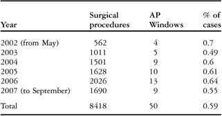
When editing their text, we were struck by the large numbers of patients with this rare lesion they had encountered over a relatively short period. From discussions with the authors, we learnt that their experienceReference Kiran, Singh, Shah, John and Maheshwari1 matches the incidence of the lesion within the overall group of patients with congenitally malformed hearts, cited at 0.59% in the previous study of Van Mierop and Kutsche.Reference Kutsche and Van Mierop3 Their centre became functional in 2001, but only in May of 2002 did they begin to offer surgical treatment for patients with congenitally malformed hearts. Since then, they have seen a huge number of patients, as shown in the Table 1. As they explained to us in ongoing correspondence, almost certainly their centre is now one of the largest in the World providing treatment for patients with congenitally cardiac malformations.
Numbers of aortopulmonary (AP) windows as a percentage of the total case load for congenital cardiac surgery at Narayana Hrudayalaya Institute of Cardiac Sciences, Anekal Taluk, Bangalore.

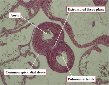
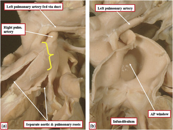
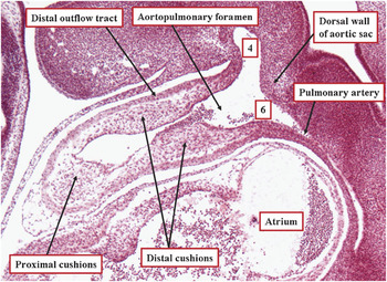
In the version of the manuscript submitted for textual editing, the authors had been describing “aortopulmonary septal defects”. In editing the text, we had converted this description to “aortopulmonary window”. This is because, in terms of the structure of the postnatal heart, and indeed beyond the initial few months of gestation, there is no such thing as an aortopulmonary septum. Almost as soon as the aortic and pulmonary pathways become separate entities within the developing heart, each of the intrapericardial arterial trunks develops its own discrete arterial wall. So, even though the two trunks are contained within a common epicardial sleeve, there is a discrete plane of extramural tissue separating the newly formed walls of the aorta and the pulmonary trunk (Fig. 1). This means that, when there is a communication between the cavities of the trunks in postnatal life (Figs 2–4), it would be incorrect to call it an aortopulmonary septal defect, since after birth there is no septum separating the lumens of the intrapericardial arterial trunks. Instead, the presence of such a hole implies failure of formation of the separate and adjacent walls of the intrapericardial arterial trunks, with persistence of the aortopulmonary foramen that certainly does exist during embryological development (Fig. 5).
The section is taken from a human embryo at Carnegie stage 20, just after closure of the embryonic interventricular foramen. Already the intrapericardial arterial trunks have developed their own discrete walls, albeit within a common epicardial sleeve. Note, however, the presence of the tissue plane between the adjacent walls.

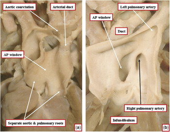
The images show a typical aortopulmonary window viewed from the front (a) and through the opened pulmonary trunk (b). Note that proximally the walls of the aorta and pulmonary trunk remain as discrete and separate entities. This is the so-called “Type I” lesion as defined by Mori and colleagues.Reference Mori, Ando, Takao, Ishikawa and Imai5

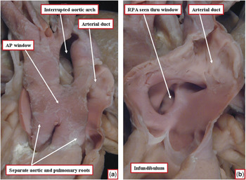
In this specimen, the aortopulmonary window is associated with interruption of the aortic arch, as shown in the frontal view (a). Opening the pulmonary trunk shows that the right pulmonary artery arises from the aortic aspect of the window (b). This is the so-called “Type II” lesion as defined by Mori and colleagues.Reference Mori, Ando, Takao, Ishikawa and Imai5

In this example of an aortopulmonary window, the pulmonary arteries are discontinuous, the left pulmonary artery being fed from the aorta through a persistently patent arterial duct (a). Opening the pulmonary trunk (b) shows that the window extends from the sinutubular junctions to the margins of the pericardial cavity. This is an example of the so-called “Type III” lesion defined by Mori and colleagues.Reference Mori, Ando, Takao, Ishikawa and Imai5

This section taken in the sagittal plane, replicating the parasternal long-axis echocardiographic cut, is from a human embryo at Carnegie stage 15. At this stage, as can be seen, the distal aorta is a common channel, feeding the aortic sac at the margin of the pericardial cavity. At this stage, the fourth (4) and sixth (6) aortic arches take origin from the sac, and the dorsal wall of the sac, therefore, at this stage, represents the aortopulmonary septum, albeit an extrapericardial structure. Note that there are cushions within the distal outflow tract, approaching each other so as to separate the outflow tract into pulmonary and aortic channels. The space between the distal ends of the cushions and the aortic sac is the aortopulmonary foramen.

It transpired that the authors were well aware of these anatomic subtleties. Indeed, when submitting the initial version of their manuscript for peer review, they had used the term “aortopulmonary window”. They had then been advised, during the process of review, to change to description of “aortopulmonary septal defect”, since this term is more frequently used elsewhere within the literature. The authors agree with us, nonetheless, that mere usage of a term by multiple authors does not necessarily make it scientifically accurate. In this instance, as with the ongoing description of the superior interatrial fold as the “septum secundum”, the usage of the incorrect term obscures the appropriate understanding of the anatomy of the malformation in question.Reference Anderson and Brown4 The authors, therefore, were more than happy to revert to their initial appropriate description of the lesion, as seen in the postnatal heart, as an aortopulmonary window.
The extensive experience of the authors shows that such windows can come in various guises, which they choose to list in descriptive fashion, rather than using the popular numerical categorisation suggested by Mori and colleagues.Reference Mori, Ando, Takao, Ishikawa and Imai5 The basic categories are small windows confined to the adjacent components of the interapericardial parts of the arterial trunks, the so-called Type I (Fig. 2), larger and spiralling lesions that involve the origin of the right pulmonary artery, the so-called Type II (Fig. 3), and more extensive defects extending from the sinutubular junctions to the margins of the pericardial cavity, the Type III (Fig. 4). The authors also note that the windows can exist in isolation, or can be associated with more severe malformations, a feature which previous authors suggested the possibility of further categorisation into simple and complex forms.Reference Jansen, Hruda, Rammeloo, Ottenkamp and Hazekamp6 In the ongoing correspondence, Kiran and his colleagues have endorsed our own doubts about the value of such a system, which like the numeric categorisation must involve a degree of Procrustean division. For those approving of such categorisations, however, all the necessary information is provided by the study from Bangalore.Reference Kiran, Singh, Shah, John and Maheshwari1
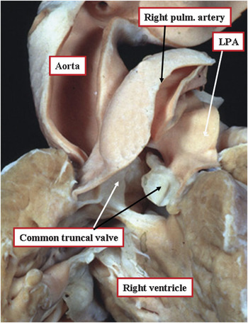
The unifying feature of the aortopulmonary windows, of course, is the separate nature of the arterial roots, with the presence of separate aortic and pulmonary valves distinguishing the entity from a common arterial trunk. When the window is large, however, as was the case in some of their patients, there is essentially a common intrapericardial component of the arterial pathways, or in other words a common intrapericardial arterial trunk. It is generally accepted, nonetheless, that it is the common arterial valve, and the common ventriculo-arterial outflow tract, that is the defining feature of common arterial trunk,Reference Crupi, Macartney and Anderson7 rather than the persistence of a common intrapericardial arterial channel. Indeed, in some forms of so-called common arterial trunk, the pulmonary trunk itself can separate from the aorta within the pericardial cavity (Fig. 6), this representing the so-called Type I variant as described by Collett and Edwards.Reference Collett and Edwards8
The picture shows a so-called common arterial trunk, defined because of the presence of a common ventriculo-arterial junction guarded by a common truncal valve. The trunk supplies directly the aortic, pulmonary, and coronary arterial systems, but note that there are virtually completely separate aortic and pulmonary trunks within the pericardial cavity, with crossed pulmonary arteries. This is the so-called “Type I” variant of common trunk as described by Collett and Edwards.Reference Collett and Edwards8

Here, therefore, we have another paradox, namely that the defining feature for so-called “persistent truncus arteriosus”, or common arterial trunk as we prefer to call it, is the presence of a common truncal valve, and common ventriculo-arterial junction, rather than a common intrapericardial arterial trunk. The defining feature, nonetheless, is itself important, since the presence of the common outflow tract points to the morphologic similarity between the entity currently described as common arterial trunk and the doubly committed and juxtaarterial interventricular communication. The pathognomonic feature of the latter lesion is failure of formation of the free-standing muscular subpulmonary infundibulum. In interventricular communications of this type, the outlet septum is often formed as a fibrous structure, known as the raphe, which is seen on the ventricular aspect of the fibrous continuity existing between the leaflets of the aortic and pulmonary valves. The presence of the common ventriculo-arterial junction, pointing to the affinity between the doubly committed interventricular communication and common arterial trunk, parallels the situation between the so-called “partial” and “complete” variants of atrioventricular septal defect, which in reality have either separate right and left valvar orifices within the common atrioventricular junction, or else have the junction guarded by a common atrioventricular valve.
The evidence accruing from these various malformations, therefore, shows that separation of the developing aortic and pulmonary pathways within the pericardial cavity can be perturbed in at least 3 fashions. In the most severe malformation, there can be failure to divide the common ventriculo-arterial junction, with persistence of a common valve guarding the common ventriculo-arterial junction, albeit that there can be virtually complete separation of the intrapericardial arterial trunks distal to the level of the valvar sinuses (Fig. 6). This is the entity we describe as common arterial trunk, or persistent truncus arteriosus. In the second malformation, as described by Kiran and associates,Reference Kiran, Singh, Shah, John and Maheshwari1 there has been appropriate separation of both the ventricular outflow tracts and arterial valves, but incomplete separation of the intrapericardial arterial trunks. This lesion, which we describe as an aortopulmonary window, can be sufficiently extensive as to produce a common intrapericardial arterial channel, albeit that we do not call it a common arterial trunk. The third lesion involving perturbation of separation of the proximal part of the outflow tract is confined to the ventricular components, with appropriate separation of the intrapericardial arterial trunks, and formation of separate aortic and pulmonary roots, but with retention of a common ventriculo-arterial junction, failure of formation of the free-standing muscular subpulmonary infundibulum, and failure of muscularisation of the outlet septum, in other words it is the doubly committed and juxtaarterial interventricular communication.
The morphology of these congenital cardiac malformations is particularly pertinent to concepts of embryonic separation of the developing outflow tracts. In most current textbooks, development of the outflow tract is still held to be based on existence of a “truncus” and a “conus”. There is, however, no current agreement on which lesions stem from maldevelopment of these parts, nor indeed on how we are to determine the components developed from these entities. Examination of the textbooks shows that it is the proximal part of the developing outflow tract that is usually described as the conus. It is, therefore, lack of division of the conus, rather than the distal part of the outflow tract, which is the embryologic perturbation producing the malformation still frequently described as persistent truncus arteriosus!
Those studying cardiac development increasingly describe the proximal and distal components of the outflow tract, rather than opting for use of “truncus” and “conus”. This is a much better approach, the more so since uncertainty remains with regard to the mechanics of transformation of these embryonic parts into the definitive structures making up the aorrtic and pulmonary channels in the postnatal heart. Those wishing to retain the concept of “conus” and “truncus” need to recognise that these 2 developmental components must give rise to 3 parts of the definitive outflow tracts, namely the intrapericardial arterial trunks, the arterial valvar leaflets and their supporting sinuses, and the ventricular outflow tracts.
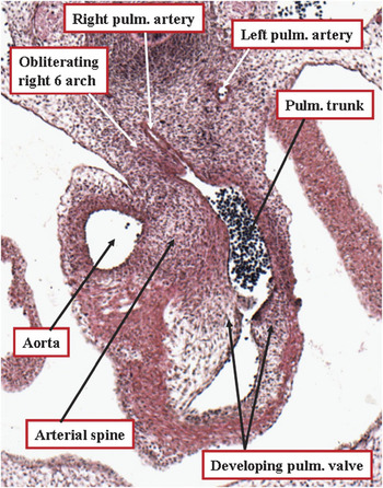
Our own ongoing studies have, we believe, cast some light on the origins of these components, but we still have work to do to resolve the precise site of formation of the definitive sinutubular junctions. This is because we know that, originally, the entirety of the embryonic outflow tract has muscular walls.Reference Anderson, Webb, Brown, Lamers and Moorman2 The mechanics of change of the initially muscular distal walls of the intrapericardial outflow tract into the arterial walls of the aorta and pulmonary trunk are remarkably complex. The separation of the cavity of the initially common distal outlet is equally complex, since when first seen, endocardial cushions or ridges extend almost to the margins of the pericardial cavity (Fig. 5). Beyond the margins of the pericardial cavity, the arteries of the pharyngeal arches extend through the pharyngeal mesenchyme, taking their origin from the so-called aortic sac. It is the dorsal wall of this sac that represents the so-called aortopulmonary septum, albeit an extrapericardial structure. At this early stage, the space between the distal ends of the cushions and the dorsal wall of the aortic sac is an aortopulmonary foramen. It is failure to close this foramen (Fig. 5) that produces the aortopulmonary window in postnatal life (Figs 2–4). The fused outflow cushions themselves then cavitate to form the arterial valves and their supporting sinuses (Fig. 7). Evidence from the structure of aortopulmonary windows would suggest that these cushions are capable also of producing the most proximal parts of the separate walls of the aorta and pulmonary trunk immediately distal to the sinutubular junction, since always these walls are discrete and separate in the setting of aortopulmonary windowReference Mert, Paker and Akcevin9 (also shown in Figs 2–4), as they are in the definitive postnatal heart (Fig. 1).
This section comes from a human embryo at Carnegie stage 16. The aortopulmonary foramen has been closed by ingrowth of an arterial spine from the back wall of the aortic sac that has fused with the distal ends of the outflow cushions, themselves now fused. The cushions themselves now cavitate to form the leaflets of the aortic and pulmonary valves, with intercalated cushions also contributing one leaflet to each valve. It remains to be determined if the cushions also contribute to the proximal walls of the aorta and the pulmonary (pulm) trunk just distal to the developing sinutubular junctions.

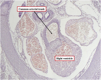
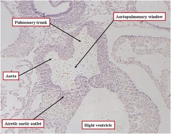
Presumptions made on the basis of the malformations perturbing the outflow tract, therefore, suggest that the cushions are intimately involved in the separation of the ventricular outflow tracts, where normally they become muscularised to form the subpulmonary infundibulum, and also in separation of the aortic and pulmonary valves and their supporting sinuses.Reference Anderson, Webb, Brown, Lamers and Moorman2 We know that, during development, the cushions themselves become richly populated by cells migrating from the neural crest. It is our belief that disappearance of some of these cells derived from the neural crest, which initially pack the central parts of the fused cushions, permits separation of the forming arterial roots one from the other, their disappearance resulting in the formation of the tissue plane between the muscularising subpulmonary infundibulum and the aortic root.Reference Anderson, Webb, Brown, Lamers and Moorman2 It is also known that many genetically modifed mice develop so-called persistent truncus arteriosus, in fact having common truncal valves, usually with the solitary arterial trunk arising exclusively from the right ventricle (Fig. 8). Most recently, we have observed genetically modified mice with aortopulmonary windows, albeit also with aortic atresia (Fig. 9). It will be ongoing studies of these normal and abnormal mice, during their embryonic development, which will clarify the mechanisms involved in separation of the intrapericardial arterial trunks. The lessons learnt from extensive clinical experiences, nonetheless, such as that described in such excellent fashion by our colleagues in Bangalore,Reference Kiran, Singh, Shah, John and Maheshwari1 will continue to guide our developmental researches.
This transverse section is from a loop-tail (Lp/Lp) embryo, at 15 days of gestation, and carrying mutations in the Vangl2 gene. It shows a common arterial trunk arising exclusively from the right ventricle.

This section is from another Lp/Lp embryo at 15 days of gestation. In this instance, there is an aortopulmonary window feeding the aorta, which is atretic at its origin from the right ventricle. The pulmonary trunk also arises from the right ventricle.