Introduction
The most common zoonotic filarial parasites in Europe that have an effect on human health are Dirofilaria (Nochtiella) repens and the heartworm Dirofilaria (Dirofilaria) immitis. These parasites utilize mosquitoes (Culicidae: Anopheles, Aedes, Culex) as intermediate hosts, thus humans are usually infected by zooanthropophilic species which fed previously on the animal with microfilaraemia. Dogs are the main reservoir hosts for D. repens, although the presence of the nematode has been reported in other species, such as cats and wild carnivores (foxes) (Magi et al., Reference Magi, Calderini, Gabrielli, Dell'Omodarme, Macchioni, Prati and Cancrini2008). In addition, humans can be accidental, though unsatisfactory, hosts for D. repens. Microfilariae are rarely produced in humans and the parasite can survive for considerable lengths of time, a few months, even years, without doing any harm. Dirofilaria repens is restricted mainly to dermal and subcutaneous tissues of the head, with the most common site being the conjunctiva and periocular region (Raccurt, Reference Raccurt1999).
Human D. repens infection is often relegated to a medical curiosity as a result of the relatively small number of reported cases. However, according to recent reports, D. repens represents an emerging infectious agent in Europe, particularly in the Mediterranean region, central and south-eastern Europe (Muro et al., Reference Muro, Genchi, Cordero and Simón1999; Pampiglione et al., Reference Pampiglione, Rivasi, Angeli, Boldorini, Incensati, Pastormerlo, Pavesi and Ramponi2001; Szénási et al., Reference Szénási, Kovács, Pampiglione, Fioravanti, Kucsera, Tánczos and Tiszlavicz2008). In Europe most human cases of D. repens infection occurred in Italy (Pampiglione et al., Reference Pampiglione, Canestri-Trotti and Rivasi1995), southern France (Raccurt, Reference Raccurt1999) and Russia (Kramer et al., Reference Kramer, Kartashev, Grandi, Morchón, Nagornii, Karanis and Simón2007). Furthermore, there are studies showing that D. repens infection appears to be spreading into the areas previously considered to be free of the disease, such as northern parts of Europe (Genchi et al., Reference Genchi, Rinaldi, Cascone, Mortarino and Cringoli2005; Mortarino et al., Reference Mortarino, Musella, Costa, Genchi, Cringoli and Rinaldi2008). Isolated human D. repens infection has also been reported in Serbia (Džamić et al., Reference Džamić, Arsić-Arsenijević, Radonjić, Mitrović, Marty and Kranjčić-Zec2004).
The clinical picture of dirofilariasis infection is usually benign; however, difficulties often arise in respect to the differential diagnosis. In clinical practice parasitic lesions are often mistaken for benign or malignant neoplasia, especially when the parasite is arrested in the orbital cavity, lungs, breast, male genitals and abdominal tissues (Hemmersbach-Miller et al., Reference Hemmersbach-Miller, Delmont, Hours and Brouqui2005). In the majority of cases, diagnosis of D. repens infection is based on macroscopic and microscopic characteristics of the worm. In the cases where this approach remains inconclusive, polymerase chain reaction (PCR) could be performed, either from the intact worm, fresh infected tissue or from paraffin-embedded tissues (Favia et al., Reference Favia, Lanfrancotti, della Torre, Cancrini and Coluzzi1997; Vakalis et al., Reference Vakalis, Vougioukas, Patsoula, Spanakos, Sioutopoulou and Vamvakopoulos2002; Rivasi et al., Reference Rivasi, Boldorini, Criante, Leutner and Pampiglione2006).
The first cases of dirofilariasis in Serbia were reported in the early seventies. Furthermore, it has been shown that the number of cases increased over the past decade. Our study of D. repens infection in Serbia during the period from 2001 to 2008, has enabled us to update the situation regarding this infection, to understand its geographical distribution and to deal with some of the diagnostic challenges. In the context of our findings, we present a critical discussion of the earlier case reports in Serbia.
Materials and methods
Patients and samples
From 2001 to 2008, 19 patients were examined by general practitioners, infectious and tropical diseases specialists, ophthalmologists, dermatologists and surgeons from different parts of Serbia, although the final diagnosis was made in our laboratory in Belgrade.
Examined samples were surgically excised nodules, intact worms, fragments of the parasite, crushed tissue with suspected parasitic elements, paraffin-embedded tissue and histopathological preparations.
Diagnostic methods
Samples were preserved in saline, glycerine-alcohol (5% glycerine in 70% ethyl alcohol) or 10% buffered formalin. Some samples were initially stored in dry sterile Petri dishes or laboratory bottles and transferred to glycerine-alcohol right after the examination. One sample (table 1, case 11) was preserved in 10% buffered formalin for more than 7 months before being sent to our laboratory.
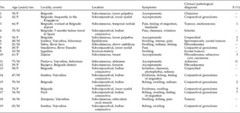
Clinical and epidemiological characteristics of human Dirofilaria repens infection in Serbia from 2001 to 2008.

Identification of the parasites was based on previously described morphological criteria (Gutierrez, Reference Gutierrez1984; Pampiglione et al., Reference Pampiglione, Canestri-Trotti and Rivasi1995; Orihel & Eberhard, Reference Orihel and Eberhard1997). During the macroscopic examination of worms, morphological characteristics such as shape, length, width, cephalic end and caudal end, were observed by naked eye and measured using the ocular micrometer of the optic microscope at 4 × magnification. For the purposes of microscopic examination, direct wet preparations were made with saline, glycerine and chloral-lactophenol. The parasites' characteristics, such as cuticle features, vaginal and anal opening, position of the digestive and genital structures, were examined using 4 × , 10 × and 20 × magnifications of the optical and inverted microscope.
In order to identify parasite species, sex and maturity and for comparison purposes, histological sections from the intact worms or fragments were made using haematoxylin-eosin (HE), Masson trichrome (MT), Movat pentachrome (MP) and Azan stains.
As per our request in 7 out of 19 cases, blood samples were taken from the patients for the detection of microfilariae. Blood samples could not be provided for the other patients since they had already been discharged from hospital or health centre. The presence of microfilariae was confirmed by the examination of direct wet blood preparations and Giemsa-stained thick and thin blood films before and after blood concentration using Knott's concentration technique and millipore membrane filter technique (Denham, Reference Denham, Gillespie and Hawkey1995).
Polymerase chain reaction
DNA was extracted from two paraffin-embedded tissues (table 1, cases 5 and 11) and from one intact worm preserved in glycerine-alcohol as a D. repens control (table 1, case 2). Sections of the paraffin block (approximately 1 mm3) containing the worm, were cut by blade and 1 cm of the intact worm (cut in 1 mm sections) was placed in a 1.5 ml Eppendorf tube. After 400 μl xylene was added to each tube (mixed using a vortex for 10 s) samples were centrifuged at 12,000 rpm for 5 min. Sediment was resuspended in 400 μl absolute ethyl alcohol and centrifuged again. Samples were suspended in lysis buffer containing 500 μg/ml proteinase K final concentration, and incubated at 56°C overnight. The next day 600 μl 25% Chelex (Chelex 100; Sigma, St. Louis, Missouri, USA) was added to the samples, followed by incubation at 56°C for 120 min before PCR was performed.
The specific PCR primer set used was: for D. repens DIR3 (GenBank accession no. L15323): 5′-CCGGTAGACCATGGCATTAT-3′ and DIR4: 5′-CGGTCTTGGACGTTTGGTTA-3′ and for D. immitis (GenBank accession no. M95173): 5′-ACGTATCTGAGCTGGCTCAC-3′ and 5′-ATGATCATTCCGCTTACGCC-3′.
PCR reaction was performed as described previously (Favia et al., Reference Favia, Lanfrancotti, della Torre, Cancrini and Coluzzi1997) resulting in a PCR product for D. repens of 246 bp. According to the standard molecular biological procedures, amplified PCR product was submitted to gel electrophoresis (2% horizontal agarose gel) and detected using ethidium bromide staining. The size of amplified PCR product was compared with a 100 bp DNA ladder and with a commercial 245 bp control marker (Pharmacia Biotech, Uppsala, Sweden).
Results
Clinical and epidemiological aspects
The mean age of our patients was 49.8 years, with 9 females and 10 males. Patients were from various parts of Serbia, including northern and southern provinces (Vojvodina, and Kosovo and Metohija) as well as from Belgrade. Five (21%) of the infected patients were from Vojvodina. Except in one case where the patient travelled to Spain (Valencia) for a short time (20 days) on vacation, 5 months before symptoms appeared, the patients had not travelled abroad during the years preceding suspicion of dirofilariasis. Some of the patients were engaged in recreational fishing, while others had summer houses on the river banks (Danube, Sava, Timok) and near forest areas. For the patient employed at Belgrade International Airport, a specialist in infectious diseases and a pathologist assumed that her initial diagnosis was onchocercosis as she worked in the immigration office and had frequent contact with immigrants from Africa and Asia (table 1, case 3). Most of investigated patients did not have dogs or cats as pet animals and could not remember having been bitten by mosquitoes or some other arthropods.
The clinical and epidemiological characteristics of patients are summarized in table 1. In all described cases the parasites and parasitic nodules occurred singly, with the most frequent site of infection in the eye and periocular region in 12 patients (63.1%). Other infection sites of great concern were epididymis, testicle and breast, where malignancies were considered in differential diagnosis. In 31.5% patients infection was asymptomatic. Recurrent migratory swellings were notable for approximately 2.5 years in one patient (table 1, case 7).
In most cases, the clinical diagnosis was mistaken, unless the parasite became visible to the naked eye, as when located under the bulbar conjunctiva. Eosinophilia was detected in only three cases, regardless of any clinical symptoms. In the seven patients who were tested for microfilariae, no larvae were detected in the patients' blood samples.
Parasitological findings
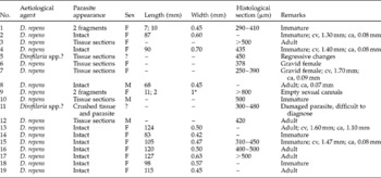
In two cases, because the morphology of the worms was insufficient for identification, parasites were determined as Dirofilaria spp. (table 2). In more than half the cases (12/19) the morphology of the filarial worms was well preserved. There was never more than one parasite inside the lesions. Intact worms from one subcutaneous nodule and two from the subconjunctival space were alive when samples arrived in the laboratory. In two cases determination of gender was impossible because of regressive changes in one (table 2, case 5) and small pieces of parasite in crushed tissue in the second case (table 2, case 11). In two cases inside the pseudocoelom, sections of the female reproductive tubes filled with clearly visible larval stages, microfilariae, were discovered.
Parasitological characteristics of human Dirofilaria repens infection in Serbia from 2001 to 2008.

F, female; M, male; cv, distance from vulva to parasites's cephalic end; ca, distance from anus to parasite's caudal end; *This value could be the result of prolonged conservation of the parasite in glycerine (cuticle swelling).
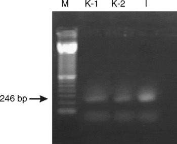
Figure 1 shows PCR-based amplification of the genomic DNA extracted from infected paraffin-embedded tissues, where diagnosis could not be made according to morphological findings. No amplification was detected with the specific primers for D. immitis, which could also be an aetiological agent of subcutaneous granuloma in humans. The amplified PCR product of 246 bp showed that the worms were D. repens.
PCR amplifications with primers DIR3 and DIR4. Lanes: M, 100 bp DNK ladder; K-1, K-2, positive results with expected PCR product of 246 bp from paraffin-embedded tissue; I, intact D. repens preserved in glycerine-alcohol used as the contol.

Discussion
Human superficial and visceral dirofilariasis, caused by several filarial species, in particular by D. repens, is an increasingly reported zoonosis in Europe. The Mediterranean area and south-eastern Europe are the endemic zones with the highest incidence of human infections. In Norway (Settnes & Engebjerg, Reference Settnes and Engebjerg1991), Germany (Jelinek et al., Reference Jelinek, Schulte-Hillen and Löscher1996; Siepmann et al., Reference Siepmann, Wannke, Neumann and Rohrbach2005), Austria (Bardach et al., Reference Bardach, Heimbucher and Raff1981) and recently Slovenia (Logar et al., Reference Logar, Novsak, Rakovec and Stanisa2001) infections by D. repens were described only in patients who had travelled to endemic regions. In some of the reported cases, infections by D. repens were most likely acquired in Serbia as patients had holidays there (Franz et al., Reference Franz, Volkner and Lenze1982).
Dogs are the main reservoir hosts for D. repens, harbouring adult parasites under the skin as well as microfilariae in the blood and other tissues. Studies conducted in Italy showed that the geographical distribution of D. repens infections in humans and dogs is very similar. A survey conducted in Piedmont (1966–67, repeated in 1991–92) clearly showed an increase in the number of infected dogs, with a fourfold increase in the prevalence of infections by D. immitis (Rossi et al., Reference Rossi, Pollono, Meneguz, Gribaudo and Balbo1996). The most recent study conducted in Italy showed spreading of D. immitis and D. repens infection in canines (Mortarino et al., Reference Mortarino, Musella, Costa, Genchi, Cringoli and Rinaldi2008). One earlier investigation in France showed that out of 5000 dogs examined, 1.4% had microfilariae in the blood (Marty, Reference Marty1997) while studies from northern Greece showed that 30% of dogs are infected by D. repens (Papazahariadou et al., Reference Papazahariadou, Koutinas, Rallis and Haralabidis1994). Apart from one report of an isolated case of D. repens infection in dogs (Simić & Vlatković, Reference Simić and Vlatković1940), there have been few veterinary surveys concerning the presence of this parasite in dogs and/or other animals in Serbia. On the other hand, reports of the presence of D. immitis in dogs have been based mainly on autopsy analyses (Kulišić & Milosavljević, Reference Kulišić and Milosavljević1994). According to investigations in the 1930s in southern Serbia, the infestation rate with D. repens was 60% in dogs and 3% in examined mosquitoes (Simić & Petrović, Reference Simić and Petrović1962). A study conducted in 2006–2007 showed that D. repens microfilariae were found in 7.2% of 193 privately owned dogs in the northern province of Serbia known as Vojvodina (Tasić et al., Reference Tasić, Rossi, Tasić, Miladinović-Tasić, Ilić and Dimitrijević2008). Until now there have been no reports of clinical cases of D. repens infection in dogs or other animals in Serbia.
It is of great importance to state that Serbia lacks national veterinary programmes for the control and protection of animals, and thereby lacks adequate accommodation for stray dogs, which often come into contact with adults or children who feed and/or play with them.
The only study concerning infection of mosquitoes with Dirofilaria spp. in Serbia was carried out in 1928, and showed that the vector of D. immitis in southern Serbia is Anopheles maculipennis (Simić, Reference Simić1928). It is plausible that A. maculipennis also serves as the intermediate host for D. repens. The majority of investigated patients lived, or stayed in, areas heavily infested by mosquitoes (near rivers and forested areas), including Vojvodina, where many natural pools and artificial water canals favour their multiplication. Nearly one-third (28%) of all reported patients with D. repens infection (tables 1 and 3) were from Vojvodina, which shares many geomorphological and climatic features with Hungary, where a recent study showed human D. repens infection as an emerging zoonosis (Szénási et al., Reference Szénási, Kovács, Pampiglione, Fioravanti, Kucsera, Tánczos and Tiszlavicz2008).
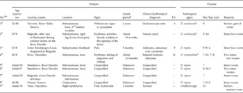
Human infection with Dirofilaria repens reported in Serbia from 1971 to 2001.

No.Ref, number of case, with reference: AStevanović et al., Reference Stevanović, Savić-Cvetojević and Petrović1971; BJoković & Nikolić, Reference Joković and Nikolić1977; CKranjčić-Zec et al., Reference Kranjčić-Zec, Mitrović, Arsić, Džamić and Radojičić1994; DObradović & Petrović, Reference Obradović and Petrović1979; EMišić et al., Reference Mišić, Stajković, Tešić, Mišić and Lesić1996; FJakšić et al., Reference Jakšić, Kujundžić and Pavlović1996; GLalošević et al., Reference Lalošević, Jeličić, Krese, Radulović and Petrović2001.
M, male; F, female; E, eosinophilia.
† Suspected time of infection by mosquito.
‡ D. acutiuscula, D. conjunctivae are synonyms for D. repens.
The first human infection with D. repens in Serbia was reported in 1971 as a case of subcutaneous nodule found on the back of a young boy (Stevanović et al., Reference Stevanović, Savić-Cvetojević and Petrović1971). Based on the published literature, concluding in mid-2001, eight additional cases were reported, and the most frequent site of the infection was subcutaneous tissue, except in two cases – one subconjunctival and one with the parasite in the epididymis (Joković & Nikolić, Reference Joković and Nikolić1977; Jakšić et al., Reference Jakšić, Kujundžić and Pavlović1996). In seven patients the diagnosis was conceived on examination of parasites that had been removed intact from the tissue (table 3). The presence of more than one worm inside the lesion is a very unusual finding, but such rare cases have been reported (Pampiglione et al., Reference Pampiglione, Canestri-Trotti and Rivasi1995; Fernando et al., Reference Fernando, Ihalamulla and De Silva2000). In Serbia, two worms were detected in the same lesion on two occasions. In the first case, a male and a female intact parasites were discovered in the same lesion (Mišić et al., Reference Mišić, Stajković, Tešić, Mišić and Lesić1996). In the second case, the sex was unspecified (Obradović & Petrović, Reference Obradović and Petrović1979) but given the size of the parasites (12 and 7–8 cm) it is plausible that they were also a female and a male (table 3). In the reported case of parasitic nodule found in the epididymis, the worm was assigned as male Dirofilaria spp. (Lalošević et al., Reference Lalošević, Jeličić, Krese, Radulović and Petrović2001), since male reproductive cells in various phases of maturation were observed. However, according to description of the worm in sections of the nodule, e.g. diameter between 0.3 and 0.4 mm, thickness of cuticle and subcuticular layer of 12 μm and presence of longitudinal ridges on its surface, the authors of the present paper consider these morphological data inconsistent with D. repens as a probable aetiological agent. The first report of subperiosteal localization of D. repens was submitted in 1994, after an olive-sized tumour was removed from the bone of a patient from Montenegro who had a nodule on his forehead (Kranjčić-Zec et al., Reference Kranjčić-Zec, Mitrović, Arsić, Džamić and Radojičić1994).
In the present review of 19 cases, the infection was not observed in children, consistent with reports from European countries where infections in children are rarely described (Marty, Reference Marty1997; Pampiglione et al., Reference Pampiglione, Rivasi, Angeli, Boldorini, Incensati, Pastormerlo, Pavesi and Ramponi2001). In contrast, in Sri Lanka the infection is most common in children under the age of nine (Dissanaike et al., Reference Dissanaike, Abeyewickreme, de S. Wijesundera, Weerasooriya and Ismail1997). A possible explanation for this distinct incidence in children could be the existence of different subspecies of D. repens and/or some separate social and biogeographical impacts.
In our study the most common localizations of D. repens infection in the 19 patients were bulbar conjunctiva, external palpebra and submucosal palpebra. Our findings are in agreement with the review of human dirofilariasis worldwide, stating that in nearly 47% of D. repens infections, the parasite was found in the region of the head, particularly in the conjunctiva (Pampiglione et al., Reference Pampiglione, Canestri-Trotti and Rivasi1995). This agrees well with data from France (Raccurt, Reference Raccurt1999) and the former Soviet Union, reporting, respectively, nearly 30% and 46% (Avdiukhina et al., Reference Avdiukhina, Supriaga, Postonova, Kuimova, Mironova, Murashov and Putintseva1997) infection of the eye and periocular tissue. Although different body sites of D. repens infection in humans are constantly reported, the ocular and periocular regions remain predominant sites of infection (Gorezis et al., Reference Gorezis, Psilla, Asproudis, Peschos, Papadopoulou and Stefaniotou2006; Estran et al., Reference Estran, Marty, Blanc, Faure, Leccia, Pelloux, Diebolt, Ambrosetti and Cardot-Leccia2007; Eccher et al., Reference Eccher, Dalfior, Gobbo, Martignoni, Brunelli, Decaminada, Bonetti, Rivasi, Barbareschi and Menestrina2008).
Dirofilaria repens and D. immitis infections of male genital organs, particularly of the testicle, epididymis and spermatic cord (Theis et al., Reference Theis, Gilson, Simon, Bradshaw and Clark2001; Supriaga et al., Reference Supriaga, Starkova and Korotkova2002; Soussi et al., Reference Soussi, Farah Klibi, Zermani, Rammeh, Ismail, Zakraoui, Atallah and Ben Jilani2004) represent a serious problem for urologists, since dirofilariasis is not often considered as a differential diagnosis. Such infections could present as neoplasia or conditions of urological emergency. Dirofilaria repens infection of the epididymis (two cases reported) and testis (one case) have been described in Serbian patients.
Pregnant D. repens are rarely encountered in human lesions. In the recent review of 60 cases of D. repens infection in Italy (Pampiglione et al., Reference Pampiglione, Rivasi, Angeli, Boldorini, Incensati, Pastormerlo, Pavesi and Ramponi2001) pregnant females were not found. In the first described infection with D. repens in Serbia (Stevanović et al., Reference Stevanović, Savić-Cvetojević and Petrović1971) a gravid parasite was found in the histological section from the subcutaneous nodule which had persisted for 2 years. In our study (table 2) two specimens of pregnant parasites have been observed. In such infections the attendance of sexually mature males in the same subject would be required, suggesting the presence of both worms in the same infective inoculum. However, we did not detect the presence of a male worm. Some authors have reported the presence of microfilariae in the blood and surrounding tissue (Nozais et al., Reference Nozais, Bain and Gentillini1994; Negahban et al., Reference Negahban, Daneshbod, Atefi, Daneshbod, Sadjjadi, Hosseini, Bedayat and Abidi2007). On the contrary, in our patients microfilaraemia was absent in the cases where pregnant parasites were found.
According to the literature, in the majority of cases the parasites have been found in histological preparations of surgically excised lesions, and they have been removed intact from the tissue less frequently (Pampiglione et al., Reference Pampiglione, Canestri-Trotti and Rivasi1995). In the present report, the diagnosis was made, in 12 out of 19 cases, on examination of parasites that had been removed intact from the tissue. Such findings are beneficial for parasitologists because important features of the parasite are more or less preserved, while diagnosis can also be confirmed by examination of histological sections using different staining techniques, which help in better visualization of a parasite's morphology. However, identification of the parasite in tissue sections only is difficult at times, particularly when regressive phenomena render the parasite's morphological characteristics unrecognizable as a result of death of the parasite caused by the host inflammatory reaction (Pampiglione et al., Reference Pampiglione, Rivasi and Canestri-Trotti1999). In our study we examined seven cases of parasites in tissue sections. In 5/7 cases diagnosis of D. repens did not pose significant problem. In 2/7 cases regressive phenomena and a crushed parasitic nodule made identification of an infectious agent very difficult, so we decided to utilize the PCR as an alternative diagnostic approach.
One of the samples tested by PCR was a paraffin-embedded tissue block prepared immediately after a periocular granuloma was extracted, and the second sample was a mix of tissue and parasitic elements originating from a subcutaneous abdominal nodule. The latter had been preserved in formalin for more than 7 months before being embedded in a paraffin block. An intact worm identified as D. repens preserved in glycerine-alcohol served as the reaction control. It has been suggested that different preservation methods prior to PCR could have inhibitory effects on the PCR reaction. The most important limitation of formalin-fixed samples, especially for a long time, e.g. months, is the breakage of DNA into fragments of about 250–350 bp. Such a problem can be surmounted by using D. repens specific primers of 246 bp (Rivasi et al., Reference Rivasi, Boldorini, Criante, Leutner and Pampiglione2006). The preservation methods used in our study did not appear to influence the amplification reaction.
The number of reported cases of human dirofilariasis in some countries or regions is usually lower than those actually occurring, and medical awareness of the infection risk is essential for correct diagnosis. Our findings concerning human D. repens infections in Serbia are consistent with the reports from other countries in the Mediterranean area with regards to the clinical presentation, age of patients and clinical diagnosis. Given the fact that the infection rate in dogs is relatively high, and that patients represented here come from different parts of the country, we can conclude that in Serbia many cases may be misdiagnosed.
Human D. repens infection in Serbia is increasing steadily. The best way to explain the difference between the numbers of new cases in the two observed periods is the significant improvement in diagnostic approach. Also, current climatic trends may be an influence on the increased prevalence of infection in reservoir hosts, vectors as well as in humans (Medlock et al., Reference Medlock, Barrass, Kerrod, Taylor and Leach2007; Mortarino et al., Reference Mortarino, Musella, Costa, Genchi, Cringoli and Rinaldi2008), but relevant studies have not been conducted in Serbia till now.
Since our laboratory became the National Reference Laboratory for Parasitic Zoonoses, two new cases of suspected dirofilariasis have come to our laboratory from neighbouring Bosnia and Herzegovina and the Republic of Macedonia for an expert opinion, thus allowing us to have better insight into human Dirofilaria spp. infections in the Balkans.
Acknowledgements
This work received partial financial support from the Ministry of Science and Technological Development of the Republic of Serbia (project no.14010).