Introduction
To date, the field of psychiatry has primarily relied on phenomenology-based disease classification systems, such as the Diagnostic and Statistical Manual of Mental Disorders (DSM). However, particular problems of heterogeneity in psychiatry present as a challenge, with the difficulty of replicating results of basic research in clinical studies, as well as with the variable long-term trajectory and treatment response of mental illness (Everling & Fischer, Reference Everling and Fischer1998; Woolf, Reference Woolf2008). Recently, the Research Domain Criteria (RDoC) has been proposed as a significant effort in the direction of moving away from the traditional phenomenology-based categorization, with the aim to re-examine the existing mental illnesses across diagnostic boundaries based on the underlying neural mechanisms (Cuthbert, Reference Cuthbert2014). Such examination of transdiagnostic, neural-based mechanisms of mental disorders is expected to provide crucial insights into the pathophysiology that generate psychiatric symptoms and to advance the development of novel therapeutic techniques.
The current lack of clear and objective grounds for psychiatric diagnoses has also led to heterogeneity problems. Obsessive–compulsive disorder (OCD) is a mental disorder with a prevalence rate of 2–3% and is defined as the presence of obsessions and/or compulsions that are time-consuming, distressing, or disabling (Gillan, Fineberg, & Robbins, Reference Gillan, Fineberg and Robbins2017). Accumulated evidence has revealed OCD to comprise a unique spectrum of diseases, and the DSM-5 acknowledged the disorder as a distinct diagnostic category that no longer falls under the anxiety disorder classification (Phillips et al., Reference Phillips, Stein, Rauch, Hollander, Fallon, Barsky and Leckman2010). Schizophrenia (SCZ), which is prevalent in 1% of the population, is a mental disorder accompanied by hallucinations and delusions that cause chronic deteriorative changes. Although the SCZ spectrum disorders are epidemiologically and biologically similar, the clinical manifestations of each patient are extremely broad, diverse, and heterogeneous (Carpenter & Kirkpatrick, Reference Carpenter and Kirkpatrick1988; Guloksuz & van Os, Reference Guloksuz and van Os2018; Wolfers et al., Reference Wolfers, Doan, Kaufmann, Alnaes, Moberget, Agartz and Marquand2018). Despite efforts to mitigate the problem of an overly wide variety of clinical manifestations of SCZ made during the evolution from DSM-IV to DSM-5, SCZ remains a heterogeneous disorder, with biological boundary also remaining blurred overlapping with that of other mental disorders (Clementz et al., Reference Clementz, Sweeney, Hamm, Ivleva, Ethridge, Pearlson and Tamminga2016; Tandon et al., Reference Tandon, Gaebel, Barch, Bustillo, Gur, Heckers and Carpenter2013). Obsession and delusion are theoretically distinct from each other in terms of reality testing, as shown by the ego-dystonic nature of the former and the ego-syntonic of the latter (Oulis, Konstantakopoulos, Lykouras, & Michalopoulou, Reference Oulis, Konstantakopoulos, Lykouras and Michalopoulou2013). However, in patients with severe OCD, especially those with poor insight or absent insight/delusional beliefs specifiers, the boundary between the two symptoms is obscured. Moreover, a higher co-occurrence rate of psychosis and OCD, clozapine-induced OC symptoms, and the presence of OCD in the prodromal stage of SCZ have all suggested a biological link between the two disorders (Bebbington & Freeman, Reference Bebbington and Freeman2017; de Haan, Linszen, & Gorsira, Reference de Haan, Linszen and Gorsira1999; Niendam, Berzak, Cannon, & Bearden, Reference Niendam, Berzak, Cannon and Bearden2009). In particular, it is unclear whether these obsessive and delusional symptoms are obsessive–delusion continuum based on the same neural correlates or distinct clinical symptoms based on different brain regions. In this context, identifying common and distinct neural correlates of obsession and delusion by utilizing neuroimaging biotypes, beyond and rather than phenotypes, could provide further insight into the pathophysiology of the disease and the establishment of a treatment strategy.
Specific brain networks characterized by resting-state functional connectivity (RSFC) have been shown associated with diverse phenotypic variability (Vaidya & Gordon, Reference Vaidya and Gordon2013). Several RSFC studies have also provided evidence of abnormalities in the frontostriatal or frontoparietal circuits of OCD and in the frontotemporal circuit of SCZ (Gursel, Avram, Sorg, Brandl, & Koch, Reference Gursel, Avram, Sorg, Brandl and Koch2018; Harrison et al., Reference Harrison, Soriano-Mas, Pujol, Ortiz, Lopez-Sola, Hernandez-Ribas and Cardoner2009; Jung et al., Reference Jung, Yucel, Yun, Yoon, Cho, Parkes and Kwon2017; Yoon et al., Reference Yoon, Yun, Jung, Cho, Kim, Lee and Kwon2015). However, most of these studies employed mass-univariate statistical approaches to connectivity estimated from a prior seed or network and considered the association of a phenotypic variable with only one functional connection at a time. Because such univariate analyses explore only a restricted set of brain networks while ignoring the concurrent contributions of other connections, the ability to identify functional networks associated with phenotypic measures beyond our prior knowledge has been somewhat constrained (Cole, Smith, & Beckmann, Reference Cole, Smith and Beckmann2010; Shehzad et al., Reference Shehzad, Kelly, Reiss, Cameron Craddock, Emerson, McMahon and Milham2014). Recently, a multivariate distance-based matrix regression (MDMR) approach for connectome-wide association studies (CWAS), which explores brain–phenotype relationships across the entire connectome at the voxel level, has been introduced as a data-driven multivariate approach that could overcome the aforementioned constraints (Shehzad et al., Reference Shehzad, Kelly, Reiss, Cameron Craddock, Emerson, McMahon and Milham2014). Unlike other multivariate methods, the MDMR-based CWAS approach also allows one to examine the association between a phenotypic variable and multivariate patterns of connectivity while controlling for covariates. Additionally, the approach is able to examine more than one predictor variable at a time, regardless of types of categorical and/or continuous variables (see Shehzad et al., Reference Shehzad, Kelly, Reiss, Cameron Craddock, Emerson, McMahon and Milham2014 for detailed description regarding the benefits of the approach). This approach has been successfully applied to studies of RSFC in association with IQ, development (age), and medication administration in healthy individuals and with functional dysconnectivity in psychiatric patients (Satterthwaite et al., Reference Satterthwaite, Vandekar, Wolf, Bassett, Ruparel, Shehzad and Gur2015; Sharma et al., Reference Sharma, Wolf, Ciric, Kable, Moore, Vandekar and Satterthwaite2017; Shehzad et al., Reference Shehzad, Kelly, Reiss, Cameron Craddock, Emerson, McMahon and Milham2014).
Here, we investigated the dimensional effects of obsession and delusion on RSFC that may be present transdiagnostically, using a large sample comprised of patients with OCD and SCZ and healthy control (HC). Specifically, we employed the MDMR-based CWAS approach to explore the multivariate patterns of RSFC across the entire brain, rather than using a prior seed or network selection. Through this approach, as described below, we provide novel evidence of functional network aberrations associated with obsession and delusion symptoms across diagnostic boundaries.
Materials and methods
Participants
A total of 236 subjects (96 OCD, 75 SCZ, and 65 HC) were included in the final analysis (Table 1). All subjects were recruited from the OCD clinic and psychosis clinic at Seoul National University Hospital (SNUH). Subjects with OCD have fulfilled the DSM-IV criteria for OCD, and subjects with SCZ were also evaluated using the Structured Clinical Interview (SCI) for DSM-IV. The HC were screened using the SCI for DSM-IV Axis I Disorders Non-Patient Edition with an additional exclusion criterion of any first- and third-degree biological relatives with a psychiatric disorder. All study procedures were approved by the Institutional Review Boards of Seoul National University Hospital, and written informed consent was obtained from all participants.
Demographic and clinical characteristics of participants

OCD, obsessive–compulsive disorder; SCZ, schizophrenia; HC, healthy controls.
a Relative root-mean-square value of the translation parameters for head motion.
Two groups showed significant between-group differences: AOCD v. SCZ; BOCD v. HC; CSCZ v. HC.
Clinical assessment
Patients with OCD were administered the Yale-Brown Obsessive-Compulsive Scale (Y-BOCS) (Goodman et al., Reference Goodman, Price, Rasmussen, Mazure, Fleischmann, Hill and Charney1989) to measure the severity of their obsessive–compulsive symptomatology, whereas patients with SCZ were administered the Positive and Negative Syndrome Scale (PANSS) (Kay, Fiszbein, & Opler, Reference Kay, Fiszbein and Opler1987) to assess a variety of symptoms related to SCZ. We used the total obsession score from the Y-BOCS as an indicator of the severity of obsession symptoms for patients with OCD. The P1 score from PANSS was used to determine the severity of delusion symptoms for patients with SCZ. For patients with SCZ (or OCD) who had no obsession (or delusion) scores, these scores were estimated through medical record review by attending psychiatrists (T.Y.L. and J.L.) who have already diagnosed and treated the patients in clinic and showed excellent inter-rater reliability (k = 0.92). There was no significant correlation between obsessive symptoms and delusional symptoms in each group.
Image acquisition and preprocessing
All participants underwent high-resolution 3D T1-weighted imaging and 7 min resting-state fMRI on a 3T scanner. After they were scanned, a simple questionnaire was administered to confirm that they had not fallen asleep.
Image preprocessing was performed using SPM12 (http://www.fil.ion.ucl.ac.uk/spm) and DPARSFA toolbox (http://rfmri.org/DPARSF/). Preprocessing included removal of the first four images, slice timing correction, motion correction, coregistration, nuisance covariate regression (head motion parameters, scrubbing regressors, five principal components estimated from both white matter and cerebrospinal fluid regions, and two polynomial trending terms), spatial normalization to the Montreal Neurological Institute space, spatial smoothing with a 6 mm full-width-at-half-maximum Gaussian kernel, and bandpass filtering (0.01–0.1 Hz) was conducted. The preprocessed data were ultimately downsampled to 4 mm isotropic voxels to allow for computational feasibility (Shehzad et al., Reference Shehzad, Kelly, Reiss, Cameron Craddock, Emerson, McMahon and Milham2014) (see online Supplementary Methods for more information).
MDMR-based CWAS and follow-up seed-based connectivity analysis
This analysis was conducted using the Connectir package (http://czarrar.github.io/connectir). Using MDMR, we examined the association between obsession (or delusion) scores and multivariate patterns of connectivity (the distances between participants connectivity maps) while controlling for age, sex, clinical group status, in-scanner motion (mean relative RMS), and delusion (or obsession) score as confounding variables. MDMR yielded a pseudo-F statistic whose significance was assessed using 5000 iterations of a permutation test. The pseudo-F statistic referred to this simulated distribution to obtain a p value, and the p value map was then converted to z value for multiple comparison corrections. Given recent issues about clusterwise inference (Eklund, Nichols, & Knutsson, Reference Eklund, Nichols and Knutsson2016), the significance threshold was set at a height threshold of z > 3.1 (corresponding to p < 0.001) and a cluster-extent threshold of p < 0.05, with family-wise error rate (FWER) corrected, using Gaussian random fields. For completeness and to compare with previous MDMR-based CWAS studies (Satterthwaite et al., Reference Satterthwaite, Vandekar, Wolf, Bassett, Ruparel, Shehzad and Gur2015; Sharma et al., Reference Sharma, Wolf, Ciric, Kable, Moore, Vandekar and Satterthwaite2017), we also examined the data at lower thresholds (z > 2.3, p < 0.05 and z > 1.64, p < 0.01). MDMR-based CWAS indicates only the presence of an association between phenotypic variables and connectivity patterns, but it does not describe the specific connections and directions of observed associations. Thus, to further characterize the connectivity associated with individual obsession (or delusion) scores, we conducted post hoc seed-based functional connectivity (FC) analysis for clusters identified by MDMR as seeds. Group-level regression analyses were performed to determine the association between the seed-based FC maps generated from each seed cluster and obsession (or delusion) score. The group-level regression analyses included the same covariates as those listed above. Statistical significance was set at the same cluster-corrected threshold as for MDMR (online Supplementary Fig. S1).
Functional connectivity between identified clusters
To further investigate whether the strength of FC between three clusters identified by MDMR was also associated with individual obsession scores, we performed partial correlation analysis (controlling for the covariates listed above) between these FC strengths and obsession scores [q < 0.05, corrected for false discovery rate (FDR) for three FC-obsession scores correlations].
Network construction and analysis
The results from the abovementioned analyses suggested that the results from MDMR-based CWAS were driven by interactions between specific functional networks. Thus, to validate this hypothesis and summarize all the observed results, we evaluated the data within a network framework. We calculated Pearson correlation coefficients between time series in 10 brain regions identified in this study (three from MDMR analysis and seven from seed-based FC analysis) as nodes. Then, the individual correlation matrices were Fisher z-transformed by z = 0.5 × ln[(1 + r)/(1 − r)], where r is a correlation coefficient, to improve normality and averaged across the entire sample. This averaged matrix was separated into three distinct network modules using the Louvain modularity algorithm (Blondel, Guillaume, Lambiotte, & Lefebvre, Reference Blondel, Guillaume, Lambiotte and Lefebvre2008). Inner-network connectivity and inter-network connectivity were defined as the average connectivity across node pairs within the same network and the average connectivity across all node-to-node connections between two networks, respectively. We then performed partial correlation analysis (controlling for the covariates listed above) between these connectivity measures (i.e. three inner-network and three inter-network connectivity strengths) and obsession scores (q < 0.05, FDR-corrected for six network connectivity–obsession scores correlations).
To confirm which network each of the modules belongs to and to validate our findings from the above modularity analysis, we further conducted additional inter-network connectivity analyses using nodes derived from alternative network definitions from previous studies (Power, Barnes, Snyder, Schlaggar, & Petersen, Reference Power, Barnes, Snyder, Schlaggar and Petersen2012; Yeo et al., Reference Yeo, Krienen, Sepulcre, Sabuncu, Lashkari, Hollinshead and Buckner2011). Based on the results from our modularity analysis, four cognitive networks, including the dorsal attention network (DAN), ventral attention network (VAN), frontoparietal control network (FPCN), and default mode network (DMN), were included in this analysis. Like the abovementioned inter-network analysis, we estimated Fisher's z-transformed inter-network connectivity strengths between these four cognitive networks and then performed partial correlation analyses between these connectivity measures and obsession scores (q < 0.05, FDR-corrected for six inter-network connectivity strengths–obsession correlations for each of atlases).
Supplementary analyses
Although the above analyses focused on the dimensional relationship with obsession (or delusion) severity across all three groups while controlling for the effects of the potential confounding variables, we conducted several supplementary analyses to confirm our findings. First, we re-evaluated clusters identified in the dimensional analysis of obsession (delusion) severity while excluding delusion scores (or obsession scores) from the covariates. Second, to evaluate whether the dimensional results were similar within the patient groups alone, we re-evaluated significant clusters while excluding HC subjects.
Results
Brain regions identified by MDMR
MDMR-based CWAS revealed that obsession severity was robustly associated with the multivariate patterns of connectivity in the right supplementary motor area [SMA; Brodmann area (BA)6] extending to the frontal eye field (BA8), dorsal anterior cingulate cortex (ACC; BA32), left precentral gyrus (PreCG), and right superior parietal lobule (SPL), while there were no regions significantly associated with delusion severity (at a joint height and cluster-extent threshold of z > 3.1 and p < 0.05, FWER-corrected; Fig. 1).
Brain regions identified by MDMR-based CWAS. Obsession severity was associated with multivariate patterns of connectivity in the right supplementary motor area (SMA), left precentral gyrus (PreCG), and right superior parietal lobule (SPL) (z > 3.1, p < 0.05). L, left hemisphere; R, right hemisphere.

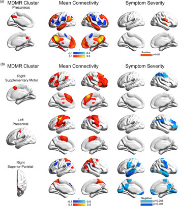
To validate our findings, we further conducted additional MDMR-based CWAS analyses by excluding other symptom severity (delusion or obsession score) or HC subjects in the models. When delusion scores were excluded from the model covariates or when HC subjects were excluded, only the SMA cluster remained significantly associated with obsession severity (at z > 3.1, p < 0.05). When the obsession score was excluded from the model covariates, delusion severity was associated with multivariate connectivity patterns in the precuneus (PreCU) across all three groups, though it was less robust than the above analysis for the delusion severity (at z > 2.3, p < 0.05; Fig. 2a); however, this threshold is more comparable to the results from previous MDMR-based CWAS studies (z > 1.64, p < 0.01) (Satterthwaite et al., Reference Satterthwaite, Vandekar, Wolf, Bassett, Ruparel, Shehzad and Gur2015; Sharma et al., Reference Sharma, Wolf, Ciric, Kable, Moore, Vandekar and Satterthwaite2017). The PreCU cluster's association with the delusion severity remained even when the same threshold used in the previous studies was applied (at z > 1.64, p < 0.01). However, when HC subjects were excluded, there were no clusters associated with the delusion severity.
Results from post hoc seed-based connectivity analyses. (a) Seed-based connectivity results for the cluster associated with delusion severity. The left column displays the precuneus cluster identified by MDMR (z > 2.3, p < 0.05). The middle column displays the mean connectivity map across all subjects from the precuneus seed. Red (blue) colors on brain surfaces indicate positive (negative) connectivity with each seed cluster. The right column displays a significant association between delusion severity and seed-based connectivity maps for the precuneus (z > 2.3, p < 0.05). (b) Seed-based connectivity results for clusters associated with obsession severity. The left column displays each cluster identified by MDMR that was used as a seed for follow-up seed-based connectivity analysis (z > 3.1, p < 0.05). The middle column displays the mean connectivity map across all subjects from each seed. Red (blue) colors on brain surfaces indicate positive (negative) connectivity with each seed cluster. The right column displays significant associations between obsession severity and seed-based connectivity maps for each seed (z > 3.1, p < 0.05).

Seed-based connectivity results for clusters from MDMR as seeds
The regression analyses with seed-based FC maps demonstrated that the multivariate results from MDMR were derived from dysconnectivity between specific functional networks (Fig. 2b). For example, with increasing obsession severity, the SMA cluster showed decreased connectivity with the areas that were positively correlated with the PreCG cluster, including the SPL, which was the area that was negatively correlated with the SMA. Conversely, the PreCG cluster showed decreased connectivity with the areas that were positively associated with the SMA cluster, including the dorsolateral prefrontal cortex and inferior parietal lobule, such as the supramarginal gyrus. For the SPL cluster, higher obsession severity was associated with decreased connectivity among the areas belonging to the DMN, including the medial prefrontal cortex (MPFC), temporal pole, middle temporal cortex, angular gyrus, and PreCG. All clusters showed significant associations between obsession severity and the areas anticorrelated with each cluster identified by MDMR (at z > 3.1, p < 0.05; Fig. 2b).
Although it was less robust (z > 2.3, p < 0.05), higher delusion severity was associated with the area negatively correlated with the PreCU cluster, particularly a larger cluster including Heschl's gyrus, posterior insula, and Rolandic operculum (Fig. 2a) for all subjects. There was no significant correlation between the scores of obsession and delusion in each group.
Connectivity and modularity between clusters from MDMR
Considered collectively, seed-based FC analyses consistently implicated the association of the interaction between the maps of the SMA and PreCG clusters. Using node-to-node FC analyses between clusters from MDMR, we confirmed that obsession severity was negatively associated with the FC strength between the SMA and PreCG clusters (r/q value = −0.178/0.021; Fig. 3a).
Results from connectivity between clusters identified by MDMR. (a) Obsession severity was associated with the connectivity between the supplementary motor area (SMA) and precentral gyrus (PreCG). (b) A modularity analysis divided 10 areas into three functional network modules. Obsession severity was associated with the connectivity between modules 1 and 2 and between modules 2 and 3.

Modularity analysis divided 10 brain areas (three from MDMR and seven from seed-based analysis) into three network modules. Additionally, obsession severity was significantly associated with inter-network connectivity strength between modules 1 and 2 (r/q value = −0.309/<0.001) and between modules 2 and 3 (r/q value = −0.200/0.007) (Fig. 3b).
Inter-network connectivity between large-scale brain networks based on prior brain atlases
The additional inter-network connectivity analyses revealed that regardless of the type of atlases we used (Power et al., Reference Power, Barnes, Snyder, Schlaggar and Petersen2012; Yeo et al., Reference Yeo, Krienen, Sepulcre, Sabuncu, Lashkari, Hollinshead and Buckner2011), higher obsession severity was consistently associated with decreased inter-network connectivity between the DAN and FPCN (r/q value = −0.161/0.048 for Yeo's atlas, Fig. 4a; r/q value = −0.179/0.036 for Power's atlas, Fig. 4b). For Yeo's atlas, higher obsession severity was also associated with decreased inter-network connectivity between the DAN and VAN (r/q value = −0.159/0.048; Fig. 4a).
Results from network connectivity analysis based on prior atlases. (a) Four functional networks (default mode network, DMN; dorsal attention network, DAN; ventral attention network, VAN; and frontoparietal control network, FPCN) were identified using the atlas of Yeo et al. Obsession severity was associated with inter-network connectivity between the DAN and VAN and between the DAN and FPCN. (b) The aforementioned four functional networks were identified using the atlas of Power et al. Obsession severity was associated with inter-network connectivity between the DAN and FPCN. L, left hemisphere; R, right hemisphere.

Discussion
In this study, we employed the MDMR-based CWAS approach to explore how the multifocal patterns of dysconnectivity relate to the dimensionally defined symptoms of obsession and delusion across diagnostic boundaries in a large sample of patients with OCD, SCZ, and HC. This data-driven approach revealed that obsession severity was robustly associated with the SMA, PreCG, and SPL, while delusion severity was associated, though less robustly, with the PreCU. Follow-up seed-based FC and modularity analyses revealed that the multivariate results from MDMR were derived from aberrant inter-network connectivity between specific functional networks, particularly from the association between obsession severity and the FPCN (module 1)–DAN (module 2) inter-network connectivity and the DAN (module 2)–DMN (module 3) inter-network connectivity. Additional inter-network analyses based on nodes from alternative atlases also confirmed the association between obsession severity and inter-network connectivity between the FPCN and the DAN. Taken together, these findings demonstrate, for the first time, a transdiagnostic pattern of large-scale network dysconnectivity associated with obsession severity commonly shared between OCD and SCZ patient groups.
Study of the spectrum disorder provides new avenues for the research of mental illness. For example, autism spectrum disorder has been found to be similar to anxiety disorders, including OCD, even in psychosis (Cochran, Dvir, & Frazier, Reference Cochran, Dvir and Frazier2013; Postorino et al., Reference Postorino, Kerns, Vivanti, Bradshaw, Siracusano and Mazzone2017). Shared and disorder-specific abnormalities in structural and functional brain findings showed disorder-differential mechanisms in autism spectrum disorder (Carlisi et al., Reference Carlisi, Norman, Lukito, Radua, Mataix-Cols and Rubia2017). In this regard, the clinical relevance of the schizo-obsessive spectrum disorder has long been a topic of interest among psychiatrists (Scotti-Muzzi & Saide, Reference Scotti-Muzzi and Saide2017). Despite numerous studies in schizo-obsessive spectrum disorder, however, the question of whether obsession and delusion share common pathophysiology remains unclear due to the inadequate literature on biologically driven studies. In this study, we identified distinct neural networks of obsession and delusion. This finding suggests that despite the phenotypical similarities shared between OCD and SCZ, they are distinct disorders with different pathophysiology rather than showing a diverse expression of psychosis or obsession.
We found that obsession severity was correlated with multivariate patterns of connectivity in the right SMA extending to the dorsal ACC, right SPL, and left PreCG. This finding somewhat differs from the results reported by previous univariate studies conducted only in OCD patients and those from studies including HC that emphasized the orbitofrontal-striatal or the dorsal ACC and amygdalo-cortical circuits (Kwon, Jang, Choi, & Kang, Reference Kwon, Jang, Choi and Kang2009; Milad & Rauch, Reference Milad and Rauch2012). The SMA constitutes a part of the brain circuit that contributes to the control of movement as well as to cognitive functions (de Wit et al., Reference de Wit, de Vries, van der Werf, Cath, Heslenfeld, Veltman and van den Heuvel2012). This region plays a role in coordinating the reaction known to be hyperactive in OCD, and findings from neuromodulation studies also suggest the crucial involvement of the SMA in OCD pathophysiology (Kibleur et al., Reference Kibleur, Gras-Combe, Benis, Bastin, Bougerol, Chabardes and David2016; Lee et al., Reference Lee, Koo, Seo, Kim, Kim and Cheon2017). Given that OCD is related to dysfunctional brain circuits mediating sensory–motor integration processes, the alterations in sensory–motor integration via the circuits centering on the SMA can be assumed to play a key role in causing obsession that go beyond the meticulousness of normal people via the deficient inhibition or overabundant of internally triggered intrusive and repetitive thoughts in patients with OCD.
In contrast to previous findings that highlight the association of delusion with the striatum, ACC, and MPFC (Huang et al., Reference Huang, Pu, Li, Greenshaw, Dursun, Xue and Liu2017; Menon et al., Reference Menon, Schmitz, Anderson, Graff, Korostil, Mamo and Kapur2011; Zhu et al., Reference Zhu, Zhuo, Liu, Qin, Xu and Yu2016), our data-driven analysis using MDMR identified the PreCU as the brain region related to delusion. The PreCU comprises the medial part of the SPL and is known to be a pivotal region of the DMN, which has been suggested to play a role in self-awareness (Utevsky, Smith, & Huettel, Reference Utevsky, Smith and Huettel2014). The DMN is one of the brain networks that have long been studied in SCZ, and its alteration was found not only to be a trait marker of SCZ but also to be related to the manifestation of psychotic symptoms (Landin-Romero et al., Reference Landin-Romero, McKenna, Salgado-Pineda, Sarro, Aguirre, Sarri and Pomarol-Clotet2015; Orliac et al., Reference Orliac, Naveau, Joliot, Delcroix, Razafimandimby, Brazo and Delamillieure2013; Sass, Reference Sass2001). Though some differences between our current study and previous studies should be considered, such as in terms of group effects and the use of predefined a priori seed-based analysis, extant findings seem to collectively account for delusion as a phenomenon caused by a discrepancy between the reference to the self and a sense of the outside world. Indeed, this explanation has already been provided by many researchers (Sass, Reference Sass2001).
Our module and network analyses revealed that the DAN–FPCN inter-network connectivity decreased according to obsession severity. These networks are known to play roles in executive control and selective attention (Vossel, Geng, & Fink, Reference Vossel, Geng and Fink2014; Zanto & Gazzaley, Reference Zanto and Gazzaley2013). A recent meta-analysis of RSFC studies comparing seed-based FC maps between OCD and HC reported dysconnectivity among the FPCN, DMN, and salience network and dysconnectivity within the DMN (Jung et al., Reference Jung, Yucel, Yun, Yoon, Cho, Parkes and Kwon2017). Wang et al. found that patients with schizo-obsessive comorbidity, compared to SCZ, OCD, and HC, to have the lowest RSFC between the DMN and subregions of the salience network, while the RSFC within subregions of DMN exhibited the highest increase (Wang et al., Reference Wang, Zou, Xie, Yang, Zhu, Cheung and Chan2019). The role of the DMN and corticostriatal tract in OCD allows us to characterize obsession as a symptom arising from the failure to coordinate the top-down processing of unwanted, repetitive inner thoughts, and OCD as the result of distressing and time-consuming emotional and cognitive responses to obsession (Beucke et al., Reference Beucke, Sepulcre, Eldaief, Sebold, Kathmann and Kaufmann2014; Hou et al., Reference Hou, Song, Zhang, Wu, Wang, Zhou and Li2013). Taken together, the decreased DAN–FPCN inter-network connectivity can be conceived to play a key role in obsession, whereas the DMN, to assume the role as a disease entity of OCD. Considering the pivotal role of PreCU in the DMN and the overlap presently found in our study for this region as associated with delusion in both OCD and SCZ, the PreCU can be speculated to affect the comorbidity between the two diseases. More precisely, this difference could further be attributed to the limitation of a syndrome-based diagnostic system that, rather than being established upon distinguishing brain circuits, simultaneously involves multiple biological mechanisms. In our follow-up seed-based connectivity analysis, higher delusion severity was associated with the area negatively correlated with the PreCU cluster, although less robustly than the association for obsession. Symptoms that cause negative emotions, such as persecutory delusions, may be considered related to the negative valence constructs of the RDoC matrix; however, it is still insufficiently described by the RDoC matrix because, in an assessment of the severity of delusions, the P1 item on the PANSS encompasses a wide variety of delusions, such as grandiose or erotic delusions, which is difficult to define as negative emotion (MacDonald, Reference MacDonald2017). These facets, also associated with manic mood, in part, may explain the weaker association reported for delusion in the seed-based analysis results.
Heterogeneity in psychiatry has long been problematic for investigators in identifying the pathophysiology of the mental illness. In psychosis, despite the same diagnosis or the same symptoms, dopamine syntheses have been shown to be different, which adds to the therapeutic resistance of antipsychotics (Demjaha, Murray, McGuire, Kapur, & Howes, Reference Demjaha, Murray, McGuire, Kapur and Howes2012; Howes et al., Reference Howes, Shotbolt, Bloomfield, Daalman, Demjaha, Diederen and Sommer2013). On the contrary, the same dopamine synthesizing capacity was shown in different diseases (Jauhar et al., Reference Jauhar, Nour, Veronese, Rogdaki, Bonoldi, Azis and Howes2017). Circuit-based classification may help to identify the pathophysiology of symptom generation. It may also help in the differential diagnosis of mental illness and even in the reclassification of heterogeneous subgroups in the same disease. In this context, existing mental illness can be thought of as being misclassified, not heterogeneous.
The present study has some limitations. First, for some patients with missing data, we estimated delusion or obsession severity through chart review. However, inter-rater reliabilities for the symptom severity of obsession and delusion demonstrated high values. Second, both obsession and delusion are heterogeneous symptoms. Future studies that include detailed subdomain information would further clarify the present findings. Third, some of our patients with SCZ were using psychiatric medication. Since there was no significant correlation between medication and RSFC in this study, we excluded the drug from the model. However, as the study shows that FC of the striatum with prefrontal and limbic regions changes with medication (Sarpal et al., Reference Sarpal, Robinson, Lencz TArgyelan, Ikuta, Karlsgodt, Gallego and Malhotra2015), antipsychotics exposure may be considered a possible confounder. Future studies should confirm our findings in unmedicated populations with short duration of illness across diagnostic groups. Lastly, we did not control the subject's IQ and education year. Future research will need to take into account these effects.
In conclusion, our study provides novel evidence for the circuit-level functional dysconnectivity transdiagnostically associated with obsession and delusion severity in a large sample comprised of HC, OCD, and SCZ patients, by employing a recently introduced data-driven approach. Our findings suggest that obsession severity is associated with the inter-network connectivity between two cognitive networks, whereas delusion severity is associated with the inter-network connectivity between self-referential/internal processing and sensory/interceptive processing. This finding suggests that current category-based diagnostic systems may be raised from a trans-diagnostic perspective. The refinement and accumulation of biomarkers from future studies, which like this study, embedded within the RDoC framework would provide useful information in the treatment of individuals who manifest a degree of obsession and/or delusion symptoms but cannot be identified solely based on clinical symptom categories.
Supplementary material
The supplementary material for this article can be found at https://doi.org/10.1017/S0033291720000057.
Acknowledgements
This research was supported by the Brain Research Program through the National Research Foundation of Korea (NRF), funded by the Ministry of Science, ICT Future Planning (grant no. 2017|M3C7A1029610, 2019R1A2B5B03100844).