Introduction
The Dendroctonus frontalis Zimmermann (Coleoptera: Curculionidae: Scolytinae) species complex includes tree-killing bark beetles that have major ecological and economic importance for pine forests (Pinus Linnaeus; Pinaceae) in North and Central America (Lanier et al. Reference Lanier, Hendrichs and Flores1988). These bark beetles are a natural clade whose taxonomy is difficult (Victor and Zuñiga Reference Víctor and Zúñiga2016) because they display a high degree of morphological similarity, broad intraspecific variation of external morphological characters, and a few reliable characters for identification (Wood Reference Wood1982; Lanier et al. Reference Lanier, Hendrichs and Flores1988). In addition, it is very common within sympatric zones that some of these species coexist in space and time on the same host trees (Zúñiga et al. Reference Zúñiga, Salinas-Moreno and Cisneros1995, Reference Zúñiga, Mendoza Correa, Cisneros and Salinas-Moreno1999).
Dendroctonus vitei Wood was described from Guatemala (Vité et al. Reference Vité, Islas, Renwich, Hughes and Kliefoth1974), and recently its taxonomic status was confirmed and geographical range (eastern Mexico to northern Central America) expanded (Lanier et al. Reference Lanier, Hendrichs and Flores1988; Armendáriz-Toledano et al. Reference Armendáriz-Toledano, Niño, Macías-Sámano and Zúñiga2014, Reference Armendáriz-Toledano, García-Román, López, Sullivan and Zúñiga2017). A comprehensive morphological analysis of D. vitei genitalia and external structures (including antennal club and sensillae) discovered additional diagnostic characters, and they used in a redescription of this species (Armendáriz-Toledano et al. Reference Armendáriz-Toledano, García-Román, López, Sullivan and Zúñiga2017).
The analysis of the antennae revealed the presence of sensillae clustered into concavities resembling pit craters only in the third and fourth antennomeres on anterior side of the antennal club. These pit craters are unique structures, and nothing similar has been documented in other species of Dendroctonus Erichson, scolytines, or curculionids (Payne et al. Reference Payne, Moeck, Willson, Coulson and Humphreys1973; Dickens and Payne Reference Dickens and Payne1977; Whitehead Reference Whitehead1981; Chen et al. Reference Chen, Zhang, Wang and Kong2010; López et al. Reference López, Armendáriz-Toledano, Macías Sámano, Shibayama-Salas and Zúñiga2014; Ranger et al. Reference Ranger, Horst, Barnett, Reding, Anderson and Krause2017). This novel structure is a reliable and useful taxonomic character to differentiate both males and females of D. vitei from other species of the D. frontalis species complex. Therefore, we describe in this study the antennal club and sensillae of D. vitei (with special attention to the pit craters) by means of phase-contrast light, environmental scanning electron microscopy, and transmission electron microscopy.
Materials and methods
Collection of specimens
Adult insects were collected directly from infested Pinus pseudostrobus Lindley (Pinaceae) near the Universidad de la Sierra de Juarez, Ixtlán de Juarez, Oaxaca, Mexico (17°17'5"N, 96°29'2"W, 2606 m), and from Pinus teocote Schiede ex Schlechtendal and Chamisso, between Laguna la Camotera and La Mesa del Oso, Santiago municipality, Nuevo León, Mexico (25°19'54"N, 100°14'4"W, 1849 m). Live insects were transported in plastic containers on a substrate of moist paper towels and maintained at 4°C. The specimens were identified by using a recently revised key for Dendroctonus species from Mexico and Central America (Armendáriz-Toledano and Zúñiga Reference Armendáriz-Toledano and Zúñiga2017). The sex of specimens was determined by the presence of frontal tubercles and stridulatory apparatus in males (Lyon Reference Lyon1958).
Phase-contrast light microscopy
Antennae of 20 specimens of each sex were removed, alcohol-preserved, and mounted on microscope-slides for examination at 100–400×. The preparation and mounting of antennae were carried out following the protocol of López et al. (Reference López, Armendáriz-Toledano, Macías Sámano, Shibayama-Salas and Zúñiga2014).
Environmental scanning electron microscopy
Five pairs of antennae of each sex were removed at the basal region of the scape and placed in 70% ethanol for one hour, and then rinsed twice with ethyl acetate. Antennae were brushed with a camel-hair paint brush to remove foreign matter before their observation under environmental scanning electron microscopy (Sigma VP, Carl Zeiss, Laboratorio de Análisis de Referencia en Sanidad Forestal de la Secretaria de Medio Ambiente, Servicio Nacional de Sanidad, Inocuidad y Calidad Agroalimentaria, Mexico).
Transmission electron microscopy
Five pairs of antennae per sex were removed at the basal region of the scape and fixed with 2.5% glutaraldehyde in 0.1 M sodium cacodylate buffer (pH 7.4) for 24 hours at room temperature. Then antennae were rinsed three times for five minutes with sodium cacodylate, and postfixed in 2% osmium tetroxide dissolved in sodium cacodylate solution for three hours at 4 °C. These were then rinsed three times for five minutes with sodium cacodylate. Subsequently, the samples were dehydrated with a graded series of ascending ethanol concentrations (50–90%) for 15 minutes each and finally in 100% ethanol for 45 minutes. They were then infiltrated with two ascending Spur’s resin concentrations with propylene oxide as the bridging solvent: six hours (1 : 1) and 12 hours (3 : 1). After they were transferred to a new Eppendorf tube with pure Spur’s resin for four hours, after this time the resin was replaced, and antennae were immersed again in pure resin for four hours. Finally they were embedded in Spur’s resin, which was allowed to polymerise for three days at 56°C. Antennal clubs were cut with a diamond knife in a Leica EM UC7 Ultra-microtome (Leica Microsystems, Wetzlar, Germany). Ultrathin sections (70–80 nm) were collected on copper grids and double stained with 5% uranyl acetate for 45 minutes. After immersion in 4% lead citrate for two minutes, the grids were rinsed three times with distilled water. The sample grids were observed under a JEM-1400 transmission electron microscope (JEOL, Tokyo, Japan) in the Centro de Investigación y Estudios Avanzados, Ciudad de México.
Results
The following descriptions pertain to both sexes and the sensilla were classified following Payne et al. (Reference Payne, Moeck, Willson, Coulson and Humphreys1973) and Dickens and Payne (Reference Dickens and Payne1977).
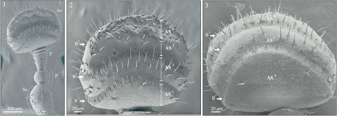
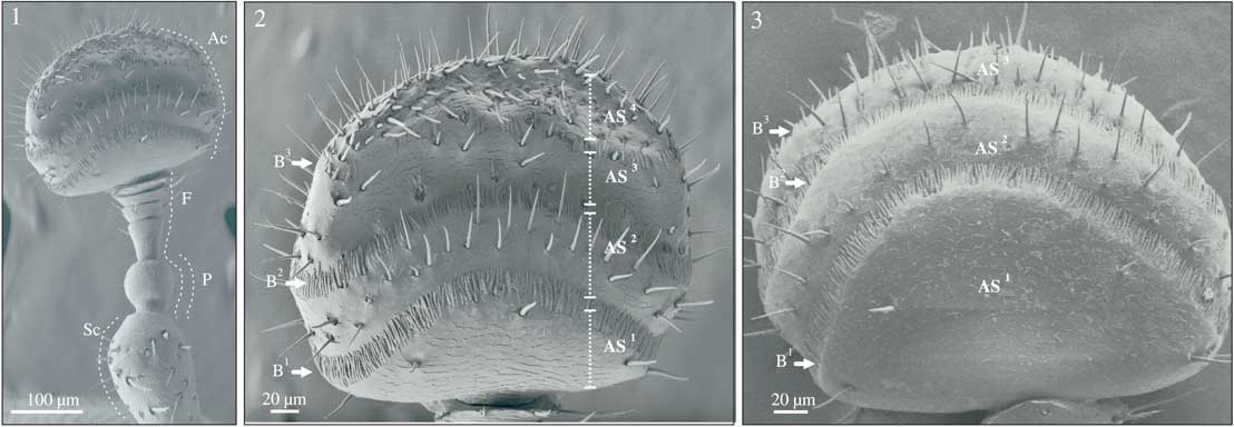
The general organisation of D. vitei antennae is similar to others species of Dendroctonus (Hopkins Reference Hopkins1909; Payne et al. Reference Payne, Moeck, Willson, Coulson and Humphreys1973; Dickens and Payne Reference Dickens and Payne1977; López et al. Reference López, Armendáriz-Toledano, Macías Sámano, Shibayama-Salas and Zúñiga2014), and it is composed of a scape, funicle, pedicel, and antennal club (Fig. 1). The club is rounded with three sensory bands that define four antennomeres (As) (Figs. 2, 3). Based on size, shape, presence or absence of pores, wall, and base type, we identified the follow sensilla on both sides of the antennal club.
Environmental scanning electron micrographs of the antenna of Dendroctonus vitei. 1, Antenna showing the scape (Sc), pedicel (P), funicle (F), and antennal club (Ac); 2, anterior face of the antennal club showing three sensory bands (B1, B2, B3) and four antennomeres (AS1 AS2, AS3, AS4); 3, posterior face of the antennal club with three sensory bands (B1, B2, B3) and three antennomeres (AS1, AS2, AS3).
Sensilla basiconica
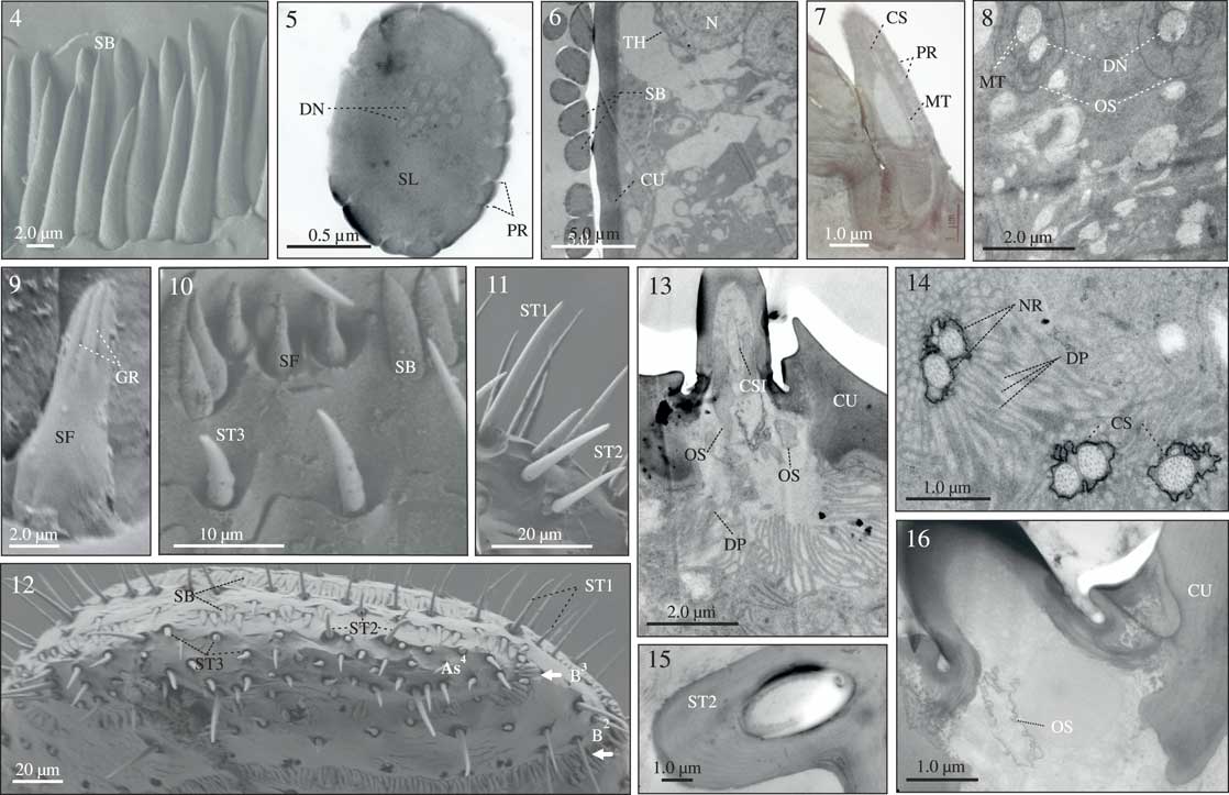
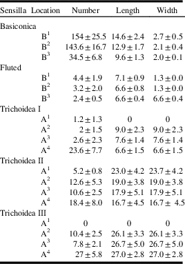
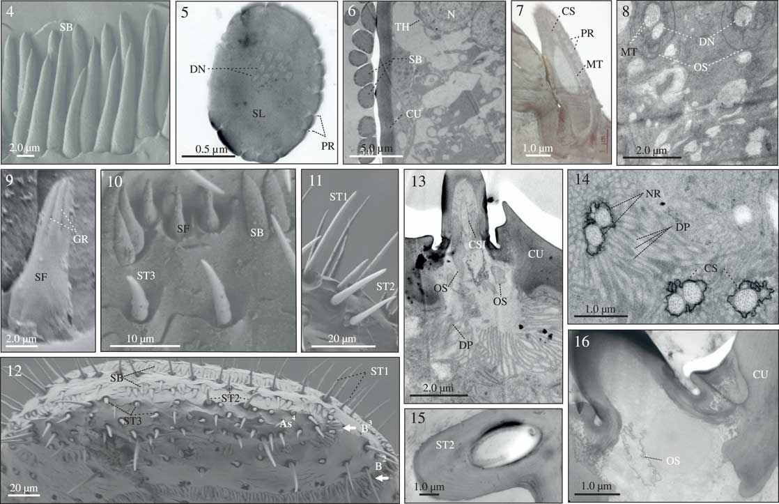
These sensilla are present in the three sensory bands, and they are much more abundant than any other sensilla type (Figs. 1–4). Their number, length, and width vary according the sensory band, being less abundant, smaller, and more discontinuously distributed (in groups of 2–10) on the third sensory band (Table 1). Within the sensory bands they tend to project distally and lie flat against the cuticle.
Environmental scanning electron and transmission electron micrographs of the antenna and types of sensilla of Dendroctonus vitei. 4, Sensilla basiconica (SB); 5, transversal section of basiconica sensillum with pores (PR) and dendrites (DN) within of the sensillar lumen (SL); 6, transversal section of antennal club: sensilla basiconica (SB), cuticle (CU), accesory cell thecogen (TH), and nucleo (N); 7, longitudinal section of sensilla basiconica: cilliar structure (SCI), microtubules (MT), and pores (PR); 8, ultrastructure of sensilla basiconica: dendrites (DN), and two outer segments (OS) integrated by microtubules (MT); 9, fluted sensilla (SF) with grooves in apical region; 10, sensilla basiconica (SB), fluted sensilla (SF), and sensilla trichodea (ST1) near the third sensory band; 11, sensilla trichodea (ST1, ST2) in the apical zone; 12, apical view of antennal club sensory bands (B2, B3) and sensilla trichodea (ST1, ST2, ST3) within AS4; 13, longitudinal section of sensilla trichodea (ST3) with its cilliar structure (SCI), two outer segments (OS), and dendritic projections (DP); 14, ultrastructure of sensilla trichodea with two bipolar neurons (NR), immersed in the cuticular structure (CS), with dendritic projections (DP) within of the sensilla lumen; 15–16, transversal section of sensilla trichodea (ST2) with cuticle (CU) and outer segment (OS).
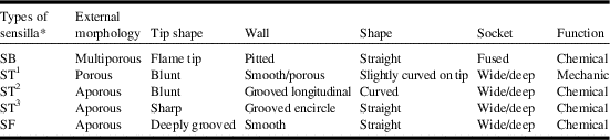
Morphological characteristics of antennal club sensilla of Dendroctonus vitei.

*(n=20 female, 20 male).
SB, sensillum basiconica; ST1, sensillum trichodea type I; ST2, sensillum trichodea type II; ST3, sensillum trichodea type III; SF, sensillum fluted.
The basiconic sensilla are characterised by having an overall peg-like shape and a flame-shaped tip, a multiporous wall, and a strongly tapered base (Table 2; Fig. 4). Transversal and longitudinal sections show the presence of two dendrites, each enclosed by two outer cilium-like segments integrated by microtubules. The lumen of the sensillar cavity is innervated by 4–12 dendritic branches, which are extensions of the dendrites towards cuticular pores. Each dendrite is connected to a sensory cell, integrated by a nucleus enclosed by three types of accessory cells: thormogen, thecogen, trichogen (Figs. 5–8).
Number, length, and location of sensilla in the antennal club of Dendroctonus vitei based on 20 females and 20 males.
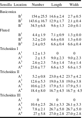
B, sensorial band 1, 2, or 3; A, antenomere 1, 2, 3, or 4.
Fluted or fluted cone-type sensilla
These sensilla are present in the three sensory bands interspersed among of the basiconic sensilla (Figs. 9–10). They are scarce and randomly distributed in the first and second sensorial bands, and in the third band they are present toward the lateral sides of the club, forming groups of two or three sensilla (Table 1; Fig. 10). These sensilla are small, straight with deeply grooved tips; their wall is smooth and without pores, and they are immersed in a deep socket (Table 2; Figs. 9–10). Due to their size, distribution, and number, it was difficult to obtain transmission electron microscopy images.
Sensilla trichodea
Three types of these sensilla were recognised (Figs. 10–11). The sensilla trichodea type I are on average longer than sensilla trichodea type II and sensilla trichodea type III (Fig. 11) and are present on antenomeres 2–4. They are in greatest abundance on the distal margin of antennomere IV (Table 1; Fig. 12). They are perpendicular to the cuticle, entirely straight and with a sharp tip; their wall has helical grooves, but they lack pores, and their base is articulated within a socket (Table 2).
The sensilla trichodea type II are randomly distributed in the four antenomeres, but they are more abundant on antennomere IV at the distal margin of the antennal club (Fig. 12). Their average length varies among antennomeres (Table 1). These sensilla are straight or curved and have a more blunt tip than sensilla trichodea type I; their wall is multiporous, and they arise from a wide, deep socket (Fig. 11). Some have a small protuberance at the base.
The sensilla trichodea type III are smallest and their average length is variable among antennomeres. They are scarce and randomly distributed in the first and third antennomere (1–3 per antennomere) but abundant in the fourth antennomere (Table 1; Fig. 12). They are slightly curved with a sharp tip, their wall is smooth and longitudinally grooved, and their base is articulate within a depth cuticular socket (Table 2; Fig. 10).
At the ultrastructural level, two types of cellular organisation of these sensilla were observed (Figs. 13–16). The first corresponds to sensilla trichodea type I, which have two bipolar neurons that are enclosed by a cuticular sheath and project distally within of sensilla lumen, with one dendritic process large and evident (Figs. 13–14). The second correspond to sensilla trichodea type II, whose ultrastructural organisation is similar to basiconic sensilla, with dendrites involved by a cuticular sheath and they branching towards to sensilla (Figs. 13–16).
Pit craters
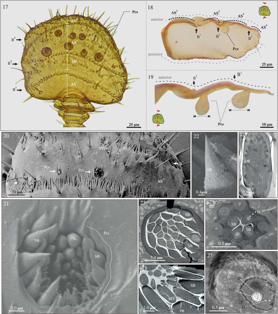
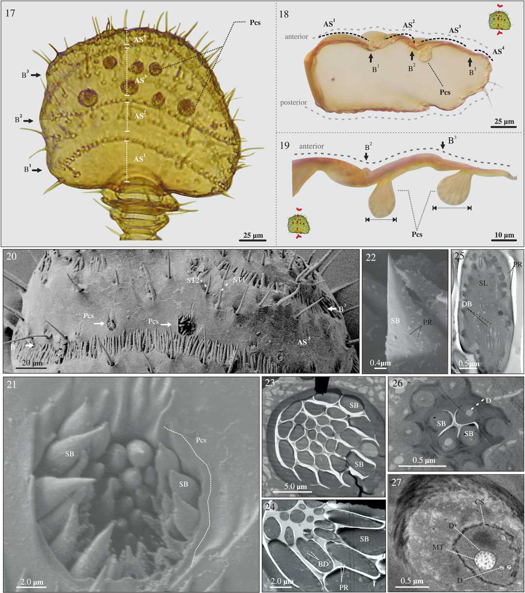
We use the name pit crater for a concavity produced by invagination of the surface of the club cuticle where several multiporous sensilla basiconica are grouped (Figs. 17–19). Pit craters are distributed on the third antenomere in two transverse lines with respect to proximal-distal axis of the antenna and are on only the anterior face of the club (Fig. 17). The proximal line of sensillae clusters located on the third antennomere and the distal line on the third sensorial band between the third and fourth antennomeres (Fig. 17). They vary from 10 to 27.5 μm (
 $$ \bar X$$
=15.2±2.1 μm) diameter within the same antenal club and among individuals. Those with greater diameter have a greater number of sensilla basiconica (
$$ \bar X$$
=21.5±2.6 μm, 15–28 sensilla) than those with smaller diameter (
$$ \bar X$$
=14.2±2.2 μm, 7–13 sensilla). In the majority of specimens, the sensilla tips do not extend above of the surface of club, whereas in other specimens the tips of at least some sensilla protrude through the opening of the pit (Fig. 20). The size and shape of the sensilla is variable (Fig. 21), however, they are typically short with a flame-shaped tip and a cone-shaped base (Fig. 22). Each sensillum is distinct (Figs. 23, 24) and their ultrastructural organisation is identical to that described for sensilla basiconica, that is, they have a multiporous wall, whose pores are innerved by dendrite branches (Figs. 25–27).
Light, environmental scanning electron and, transmission electron micrographs of antenal club of Dendroctonus vitei. 17, Antennal club showing three sensory bands (B1, B2, B3), four antennomeres (AS1, AS2, AS3, AS4), and pit craters (Pcs); 18–19, longitudinal sections of the antennal club with indicated position, size, and shape of the pit crater in the antennal club of D. vitei; 20, third antennomere (AS3) of the antennal club with pit craters (Pcs) between B2 and B3 also ST1 and ST2; 21, sensilla basiconica inside and outside of pit craters (Pcs); 22, sensilla basiconica with a terminal pore inside of the pit crater; 23–24, transversal and longitudinal sections of the pit craters and sensilla basiconica (SB) with pores (PR) and dendrite branches (BD); 25, ultrastructure of the sensilla basiconica with pores (PR) and dendrites branches (DB) within sensillar lumen (SL); 26–27, sensilla basiconica (SB) and dendrites (D) with microtubules (MT) and cuticular structure (CS) at higher magnification.
Discussion
The chemical communication system in bark beetles is fundamental to their survival and reproductive success because they locate their host trees and mates through olfactory signals such as pheromones and host-produced kairomones (Matthew and Mark Reference Matthew and Mark2004; Reinhard Reference Reinhard2004). These odorants, as well as other variables as wind speed, temperature, and moisture, are detected by sensillae concentrated in the bark beetle antenna (Reinhard Reference Reinhard2004).
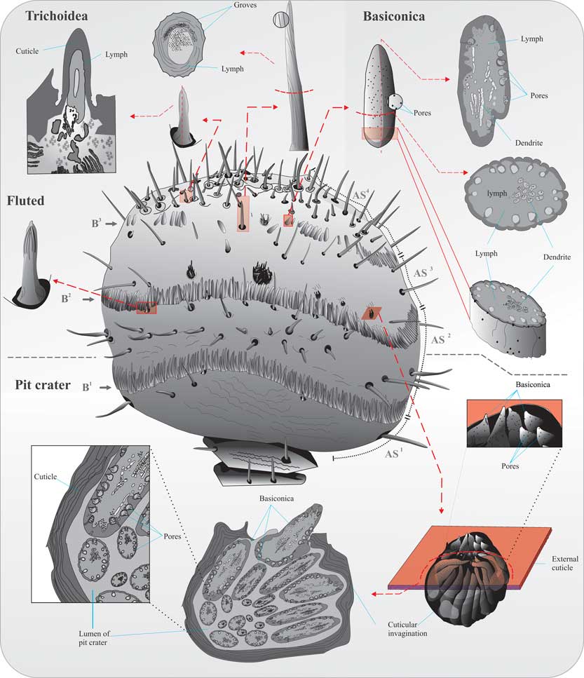
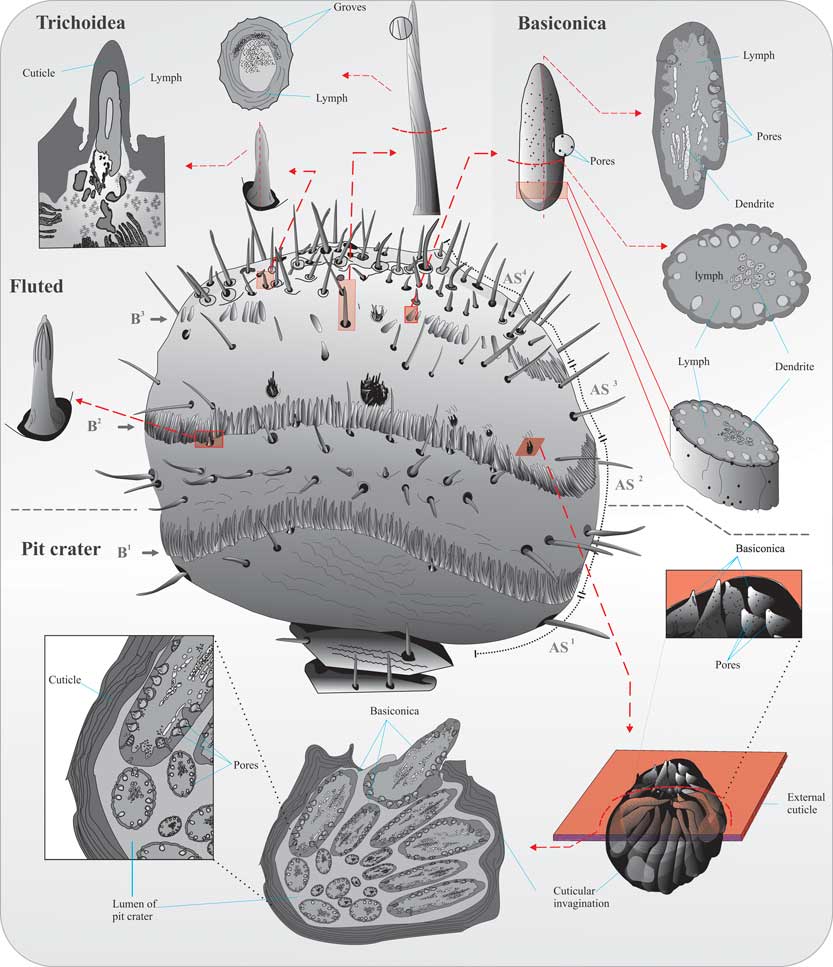
At least five types of sensilla are present in the antennal club of Dendroctonus vitei: multiporous sensilla basiconica (either arising from the cuticle surface or clustered within pit craters), aporous fluted sensilla, aporous sensilla trichodea (types I, II), and porous sensilla trichodea (type III) (Fig. 28), which by their morphology are associated with chemoreceptor and mechanoreceptor functions (Zacharuk Reference Zacharuk1985).
Illustration of the antennal club of Dendroctonus vitei and different types of sensilla: fluted (SF), basiconica (SB), trichodea (ST1, ST2, ST3), pit craters (Pcs), and transversal and longitudinal sections of these sensilla.
All sensilla basiconica examined in the antennal club of D. vitei have morphological characteristics similar to those found in other investigated bark beetles (Payne et al. Reference Payne, Moeck, Willson, Coulson and Humphreys1973; Dickens and Payne Reference Dickens and Payne1977; Chen et al. Reference Chen, Zhang, Wang and Kong2010; López et al. Reference López, Armendáriz-Toledano, Macías Sámano, Shibayama-Salas and Zúñiga2014). However, the sensilla basiconica in D. vitei are more abundant and less variable in length than in other Dendroctonus species such as D. valens LeConte, D. rhizophagus Thomas and Bright, and D. frontalis Zimmerman (Dickens and Payne Reference Dickens and Payne1977; López et al. Reference López, Armendáriz-Toledano, Macías Sámano, Shibayama-Salas and Zúñiga2014). The presence of a multiporous wall in these sensilla and two dendritic branches inside the sensilla lumen suggests that these sensilla have a chemosensory or olfactory function (Altner and Prillinger Reference Altner and Prillinger1980; Zacharuk Reference Zacharuk1985).
In other insects including bark beetles, it has been documented that sensilla basiconica function in the detection of pheromones (Payne et al. Reference Payne, Richerson, Dickens, West, Mori, Berisford and Blum1982; Gao et al. Reference Gao, Luo and Hammond2006; Sureda et al. Reference Sureda, Quero, Bosch, Avilés, Coll, Renou and Guerrero2006; Yang et al. Reference Yang, Yan and Liu2009) because their pores allow these odorant molecules to enter into the lumen of the sensillum (Keil and Steinbrecht Reference Keil and Steinbrecht1984; Steinbrecht Reference Steinbrecht1997). In bark beetles, D. brevicomis LeConte, D. frontalis, and D. ponderosae Hopkins, olfactory cells associated with sensilla basiconica respond to pheromone components (e.g., frontalin, endo/exo-brevicomina, verbenone, cis- and trans-verbenol) and host-produced terpenes (e.g., α-pinene and 3-carene) (Dickens and Payne Reference Dickens and Payne1977, Reference Dickens and Payne1978; Dickens et al. Reference Dickens, Payne, Ryker and Rudinsky1985).
The morphological characteristics, low abundance, and random distribution of fluted sensilla on the three sensorial bands of the antennal club of D. vitei suggest that their function might not be chemosensory. Sensilla with similar morphological characteristics as those in this study have been described in other species of Dendroctonus (Payne et al. Reference Payne, Moeck, Willson, Coulson and Humphreys1973; Dickens and Payne Reference Dickens and Payne1977; Whitehead Reference Whitehead1981; Chen et al. Reference Chen, Zhang, Wang and Kong2010; López et al. Reference López, Armendáriz-Toledano, Macías Sámano, Shibayama-Salas and Zúñiga2014). However, nothing is known about their function in Dendroctonus bark beetles. Studies of Coleoptera, Hemiptera, and Lepidoptera have reported that sensilla with this morphology could function as hygroreceptors (Waldow Reference Waldow1970; Bay and Pitts Reference Bay and Pitts1976; Jensen and Zacharuk Reference Jensen and Zacharuk1991; Diehl et al. Reference Diehl, Vlimant, Guerenstein and Guerin2007), thermo-chemical receptors (Hallberg Reference Hallberg1982; Zacharuk Reference Zacharuk1980), or highly selective pheromone receptors (Zacharuk Reference Zacharuk1985).
The classification of sensilla trichoidea is not consistent among different studies of Dendroctonus species (Payne et al. Reference Payne, Moeck, Willson, Coulson and Humphreys1973; López et al. Reference López, Armendáriz-Toledano, Macías Sámano, Shibayama-Salas and Zúñiga2014), which makes comparison difficult. Based on their length, shape, and wall structure, we identified three types of sensilla trichodea whose transmission electron microscopy images do not show ultrastructural differences. The absence of pores in sensilla trichodea type I and its location on the antennomere IV suggest thtat them likely play a role in mechanoreception. These type of sensilla are common in other bark beetles, and they have been linked to the same function (Zacharuk Reference Zacharuk1985).
In contrast, the pitted wall and the presence of a pore in the apical part of sensilla trichodea type II, suggest a chemoreceptor role for these sensilla in D. vitei. The same sensilla were found in Ips paraconfusus Lanier and linked to an olfactory function in this species (Lanier et al. Reference Lanier, Hendrichs and Flores1988). Other authors have suggested that these sensilla might have a gustatory (Zacharuk Reference Zacharuk1985; Gao et al. Reference Gao, Luo and Hammond2006; Yang et al. Reference Yang, Yan and Liu2009) or chemo-mechanoreceptor (Zacharuk Reference Zacharuk1985) function. This dual function might also be possible for these sensilla in D. vitei, given their size, random distribution in antennomere IV, and their location along margins of the club.
Lastly, based on their morphological characteristics, sensilla trichodea type III could have an olfactory function in D. vitei. Moeck (Reference Moeck1968) and Payne et al. (Reference Payne, Moeck, Willson, Coulson and Humphreys1973) described sensilla trichoidea in Typodendron lineatum Olivier and D. ponderosae that were similar to sensilla trichodea type III of D. vitei, and these authors inferred their function to be proprioception or olfaction, respectively. These sensilla are randomly distributed in all antennomeres of the D. vitei club, and they are not mixed with sensilla basiconica.
We can offer no immediate explanation for why multiporous sensilla basiconica are clustered into pit craters. To our knowledge, a similar arrangement of sensilla has not been reported in other curculionids or scolytines including Dendroctonus species (Payne et al. Reference Payne, Moeck, Willson, Coulson and Humphreys1973; Dickens and Payne Reference Dickens and Payne1977; Chen et al. Reference Chen, Zhang, Wang and Kong2010; López et al. Reference López, Armendáriz-Toledano, Macías Sámano, Shibayama-Salas and Zúñiga2014). This distinctive feature is unique and a diagnostic character useful for distinguishing D. vitei from other Dendroctonus species, particularly those D. frontalis complex, which are very difficult to identify. Based on Dendroctonus phylogeny (Victor and Zúñiga Reference Víctor and Zúñiga2016), the pit crater represents an autapomorphy in D. vitei, fixed in individuals of the populations included in this study. The clustering of sensilla basiconica into these concavities might reflect adaptations of their semiochemical system. Given that D.vitei is a species that can occur syntopically with other species in the D. frontalis complex, there may be strong selective pressures on these species to evolve traits that reduce potential for interference among pheromone signals when species are occupying the same host. It may be relevant for Dendroctonus vitei, because it is the only species of Dendroctonus that produces significant quantites of the pheromone component 1-phenylethanol, which is produced in large quantities by emergent males (Vité et al. Reference Vité, Islas, Renwich, Hughes and Kliefoth1974; Armendariz-Toledano et al. Reference Armendáriz-Toledano, Niño, Macías-Sámano and Zúñiga2014, Reference Armendáriz-Toledano, García-Román, López, Sullivan and Zúñiga2017).
Another possible function of these structures is protection of sensilla basiconica lining their interiors from physical contact with liquid resin during periods when the beetles are completely immersed (Brian Sullivan, United States Forest Service, Pineville Louisiana, United States of America, personal communication). Complete immersion commonly occurs in Dendroctonus during initial attack of vigorous hosts, and the layer of resin that forms over the surface of the antennal club and its sensilla presumably impedes olfaction by forming a physical barrier to the air. Olfactory habituation might also occur due to physical contact between sensilla and olfactory stimulants in the resin (e.g., host monoterpenes). Our preliminary observations indicate that the pits trap air in their interior when the beetles are immersed in resin and other liquids. If so, the “bubble” formed within the pits may allow beetles to maintain greater olfactory capacity during immersion and more rapid recovery afterward when these bubbles re-open. However, we know of no unique selective pressures on D. vitei that would promote acquisition of this trait by this species but not other Dendroctonus.
Acknowledgements
The author are grateful to Brian T. Sullivan for his comments and suggestions on this manuscript. The authors would like to thank María de Lourdes Rojas (Transmission Electronic Microscopy – Laboratorio Nacional de Servicios Experimentales-Centro de Investigación y Estudios Avanzados-Instituto Politécnico Nacional) and Ariana Guadalupe Robles and Jessica B. Valencia (Servicio Nacional de Sanidad Inocuidad y Calidad Agroalimentaria) for their technical assistance with scanning and environmental electron microscopes. The authors appreciate the field assistance of Luis Gerardo Cuellar (Universidad de Nuevo León) and Julio César Concha (Universidad de la Sierra de Juárez). This work was supported by the Secretaria de Investigación y Posgrado Instituto Politécnico Nacional (20161790). F.A.T. was a Consejo Nacional de Ciencia y Tecnología (CONACyT) fellow (267436) and member of the Programa Institucional de Formación de Investigadores of Instituto Politécnico Nacional.