Introduction
Social anxiety disorder (SAD) is characterized by a persistent fear of possible scrutiny or negative evaluation from others in social or performance situations, leading to the avoidance of social situations or enduring them with intense distress or anxiety (American Psychiatric Association, 2013). Approximately 80% of SAD patients met the diagnostic criteria of SAD before age 18 and the median age of onset is 13 years (Kessler et al., Reference Kessler, Berglund, Demler, Jin, Merikangas and Walters2005; Otto et al., Reference Otto, Pollack, Maki, Gould, Worthington, Smoller and Rosenbaum2001). Therefore, SAD has been viewed as an ‘adolescent mental disorder’ given its typical age of onset (Haller, Cohen Kadosh, Scerif, & Lau, Reference Haller, Cohen Kadosh, Scerif and Lau2015). This disorder shows a rather chronic course, with 57% of SAD cases revealed stability at 10-year follow-up (Beesdo-Baum et al., Reference Beesdo-Baum, Knappe, Fehm, Höfler, Lieb, Hofmann and Wittchen2012). Avoidance of social interactions and its impact are even more disruptive during adolescence (Miers, Blöte, Heyne, & Westenberg, Reference Miers, Blöte, Heyne and Westenberg2014).
Theories have suggested a close link between neurodevelopmental dysfunction at the adolescent juncture and SAD, but direct evidence is rare. Compared with adults, adolescents have far more social concerns about negative feedback from peers, leading to a higher risk of excessive social anxiety (Leigh & Clark, Reference Leigh and Clark2018). More importantly, adolescence is a sensitive period of brain maturation (Fuhrmann, Knoll, & Blakemore, Reference Fuhrmann, Knoll and Blakemore2015; Gogtay et al., Reference Gogtay, Giedd, Lusk, Hayashi, Greenstein, Vaituzis and Thompson2004). Pervasive social-affective development in adolescence is mediated by prolonged maturation of structural and functional brain networks. For instance, researchers have reported age-related brain maturation in subcortical regions such as the amygdala and the prefrontal cortex (PFC) dedicated to the processing of emotional and social stimulations (Blakemore, Reference Blakemore2008). Studying SAD at the adolescent juncture helps to reveal the pathogenesis of SAD, as well as its early detection and intervention.
To date, a handful of neuroimaging studies have investigated the brain dysfunction during anxiety-provoking stimulus of pediatric SAD (Killgore & Yurgelun-Todd, Reference Killgore and Yurgelun-Todd2005; Guyer et al., Reference Guyer, Lau, McClure-Tone, Parrish, Shiffrin, Reynolds and Nelson2008). As suggested by a recent meta-analysis, there should be neuroanatomical abnormalities in adolescents with SAD (Wang, Cheng, Luo, Qiu, & Wang, Reference Wang, Cheng, Luo, Qiu and Wang2018), yet the brain structural deficits among adolescent SAD sample have not been revealed in previous studies (Blackford & Pine, Reference Blackford and Pine2012). Some studies have reported inconsistent findings in youth with a mixed anxiety disorder or symptoms, for example, with both increased (Qin et al., Reference Qin, Young, Duan, Chen, Supekar and Menon2014) and decreased (Strawn et al., Reference Strawn, Hamm, Fitzgerald, Fitzgerald, Monk and Phan2015) amygdala volume, as well as nulling findings (Merz, He, & Noble, Reference Merz, He and Noble2018). In this study, we aim to examine the brain structural abnormalities in medication-free adolescents with SAD for a better understanding of its pathology.
Previous studies of adult SAD have revealed abnormalities in the ‘fear circuit’ in the brain, i.e. the PFC, insula, and amygdala (Brühl, Delsignore, Komossa, & Weidt, Reference Brühl, Delsignore, Komossa and Weidt2014; Etkin & Wager, Reference Etkin and Wager2007). It is well established that the amygdala hyperactivation is related to social threat in processing emotional faces and social stimulations (Miskovic & Schmidt, Reference Miskovic and Schmidt2012). Among the prefrontal regions, a dysfunctional orbitofrontal cortex (OFC) circuit may play a key role in social behavior and social anxiety (Beer, John, Scabini, & Knight, Reference Beer, John, Scabini and Knight2006; Milad & Rauch, Reference Milad and Rauch2007). Previous studies have found abnormal orbitofrontal–amygdala functional connectivity in SAD patients (Hahn et al., Reference Hahn, Stein, Windischberger, Weissenbacher, Spindelegger, Moser and Lanzenberger2011; Sladky et al., Reference Sladky, Höflich, Küblböck, Kraus, Baldinger, Moser and Windischberger2015). Furthermore, animal studies have suggested an OFC–insula–amygdala neural pathway in negative emotion regulation, emphasizing that anxiety was enhanced by OFC inactivation due to uncertainty (Shiba et al., Reference Shiba, Oikonomidis, Sawiak, Fryer, Hong, Cockcroft and Roberts2017).
Inspired by these accumulating findings, we focus on examining the structural abnormality of the core regions in the ‘fear circuit’ in medication-free adolescents with SAD. We first hypothesized that the OFC, insula, and amygdala could exhibit abnormal morphometric characteristics in adolescents with SAD. Furthermore, since the ‘fear circuit’ is an interconnected network instead of separated regions, we hypothesized that the coordination of the development of these brain regions may be disrupted in adolescents with SAD. Brain developmental theories have interpreted the dysregulation of heightened emotional reactivity in adolescence as a mismatch in the timing of structural maturation between prefrontal regions and subcortical regions (Casey, Glatt, & Lee, Reference Casey, Glatt and Lee2015; Mills, Goddings, Clasen, Giedd, & Blakemore, Reference Mills, Goddings, Clasen, Giedd and Blakemore2014). As an extreme case of unsuccessful control of social anxiety, an adolescent with SAD may reflect abnormal coordination of development in the fronto-limbic regions. Communities of brain regions co-vary in their morphological properties, so the developmental coordination or synchronized maturation of different brain regions can be characterized by structural co-variance (Alexander-Bloch, Giedd, & Bullmore, Reference Alexander-Bloch, Giedd and Bullmore2013). This approach has been applied to reveal a distributed network organization in patients with Attention-Deficit/Hyperactivity Disorder (Li et al., Reference Li, Cao, Pu, Li, Fan, An and Wang2015). Studies on structural co-variance in SAD are rare.
To examine these hypotheses, we acquired and analyzed high-resolution brain structural images of a relatively large medication-free adolescent SAD sample and an age- and sex-matched typical developing sample. We examined the first hypothesis by adopting a region of interest (ROI) restricted voxel-based morphometry (VBM) approach to examine the local structural alterations. Further, we characterized the disrupted coordination of these regions using structural co-variance analysis.
Methods
Participants and procedures
In total, 73 participants were included in the study, comprising 31 adolescents with SAD and 42 age- and sex-matched healthy controls (HC). The 31 adolescents with SAD ranging from 15 to 17 years old were consecutively recruited through advertisements or referred by psychiatrists from the outpatient department of child and adolescent psychiatry in Shanghai Mental Health Center, Shanghai Jiao Tong University School of Medicine. The Ethical Committee of the Shanghai Mental Health Center approved the study design (2018-46) and all participants provided written informed consent.
Trained clinicians first conducted face-to-face interviews with the patients and at least one of their primary caregivers by using the child version (Liu, Liu, & Wang, Reference Liu, Liu and Wang2011) and parent version (Liu, Liu, & Wang, Reference Liu, Liu and Wang2010) of Mini-International Neuropsychiatric Interview for Children and Adolescents (MINI-Kid) to determine the diagnosis of SAD. A senior pediatric psychiatrist further verified the diagnosis according to the Diagnostic and Statistical Manual of Mental Disorders (DSM-5) through a clinical interview. The patients did not receive any systematic medication or psychotherapy before enrollment and did not receive any medication 2 weeks before the MRI scanning. Patients who completely met the diagnostic criteria of other mental disorders, including the other subtypes of anxiety disorders, were excluded from the SAD group.
Healthy participants were recruited from schools and communities through advertisements. To exclude participants' past or present mental disorders, the participants and at least one of their primary caregivers were separately assessed with the child version and parent version of the MINI-Kid. For those participants who were judged to have no mental disorder or have suspected diagnosis in the MINI-Kid interview, a senior psychiatrist further interviewed the participants and their primary caregivers to exclude any of the DSM-5 diagnoses of mental disorders. For both groups, participants with organic brain disorders, a history of severe physical illnesses, or severe suicidal tendency were excluded.
We collected demographical information of the participants and their families, such as parental education level and family monthly income (per capita). The parental education level was represented by the highest education level of the father and mother. Parental education level was encoded by 1–7 to represent the education levels: elementary school or under, secondary school, high school, college, bachelor's degree, master's degree, and doctorate. The family monthly income was reported by the father and mother separately and the maximum value was chosen when there is an inconsistency between the father and mother. The family monthly income was encoded by 1–5 to represent the income ranges (unit: Chinese yuan): less than 1000, 1000–3000, 3000–5000, 5000–10 000, and more than 10 000. We assessed clinical symptoms using the Social Anxiety Scale for Children (SASC) (La Greca & Stone, Reference La Greca and Stone1993) and the Screen for Child Anxiety Related Emotional Disorders (SCARED) (Birmaher et al., Reference Birmaher, Khetarpal, Brent, Cully, Balach, Kaufman and Neer1997).
Image acquisition
High-resolution T1-weighted images were acquired with magnetization-prepared rapid gradient echo (MP-RAGE) sequence on a 3.0-Tesla magnetic resonance scanner (Siemens 3.0 Trio Tim, Germany), and the acquisition parameters included: repetition time (TR) = 2.53 s, echo time (TE) = 3.65 ms, inversion time (TI) = 1.1 s, flip angle = 7°, FOV = 224 × 256 mm2, matrix = 224 × 256, 256 coronal slices, and slice thickness = 1.0 mm.
Voxel-based morphometry
T1-weighted structural images were processed using CAT12 toolbox (Version 12.6; Gaser & Dahnke, Reference Gaser and Dahnke2016) and SPM12 (Wellcome Trust Centre for Neuroimaging, London, UK, http://www.fil.ion.ucl.ac.uk/spm/). We visually checked the raw T1-weighted images to exclude any obvious artifacts. After the preprocessing, the CAT12 toolbox would generate an image quality score for each subject based on the image resolution, bias, and noise. All subjects with a quality level lower than ‘B−’ (good quality) were excluded from further analyses.
The raw images were corrected for the bias field and segmented into gray matter, white matter, and cerebrospinal fluid. Then the gray matter maps were normalized into MNI space using DARTEL (Ashburner, Reference Ashburner2007) and modulated by Jacobian determinants of warp field to compensate the local volume changes caused by non-linear registration. The normalized and modulated gray matter maps were smoothed by an 8-mm FWHM isotropic Gaussian kernel to account for the potential registration inaccuracy and individual anatomical variability. These smoothed maps represented the voxel-wise gray matter volume estimations for all subjects and were used for statistical analysis.
To reveal the potential gray matter difference between HC and SAD groups in the ‘fear circuit’ regions, we utilized an ROI-restricted voxel-wise analysis strategy. Compared to whole brain voxel-wise analysis, in which all brain regions were treated equally, the ROI-restricted method could be more sensitive to potential differences by incorporating prior knowledge from previous studies. Instead of comparing the mean volume in each ROI, a voxel-wise analysis could be more sensitive to find local changes inside an ROI, as an ROI was generally composed of a large number of brain structures. This strategy has been applied in other studies (e.g. Kawaguchi et al., Reference Kawaguchi, Nemoto, Nakaaki, Kawaguchi, Kan, Arai and Akechi2016; Månsson et al., Reference Månsson, Salami, Frick, Carlbring, Andersson, Furmark and Boraxbekk2016). The OFC, insula, and amygdala masks were extracted from the AAL parcellation atlas (Tzourio-Mazoyer et al., Reference Tzourio-Mazoyer, Landeau, Papathanassiou, Crivello, Etard, Delcroix and Joliot2002). Inside each ROI mask, voxel-wise statistical analysis was performed using a permutation test and threshold-free cluster enhancement (TFCE) based multiple comparison correction (Smith & Nichols, Reference Smith and Nichols2009) with age and sex as covariates. Family-wise error rate (FWE) corrected p < 0.025 was set as the significance level for two-tailed testing. The associations between gray matter volume alterations and clinical scales were further examined within the SAD group using Pearson's correlation.
We corrected the effect of total intracranial volume (TIV) by modulating the local volume changes caused by non-linear registration and preserving the gross volume changes caused by affine registration during the spatial normalization. This approach is equivalent to proportionally scaling the individual gray matter volume estimations by TIV, and it has been commonly used in previous studies (Malone et al., Reference Malone, Leung, Clegg, Barnes, Whitwell, Ashburner and Ridgway2015). We also examined the correlations between the TIV and the mean gray matter volumes within regions showing a significant group difference. In addition, we repeated the analysis by adding the TIV as a covariate in the statistical model to validate the robustness of any findings.
Another potential confounding factor is the family socioeconomic status (Noble et al., Reference Noble, Houston, Brito, Bartsch, Kan, Kuperman and Sowell2015). Parental education level and family monthly income were reported by both the father and mother of the participant. As a considerable proportion of socioeconomical data were unavailable (some families refused to report their income levels), we examined the potential influence using a post-hoc method. We extracted the mean volumes of the regions showing a significant group difference and examined whether the difference still existed after including the family monthly income and parental education level as covariates.
Structural co-variance analysis
To characterize the structural co-variance between the ROIs, we extracted the mean gray matter volumes from the clusters with significant group difference. We then calculated the partial correlations between any two clusters, with age and sex as covariates. This computation was conducted for HC and SAD groups separately. We first determined the significance of the co-variance within each group and corrected multiple comparison error using the false discovery rate (FDR). For the co-variance that was significant in only one of the two groups, we further examined their between-group difference. Specifically, the correlation coefficients representing the co-variance were first converted into Fisher's Z, and a Z test statistic with its accompanying p value were derived following the formula: Z statistic = $( Z\_{\rm HC}-Z\_{\rm SAD}) /\sqrt {( 1/( N\_{\rm HC}- 3) ) + ( 1/( N\_{\rm SAD} - 3) ) } , \;$ in which N means the sample size for each group. The multiple comparison was corrected using the FDR. To examine that the potential difference in structural co-variance could be due to the variation in symptom severity, we repeated the co-variance analysis by using the symptom severity measure (SCARED or SASC total scores) as a covariate. The analysis was performed using R (R Core Team, 2018).
Results
Demographic and clinical data
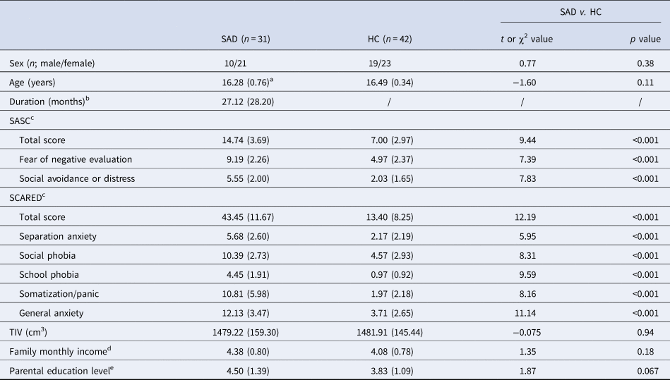
Table 1 presents the group demographic characteristics and clinical scores. SAD (n = 31) and HC (n = 42) did not differ significantly in age and sex (p > 0.11), while adolescents with SAD have significantly higher scores of SASC and SCARED than HC on anxiety symptoms (p < 0.001). There was no significant difference in family monthly income (p = 0.18) and parental education level (p = 0.067), although the SAD group showed a higher trend in the parental education level.
Demographic data and clinical scale scores

a The data were presented as mean (standard deviation).
b Duration of illness was reported by the father or/and mother of the participant. The average duration was taken if there was an inconsistency between the father and mother. These data were not available for five subjects in the SAD group.
c The SASC/SCARED data were not available for seven subjects in the HC group.
d The family monthly income data were only available for 50 subjects (26 SAD and 24 HC).
e The parental education level data were only available for 50 subjects (26 SAD and 24 HC).
VBM results
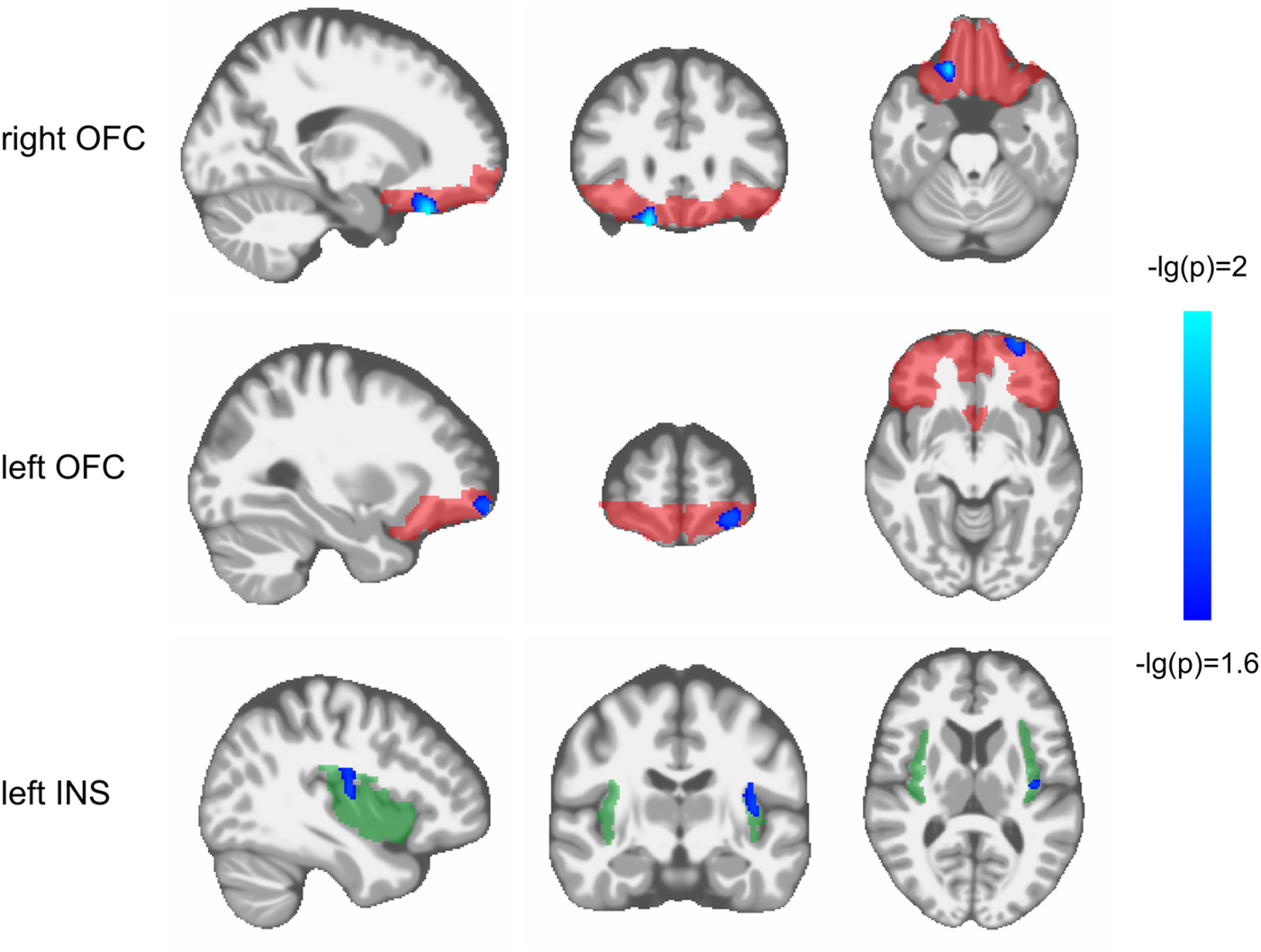
No data were excluded from the analysis due to poor image quality. As shown in Table 2, the VBM analysis revealed that compared with HC, adolescents with SAD exhibited significantly higher gray matter volume in the right superior OFC (p = 0.010), left middle/superior OFC (p = 0.019), and left insula (p = 0.020; FWE corrected for multiple comparison; see Fig. 1). There was no significant gray matter volume difference in the bilateral amygdala.
The increased gray matter volume of the right superior OFC, left superior/middle OFC, and left insula in adolescents with SAD. The significant clusters (p < 0.025, FWE corrected) are placed above the standard template. The corrected p value is logarithmic-transformed for visualization. The ROI masks for OFC and insula are shown semi-transparently in red and green. The brain is displayed in the radiological orientation (i.e. left is right). OFC, orbitofrontal cortex; INS, insula.

The significant clusters in voxel-based morphometry analysis between SAD and HC

a The anatomical labels of clusters were based on the AAL parcellation. OFC, orbitofrontal cortex; R, right; L, left.
b The effect size (measured by Cohen's d) and its confidence interval were calculated using the mean gray matter volumes within the clusters.
There were no significant correlations between TIV and the right superior OFC (r = −0.047, p = 0.69), left middle/superior OFC (r = 0.15, p = 0.20), and left insula (r = −0.043, p = 0.72). For the analysis that included the estimated TIV as a covariate in the comparison, the results showed similar group differences and locations (see online Supplementary Table S1). After regressing out the effects of family monthly income and parental education level, the significant group difference still existed in the right superior OFC (t = 4.60, p < 0.001), left middle/superior OFC (t = 2.50, p = 0.016), and left insula (t = 2.85, p = 0.0067). There was no significant correlation between gray matter volume abnormalities and clinical variables in the SAD group.
Structural co-variance results
We included all the clusters with significant group differences as well as the amygdala in the structural co-variance analysis. Although the amygdala did not exhibit a significant group difference in gray matter volume, it plays an important role in threat detection and emotion regulation (Kim et al., Reference Kim, Loucks, Palmer, Brown, Solomon, Marchante and Whalen2011). SAD patients exhibited hyperactivity in the amygdala and disrupted amygdala–prefrontal circuitry (Boehme et al., Reference Boehme, Ritter, Tefikow, Stangier, Strauss, Miltner and Straube2014; Goldin, Manber, Hakimi, Canli, & Gross, Reference Goldin, Manber, Hakimi, Canli and Gross2009). Studies have revealed anatomical connections between the amygdala, OFC, and insula (Mesulam & Mufson, Reference Mesulam and Mufson1982; Phan et al., Reference Phan, Orlichenko, Boyd, Angstadt, Coccaro, Liberzon and Arfanakis2009). Thus, even though it did not exhibit abnormality per se, it may take part in the interaction of the OFC and insula. Moreover, the volume of the amygdala is comparable to the clusters showing a significant group difference, allowing us to use the mean volume of the amygdala in the structural co-variance analysis to examine the possibilities.
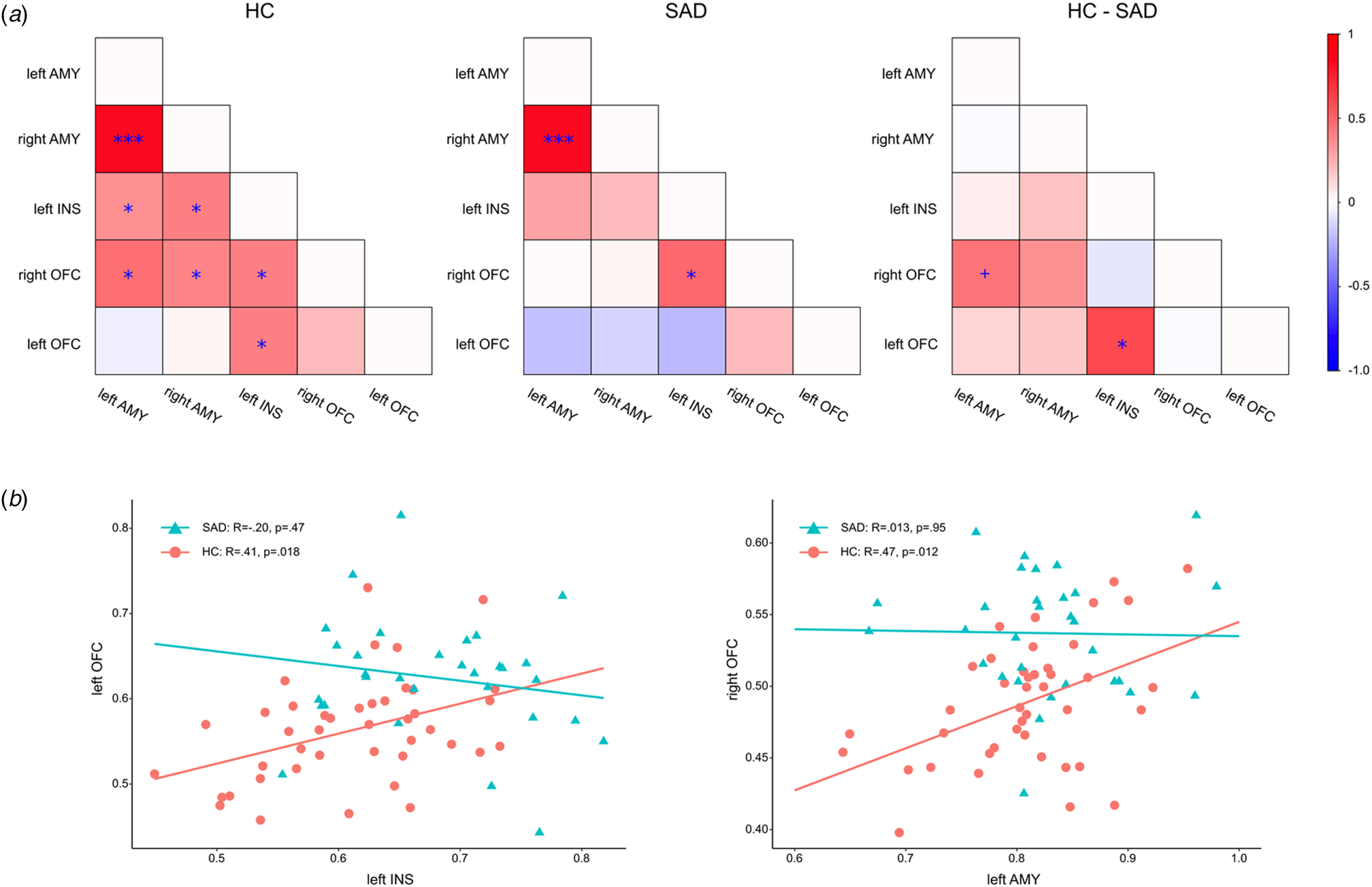
The full co-variance matrix and the statistical comparison results are presented in Fig. 2. For the HC group, there were widespread significant co-variances between the left and right amygdala (r = 0.82, FDR-corrected p < 0.001), between the left amygdala and left insula (r = 0.35, FDR-corrected p = 0.038), between the left amygdala and right superior OFC (r = 0.47, FDR-corrected p = 0.012), between the right amygdala and left insula (r = 0.41, FDR-corrected p = 0.018), between the right amygdala and right superior OFC (r = 0.38, FDR-corrected p = 0.025), between the left insula and right superior OFC (r = 0.41, FDR-corrected p = 0.018), and between the left insula and left middle/superior OFC (r = 0.41, FDR-corrected p = 0.018). For the SAD group, there were only significant co-variances between the bilateral amygdala (r = 0.83, FDR-corrected p < 0.001), and between the left insula and right superior OFC (r = 0.49, FDR-corrected p = 0.038). For the between-group difference, there was a significant positive correlation between the left middle/superior OFC and left insula in HC (r = 0.41, FDR-corrected p = 0.018), which was attenuated in adolescents with SAD (r = −0.20, FDR-corrected p = 0.46). The group difference was significant (Z = 2.61, FDR-corrected p = 0.046). In addition, there was a significant positive correlation between the left amygdala and right superior OFC in HC (r = 0.47, FDR-corrected p = 0.012), which was not significant in adolescents with SAD (r = 0.013, p = 0.95). The structural co-variance was only significantly different between the two groups without multiple comparison correction (Z = 1.99, uncorrected p = 0.046, corrected p = 0.12). After controlling the influence of symptom severity, the structural co-variance within each group and the difference between groups were similar (see online Supplementary Material).
Results of structural co-variance analysis. (a) The full co-variance matrix of gray matter volume of the ROIs in the SAD group, HC group, and the difference between the two groups (*FDR-corrected p < 0.05, ***FDR-corrected p < 0.001, + uncorrected p < 0.05). (b) Loss of structural co-variance between the left insula and left superior/middle OFC, and between the left amygdala and right superior OFC in adolescents with SAD. There are significant structural co-variances in the HC group (red), but the co-variance diminished in the SAD group (green). OFC, orbitofrontal cortex; INS, insula; AMY, amygdala.

Discussions
The present study provides direct evidence on brain structural abnormality in human adolescents with SAD. We found significantly higher gray matter volume in the insula, medial, and lateral OFC in adolescents with SAD. Considering that the gray matter volume reduction is the main trend in brain development in adolescence (Gogtay et al., Reference Gogtay, Giedd, Lusk, Hayashi, Greenstein, Vaituzis and Thompson2004; Lenroot & Giedd, Reference Lenroot and Giedd2006), these findings suggest a delayed development in the ‘fear circuit’ regions in adolescents with SAD. Furthermore, we observed a structural co-variance pattern among the OFC, insula, and amygdala in typically developing adolescents, but these co-variances diminished in adolescents with SAD. This finding provides the first evidence that the brain abnormalities in adolescents with SAD are not only in localized regions, but also reflected in the cortical and subcortical coordination in brain development. Since the abnormal development of brain circuit may occur before the explicit abnormal behavior, this study provides detailed targets in brain structure for early detection and intervention of adolescent SAD.
Structural abnormality in the ‘fear circuit’ regions in adolescents with SAD
Our findings agree with previous studies that indicated structural deficits in fronto-limbic regions in the adult SAD sample (Brühl et al., Reference Brühl, Delsignore, Komossa and Weidt2014; Etkin & Wager, Reference Etkin and Wager2007). These findings are further supported by neuroimaging studies on the effects of treatment. Researchers reported reductions in cortical volume in the dorsomedial PFC and a significant increase of structural connectivity in a fronto-limbic network after group cognitive behavioral therapy in SAD patients (Steiger et al., Reference Steiger, Brühl, Weidt, Delsignore, Rufer, Jäncke and Hänggi2017).
Previous studies in adult patients with SAD found significant gray matter volume reduction in the OFC (Talati, Pantazatos, Schneier, Weissman, & Hirsch, Reference Talati, Pantazatos, Schneier, Weissman and Hirsch2013) and insula (Kawaguchi et al., Reference Kawaguchi, Nemoto, Nakaaki, Kawaguchi, Kan, Arai and Akechi2016), as well as a significant cortical thinning in insula (Syal et al., Reference Syal, Hattingh, Fouché, Spottiswoode, Carey, Lochner and Stein2012), which were different from our findings where gray matter volume in these regions was increased. Given that this study is merely a snapshot of a rapid dynamic of brain development, we cannot draw the conclusion that the results of adolescents are opposite to those of adult sample. As suggested by the abnormal co-variance of the key regions in the limbic system, we believe that ‘abnormal developing trajectory’ is the core problem of the adolescents with SAD, and the abnormally larger gray matter volume found in this snapshot is far from sufficient to reveal the full picture of the abnormal brain development trajectory in adolescents with SAD. The identified larger gray matter volume is only meaningful when compared to the typically developing controls at the same age period. Future longitudinal studies are expected to better resolve the abnormal brain development trajectories of SAD. In addition, the adult patient samples in the above-mentioned studies were generalized SAD with comorbidities such as other anxiety disorder, major depression disorder, obsessive–compulsive disorder, bulimia nervosa, alcohol use disorder, and so on. To varying degrees, those patients received antidepressants, benzodiazepines, and antipsychotics, as well as cognitive behavior therapy. Yet comorbidity and medication might be the potential confounders of SAD at the neuroanatomical level (Wang et al., Reference Wang, Cheng, Luo, Qiu and Wang2018), which partly explained the inconsistent results.
During adolescence, different regions of the brain develop at unsynchronized schedules, bringing the risk of dysregulation of excessive social anxiety in all teenagers. For a subgroup of youngsters, the protracted maturation of fronto-limbic regions involved in regulating reactivity to social-affective information might exaggerate the dysregulation of threatening social cues. Further, the protracted maturation, in turn, could reciprocally affect the trajectories of functional brain development by biasing both the nature of incoming information and the ways in which this information is processed and subsequently consolidated. SAD can constrain normative development in a life stage typically marked by changes in neural circuitry, including synaptic pruning and reorganization of the PFC and enhanced modulation of subcortical structures by the PFC (Caouette & Guyer, Reference Caouette and Guyer2014). Therefore, we speculate that the delayed structural development of these regions (higher gray matter volume than typical developing brains) may accompany the onset of SAD symptoms, and the long-term effect of the disorder may affect the health of the neural system in adulthood (lower gray matter volume than HC).
A diminished structural co-variance pattern in adolescents with SAD
A novel finding in this study is the diminished structural co-variance between the medial OFC and the amygdala, as well as the lateral OFC and insula in adolescents with SAD. The biological interpretation of brain structural co-variance remains controversial, but it appears to be indicative of developmental coordination or synchronized maturation between brain regions (Alexander-Bloch et al., Reference Alexander-Bloch, Giedd and Bullmore2013). Structural co-variance is thought to partially reflect white matter tracts or functional connectivity of synchronous neuronal activation. The only study comparing physical connections with cortical thickness co-variance found that white matter tracts account for 30–40% of structural co-variance (Gong, He, Chen, & Evans, Reference Gong, He, Chen and Evans2012). In studies directly comparing structural co-variance networks with functional connectivity, researchers found that insula-based structural co-variance network exhibits strong correspondence with the intrinsic functional connectivity as well as task-based co-activation (Clos, Rottschy, Laird, Fox, & Eickhoff, Reference Clos, Rottschy, Laird, Fox and Eickhoff2014; Kelly et al., Reference Kelly, Toro, Di Martino, Cox, Bellec, Castellanos and Milham2012). But beyond that, the structural co-variance network revealed an exclusive network devoted to negative emotion processing and social cognition (Clos et al., Reference Clos, Rottschy, Laird, Fox and Eickhoff2014). Both resting-state and task-based functional connectivity capture the transient states of the brain network, while structural co-variance represents the accumulated and long-term shaping of the social world. Future studies could further examine our findings and these interpretations by using diffusion tensor imaging to detect abnormalities in the white matter connections in adolescents with SAD.
Our findings are supported by recent research that identified a medial OFC–amygdala projection that modulates social approach behavior and influences susceptibility to social anxiety in both mouse and human (Li et al., Reference Li, Jing, Dellarco, Hall, Yang, Heilberg and Lee2019). Indeed, SAD patients exhibited significantly lower white matter connectivity of uncinate fasciculus (Phan et al., Reference Phan, Orlichenko, Boyd, Angstadt, Coccaro, Liberzon and Arfanakis2009), decreased functional connectivity (Hahn et al., Reference Hahn, Stein, Windischberger, Weissenbacher, Spindelegger, Moser and Lanzenberger2011) between the amygdala and the OFC. The efferent projections and functional connectivity between the amygdala and medial PFC have been found to support both conditioning and extinction of aversive learning in animals and emotion regulation in humans (Kim et al., Reference Kim, Loucks, Palmer, Brown, Solomon, Marchante and Whalen2011). The interrupted coordination can be attributed to multiple factors such as genetics. For instance, BDNF Val66Met SNP may lead to altered anatomy and functionality of the OFC–amygdala circuit specifically during adolescence (Li et al., Reference Li, Jing, Dellarco, Hall, Yang, Heilberg and Lee2019). Diminished structural co-variance between the ventromedial OFC and the amygdala may indicate the failure of OFC to downregulate the amygdala.
The previous study found that gray matter volume of the OFC co-vary mainly with the insula and amygdala in healthy young subjects (Liu, Qin, Qi, Jiang, & Yu, Reference Liu, Qin, Qi, Jiang and Yu2015), which is consistent with our findings in the HC sample. Anatomical tracer studies in monkeys found strong reciprocal interconnectivity between the insula and the OFC (Mesulam & Mufson, Reference Mesulam and Mufson1982; Saleem, Kondo, & Price, Reference Saleem, Kondo and Price2008). Non-invasive diffusion imaging similarly identified anatomical connections between the insula, OFC, and inferior frontal regions (Cloutman, Binney, Drakesmith, Parker, & Lambon Ralph, Reference Cloutman, Binney, Drakesmith, Parker and Lambon Ralph2012). Importantly, the OFC and insula encode reward function, causing motivation to approach food (Castro & Berridge, Reference Castro and Berridge2017) and produce social-affiliative behaviors (Caruana, Jezzini, Sbriscia-Fioretti, Rizzolatti, & Gallese, Reference Caruana, Jezzini, Sbriscia-Fioretti, Rizzolatti and Gallese2011). Inactivation of the OFC would lead to impairment on the performance of an approach–avoidance decision-making task due to elevated uncertainty in anxiety (Clarke, Horst, & Roberts, Reference Clarke, Horst and Roberts2015). Taken together, we speculate that diminished structural co-variance between the OFC and insula may associate with decreased social motivation and avoidance behavior in SAD.
In addition, high phenotypic co-variance is generally interpreted as evidence of developmental coordination or integration. Researchers reported that the brain matures with a substantial but transient shift toward greater integration and less local segregation in late childhood between 5 and 18 years old (Khundrakpam et al., Reference Khundrakpam, Reid, Brauer, Carbonell, Lewis, Ameis and O'Neill2013). Resting-state functional MRI study also suggested that functional integration within networks increases across adolescence (Fair et al., Reference Fair, Dosenbach, Church, Cohen, Brahmbhatt, Miezin and Schlaggar2007). Taken together with our findings, we speculate that the protracted maturation of structural and functional integration among fronto-limbic regions may hinder the top-down control of excessive social anxiety in terms of both emotion regulation and social motivation.
Relating our findings to previous reports
The OFC plays an important role in social behavior including self-monitoring and social emotion–cognition interactions (Beer et al., Reference Beer, John, Scabini and Knight2006). An animal study showed that neurons in the OFC are more complex when exposed to multiple peer play group during development (Bell, Pellis, & Kolb, Reference Bell, Pellis and Kolb2010). We speculate that the increased gray matter volume of the OFC in our findings could be related to insufficient synaptic pruning in adolescents with SAD, due to both genetic coding and peer experiences. Social-affective processing and social behavior undergo profound development in adolescence, which could be mediated by the maturation of the OFC. This indicated insufficient maturation of the OFC, leading to deficits in emotion processing and regulation of excessive social anxiety.
We also found higher insular gray matter volume in adolescents with SAD. The insula has been implicated in the integration of information from the environment and the interoceptive sensibility into emotional, cognitive, and motivational functions. According to the cognitive model of SAD, socially anxious individuals have elevated interoceptive sensitivity, perceived physical arousal (e.g. heartbeat, blush), and engagement in self-referential somatic–cognitive interpretations (Clark & Wells, Reference Clark, Wells, Heimberg, Liebowitz, Hope and Schneier1995). Insular activation was shown to mediate the activity of neural correlates of interoceptive sensibility and social anxiety (Terasawa, Shibata, Moriguchi, & Umeda, Reference Terasawa, Shibata, Moriguchi and Umeda2013). Functional MRI study found that increased activation of the insula was related to anticipatory anxiety when SAD patients expect a performance situation (Boehme et al., Reference Boehme, Ritter, Tefikow, Stangier, Strauss, Miltner and Straube2014). The higher gray matter volume in the insula in adolescents with SAD could be interpreted as a deficit regarding the link between the bodily state and excessive social anxiety.
We did not find a significant group difference in the gray matter volume of the amygdala, which is inconsistent with our hypothesis and some of the previous studies in adults (Meng et al., Reference Meng, Lui, Qiu, Qiu, Lama, Huang and Zhang2013). The inconsistency may result from several reasons. The maturation of subcortical regions including the amygdala takes place before that of the PFC (Mills et al., Reference Mills, Goddings, Clasen, Giedd and Blakemore2014). The difference of the amygdala between socially anxious adolescents and HC may not directly lie in gray matter volume, but in response to negative-valence stimuli in social threatening contexts, such as peer rejection and negative evaluation (Guyer et al., Reference Guyer, Lau, McClure-Tone, Parrish, Shiffrin, Reynolds and Nelson2008; Killgore & Yurgelun-Todd, Reference Killgore and Yurgelun-Todd2005).
Limitations
The reported findings should be interpreted within the context of the following limitations. First, since this is a cross-sectional study, it could not reveal the causal relationship between brain structure and diseased state, as the identified gray matter differences could either predispose to, or be a result of the disorder. Disentangling causal pathways from compensatory pathways would require more complex epidemiological approaches: e.g. selecting subjects who are at high-risk but asymptomatic at recruitment, then tracking brain changes longitudinally as a subset go on to develop the syndrome. Furthermore, we did not compare the structural data of SAD with that of generalized anxiety disorder or other anxiety disorders, so the level of specification of our findings in SAD still requires further examination.
Conclusions
In conclusion, our study reveals higher regional gray matter volume in the OFC and insula, as well as diminished structural co-variance in the core regions of the fronto-limbic system in adolescents with SAD. These findings provide evidence for a delayed and unsynchronized development pattern of the fronto-limbic system in SAD, which is a critical neural system that underlies emotion regulation and cognitive control. This study further supports SAD as an adolescent-sensitive developmental mental disorder. Future studies could adopt longitudinal designs to investigate this argument further.
Supplementary material
The supplementary material for this article can be found at https://doi.org/10.1017/S0033291720000495.
Acknowledgements
This work was supported by the National Key R&D Program of China (Z.Y., 2018YFC2001600); National Natural Science Foundation of China (Z.Y., 81971682, 81571756, 81270023); Project of Shanghai Children's Health Service Capacity Construction (W.C., GDEK201702); Clinical Research Project of Shanghai Mental Health Center (W.C., CRC2019ZD04); Shanghai Municipal Commission of Education – Gaofeng Clinical Medicine Grant Support (Z.Y., 20171929); Hundred-Talent Fund from Shanghai Municipal Commission of Health (Z.Y., 2018BR17); Research Funds from Shanghai Mental Health Center (Z.Y., 13dz2260500; Y.H., 2018-YJ-02; Y.Z., 2018-YJ-03); and Shanghai Jiao Tong University School of Medicine Child Developing Brain Research Center Construction Funds. We thank all of the participants and their families for participating in the study and the clinicians who referred patients to the study.
Conflict of interest
None.
Ethical standards
The authors assert that all procedures contributing to this work comply with the ethical standards of the relevant national and institutional committees on human experimentation and with the Helsinki Declaration of 1975, as revised in 2008.