INTRODUCTION
As triploblastic acoelomates, schistosomes are made up of tissues in organ systems including gut, nerve, muscle and the reproductive tract. This complexity extends from the smallest larval stages, such as miracidia and cercariae containing a few hundred cells, to the macroscopic adult worms. The solid body plan (and size) means that individual tissues/organs cannot readily be dissected out for separate study. As a result, the messenger RNA (mRNA) or proteins extracted from the intact parasite represent genes expressed in multiple tissues. Consequently, although it may be possible to predict the subcellular destination of a given gene product by bioinformatics, the actual tissue(s) in which it is expressed cannot be determined via the homogenization of the whole organism. This poses a significant obstacle to the formulation of hypotheses about protein function from knowledge of transcript sequence alone. For example, knowing a protease sequence would not be fully informative, unless its expression could be localized to the cercarial acetabular glands, implicating it in the penetration of host skin (Knudsen et al. Reference Knudsen, Medzihradszky, Lim, Hansell and McKerrow2005; Curwen et al. Reference Curwen, Ashton, Sundaralingam and Wilson2006). In the context of protective immunity, knowledge of function is secondary to localization of a gene product at the parasite-host interface, a pre-requisite for promising vaccine candidates (Wilson and Coulson, Reference Wilson and Coulson2006). Given the value of determining the gene expression patterns of schistosomes, as one of the first steps towards understanding function, it is surprising that only a tiny fraction of molecules has been assigned to specific tissues.
When defining where a gene is expressed, the ideal strategy would be the direct localization of the protein product. However, cloning, expression and immunolocalization require a substantial investment in time and resources, so much so that it would be a Herculean task to localize the products of the estimated 17 250 genes in the Schistosoma mansoni genome (Wilson et al. Reference Wilson, Ashton, Braschi, Dillon, Berriman and Ivens2007). This problem may be partially mitigated by using bioinformatics, proteomics and microarray experiments to direct a researcher towards particular subsets of proteins. Unfortunately, neither a list of proteins nor assignment to a subcellular compartment are sufficient to supply much meaningful biological information, particularly in the absence of annotation; approximately 55% of schistosome genes have no homologue outside of the genus (Verjovski-Almeida et al. Reference Verjovski-Almeida, DeMarco, Martins, Guimaraes, Ojopi, Paquola, Piazza, Nishiyama, Kitajima, Adamson, Ashton, Bonaldo, Coulson, Dillon, Farias, Gregorio, Ho, Leite, Malaquias, Marques, Miyasato, Nascimento, Ohlweiler, Reis, Ribeiro, Sa, Stukart, Soares, Gargioni, Kawano, Rodrigues, Madeira, Wilson, Menck, Setubal, Leite and Dias-Neto2003). Furthermore, even when these techniques inform a line of investigation, the list of genes can still run into the hundreds (Dillon et al. Reference Dillon, Feltwell, Skelton, Ashton, Coulson, Quail, Nikolaidou-Katsaridou, Wilson and Ivens2006). Where so many genes are indicated, in situ hybridization (ISH) provides an alternative to immunocytochemistry as a useful pre-filter to localize gene expression to tissues of interest. Indeed, excellent work has already been performed on pre-sectioned worm tissues (Koster et al. Reference Koster, Dargatz, Schroder, Hirzmann, Haarmann, Symmons and Kunz1988; Osman et al. Reference Osman, Niles and LoVerde2001; Knobloch et al. Reference Knobloch, Rossi, Osman, LoVerde, Klinkert and Grevelding2004), but performing ISH on sections is still a labour-intensive task. Sectioning a group of adult worms to obtain full coverage of all tissues would extend across many slides, whilst at the other end of the scale, sections from microscopic larvae are difficult to orientate and interpret. This problem is most acute where there is a very discrete pattern of expression in a few cells.
Whole mount in situ hybridization (WISH) represents a comparatively high throughput ‘screen’, capable of processing tens of targets a week once completementary DNA (cDNA) clones have been isolated. The method ‘pin-points’ the precise cells or tissues where a gene of interest is expressed in an intact organism, provided it is small enough to examine under a dissection or compound microscope; schistosomes are in this size range. WISH is a technique used in areas of developmental biology to explore spatial changes in gene expression during embryogenesis (Pownall et al. Reference Pownall, Tucker, Slack and Isaacs1996). We have adapted the method to identify sites of gene expression in fully differentiated schistosomes, which is the first application to a parasitic flatworm. As a proof-of-principle we selected genes, both with projected and unknown expression patterns for localization in lung schistosomula and adult worms. The ability to determine the expression patterns for substantial numbers of genes should serve as a powerful tool for testing hypotheses regarding the function of specific schistosome gene products.
MATERIALS AND METHODS
Life-cycle stages and fixation
A Puerto Rican isolate of Schistosoma mansoni was maintained by passage through NMRI strain mice and Biomphalaria glabrata snails. Adult worms were obtained by portal perfusion of mice 7 weeks after infection with 180 cercariae. Schistosomula (10 days old) were derived from cercariae, obtained by exposing snails with a patent infection to a bright light, mechanically transformed and cultured in vitro (Dillon et al. Reference Dillon, Feltwell, Skelton, Ashton, Coulson, Quail, Nikolaidou-Katsaridou, Wilson and Ivens2006). Parasite material was then subjected to the following fixation technique, modified from the work of Urieli-Shoval et al. (Reference Urieli-Shoval, Meek, Hanson, Ferguson, Gordon and Benditt1992). Schistosomes were incubated in Carnoy's fixative on ice for 2 h and washed in 100% ethanol prior to fixation for 1 h in MEMFA (0·1 m MOPS, pH 7·4, 2 mm EGTA, 1 mm MgSO4, 3·7% formaldehyde). All fixed specimens were washed for 5 min in 100% ethanol and stored in 100% ethanol at −20°C until required.
Production of digoxigenin-labelled probes
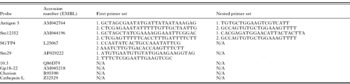
In order to establish whether WISH could reliably localize expression, 2 categories of genes, with projected and unknown patterns, were selected. The first group comprised cathepsin L as a gut marker (Bogitsh et al. Reference Bogitsh, Dalton, Brady and Brindley2001), Gp18-22 (el-Sherbeini et al. Reference el-Sherbeini, Ramadan, Bostian and Knopf1991), SGTP4 (Skelly et al. Reference Skelly, Kim, Cunningham and Shoemaker1994), Sm29 (Cardoso et al. Reference Cardoso, Pacifico, Mortara and Oliveira2006), all in the tegument cell body and the chorion gene for vitellaria (Koster et al. Reference Koster, Dargatz, Schroder, Hirzmann, Haarmann, Symmons and Kunz1988). The second group comprised 2 genes, identified in a microarray study of the larval transcriptome, Antigen 5 and a hypothetical protease inhibitor Sm12352 (Dillon et al. Reference Dillon, Feltwell, Skelton, Ashton, Coulson, Quail, Nikolaidou-Katsaridou, Wilson and Ivens2006) as well as a third gene encoding a putative adult protein antigen 10.3 (Davis et al. Reference Davis, Davis, Carroll, Rajkovic and Rottman1988). Antigen 10.3 is also an example of a protein with repetitive sequence, difficult to clone in its entirety and express. In addition to these 2 categories of gene/probe, the sense-strand of the chorion gene was used as an off-target control for both life-cycle stages. Sequences of interest were cloned into the plasmid vector pGEM-T Easy (Promega, Southhampton, UK) using primers indicated in Table 1, according to the manufacturer's instructions. Antigen 10.3 was transferred from an existing expression vector (containing only a single polymerase start site) into pGEM-T Easy, using M13 primers flanking the original clone, thus avoiding the need for the amplification of this highly repetitive gene by the polymerase chain reaction (PCR). The chorion clone was the kind gift of Dr David Johnston, Natural History Museum, London; the cathepsin L was provided by Dr Leonardo Farias, Centro de Biotecnologia, Instituto Butantan Sao Paulo, Brazil; the remainder were pre-existing clones used to manufacture the microarray described by Dillon et al. (Reference Dillon, Feltwell, Skelton, Ashton, Coulson, Quail, Nikolaidou-Katsaridou, Wilson and Ivens2006).
Primers and data base Accession numbers

Digoxigenin (DIG)-labelled antisense probes were synthesized from their template DNA using the modified method of Harland (Reference Harland1991), specifically with the following reaction mix: 10 μl of 5× transcription buffer; 2·5 μl of 10× DIG RNA labelling mix (Roche, Burgess Hill, UK); 5 μl of 100 mm dithiothretiol (DTT); 2 μl of RNase inhibitor (50 units); 3 μl of 50 units/μl SP6/T7/T3 polymerase, as appropriate (Promega); 6μl of 0·42 μg/μl linear DNA template (2·5 μg total) and 21·5 μl of RNase-free water (to a total volume of 50 μl). The reaction was incubated at 37°C for 2 h, after which time another 50 units (1 μl) of polymerase were added before a further incubation of 2 h. Then 1 μl of RNAse-free DNAse I (Promega) was added and incubated for 20 min at 37°C, and a 2 μl aliquot checked (for the absence of DNA) on a 2% agarose gel prior to precipitation of the probe. Each labelled probe was precipitated by addition of 50 μl of water and 50 μl of 7·5 m LiCl, 50 mm EDTA to the reaction and incubating it overnight at −20°C. The following day, probes were subjected to centrifugation at 12 000g in a refrigerated microcentrifuge (4°C) for 15 min, the supernatant was removed and the pellet washed with 70% ethanol. Washed pellets were centrifuged a second time for 5 min, the supernatant removed and the pellet air-dried for 5 min, before being resuspended in 50 μl of RNAse-free water. Probes were stored at −80°C until use.
Where necessary, long transcripts (i.e. >600 bp) were hydrolysed to produce fragments of ∼250 bp in length. Hydrolysis was carried out by resuspending probe pellets in a solution of 40 mm NaHCO3/60 mm Na2CO3 rather than water, after the precipitation. Probes in hydrolysis solution were then incubated at 60°C for the time t in min, determined by the following formula (available from http://www.rodentia.com/wmc/docs/Big_In_Situ.html),

where the starting length was the approximate insert size, as determined by agarose gel electrophoresis, and the desired length was 250 bp. After hydrolysis, probe sizes were confirmed by electrophoresis. The probes were then placed on ice, precipitated overnight and resuspended in water (as described previously).
Whole mount in situ hybridization
In situ hybridization was carried out according to the method described by Harland (Reference Harland1991), with the modifications described by Pownall et al. (Reference Pownall, Tucker, Slack and Isaacs1996). Briefly, worms were warmed to room temperature (22–24°C) and rehydrated by 2×5 min washes, the first in 75% ethanol/25% phosphate-buffered saline (PBS; pH 7·4) containing 0·1% Tween 20 (PBSAT) and the second in 50% ethanol/PBSAT. Worms were transferred to 100% PBSAT for 3×5 min washes, partially digested at room temperature in a 10 μg/ml solution of PCR-grade proteinase K (Roche) dissolved in PBSAT (see Results for incubation times). Digestion was halted by 2×5 min washes in 0·1 m aqueous triethanolamine. On completion of the second wash, 12·5 μl of acetic anhydride was added to the triethanolamine solution, and the worms washed for 5 min, followed by addition of 12·5 μl of acetic anhydride and a 5 min wash. After 2×5 min washes in PBSAT, the worms were re-fixed for 20 min in 4% formaldehyde-PBSAT and washed 5 times for 5 min in PBSAT. The permeabilized worms were then incubated at 60°C for 2 h in hybridization buffer (50% formamide, 5× SSC, 100 μg/ml heparin, 1× Denhardt's solution, 0·1% Tween 20, 0·1% CHAPS and 5 mm EDTA) with 1 mg/ml total yeast RNA added to block non-specific hybridization.
After 2 h the solution was replaced with fresh (pre-warmed) total RNA/hybridization buffer containing 1 μg/ml of synthesized DIG-labelled probe. Hybridization was performed at 60°C overnight. Worms were then washed in the following: hybridization buffer at 60°C, 2×10 min washes; 2× SSC+0·1% Tween 20 at 60°C, 3×20 min washes; 0·2× SSC+0·1% Tween at 60°C, 3×30 min washes and finally maleic acid buffer MAB (0·1 m maleic acid, 0·15 m NaCl, 0·1% Tween 20, pH 7·8) at room temperature, 2×15 min washes. Blocking prior to addition of anti-DIG antibody was carried out via a pre-incubation in blocking solution (MAB+2% Blocking Reagent (Roche)+20% heat-treated lamb serum) for 2 h at room temperature. Hybridized probes were detected using anti-DIG alkaline phosphatase-coupled antibodies (Roche). The anti-DIG antibody (Roche) was added to fresh blocking solution at a 1/2000 dilution and left at 4°C overnight. Excess antibody was then removed by extensive washes in MAB at room temperature (3×5 min, followed by 3×1 h washes), prior to a final wash in 1 mm levamisole (Sigma) in alkaline phosphatase buffer (100 mm Tris, 50 mm MgCl2, 100 mm NaCl, 0·1% Tween 20, pH 9·5) (Harland, Reference Harland1991; Pownall et al. Reference Pownall, Tucker, Slack and Isaacs1996).
Staining techniques
Hybridized probes were detected by addition of 2 substrates, BM Purple (Roche) and Fast Red TR (Sigma). BM Purple substrate with 1 mm levamisole was used according to manufacturer's instructions for standard light microscopy; alkaline phosphatase activity results in the deposition of an insoluble blue precipitate. BM Purple colour development was assessed visually and took from 30 min to several days, depending on transcript abundance. However, embedding and sectioning to identify precise cellular localization with this stain resulted in an unacceptably high level of leaching. Therefore, Fast Red TR was used for fluorogenic localization; development was monitored using a fluorescent microscope, as recommended by Cox and Singer (Reference Cox and Singer1999), and halted usually after ∼15 min. For both substrates, the development was stopped by washing worms twice in PBSAT for 15 min, followed by re-fixation and storage in 10% buffered formalin. For morphological analysis, adult worms were fixed in AFA (11·5% glacial acetic acid, 22% formalin, 33% ethanol and 33·5% distilled water), washed with 70% ethanol and stained with Langeron's Carmine for 30 min, as described by Machado-Silva et al. (Reference Machado-Silver, Pelajo-Machado, Lenzi and Gomes1998). Worms were then dedifferentiated in acid alcohol, dehydrated in 90% and absolute ethanol, cleared in Histoclear (National Diagnostics) and mounted in DPX (VWR, Lutterworth, UK). BM Purple-stained worms were photographed using a 18·2 Color Mosaic camera (Diagnostic Instruments Inc.) and SPOT software with a Leica (Wetzlar) DM2500 microscope. Confocal imaging was used to detect Fast Red TR or Langeron's Carmine staining using a LSM 510 meta on an Axiovert 200M (Zeiss), with a 543 nm laser and a 560 nm long pass filter.
Embedding and sectioning of treated worms
Fixed samples were removed from the primary fixative, 10% buffered formalin, and washed 3×20 min in 100 mm sodium phosphate buffer (pH 7). Washed samples were then placed in a secondary fix of 1% osmium tetroxide for 1 h on ice prior to 2 further 20 min washes in the same sodium phosphate buffer. The samples were dehydrated in a succession of 20 min washes of increasing acetone concentration (20%, 50%, 70%, 90% through to 100%), followed by an additional wash in 100% acetone. Dehydrated worms were then embedded in a succession of 30 min incubations of increasing Spurr resin concentration (Spurr:acetone, 25%:75%, 50%:50% and 75%:25%), followed by an overnight incubation in 100% Spurr resin (10 g of 3,4-epoxycyclohexylmethyl-3,4-epoxycyclohexylcarboxylate, ERL 4221-d; 6 g of diglycidyl ether of polypropylene glycol, DER 736; 26 g of nonenylsuccinic anhydride, NSA; 0·4 g of 2-diethylaminoethanol, S-1/DMAE). Samples were placed in moulds and the Spurr resin polymerized by baking for 24 h at 70°C. Sections were cut to 1 μm on an ultramicrotome and stained with a 0·6% toluidine blue solution.
RESULTS
Establishing the parameters for WISH
A range of fixation procedures was tested on adult worms, with the aim of minimizing endogenous alkaline phosphatase activity to prevent interference with DIG detection. The dual treatment with Carnoy's fixative and MEMFA proved the most effective, whilst maintaining the integrity of the RNA targets. The duration of worm incubation with proteinase K to permeablize tissues is a second critical variable. Batch testing of the enzyme is essential, with digestion time typically varying from 10–20 min for adults and 5–10 min for larvae. Too short a digestion with proteinase K results in erratic permeablization, whereas over-digestion degrades cells and tissues to the point where they cannot be identified. WISH was always performed on hundreds of larvae and 30–50 mixed adults to establish the reproducibility in the detection pattern. The final important variable is the colour development time after probing with anti-DIG antibody. Depending on transcript abundance, reaction product can be observed from as soon as 30 min and up to several days. The reaction needs to be monitored at regular intervals to prevent over-development. Both positive and negative (sense or irrelevant) controls are essential to ensure that probe and antibody have infiltrated the tissues, whilst allowing false positive results to be identified. False positives are a more significant issue for adults where reagents can pool in internal compartments or along the male gynaecophoric canal, if washing is inadequate; endogenous alkaline phosphatase activity is also higher than in the larvae.
WISH localizes gene expression to specific adult worm tissues
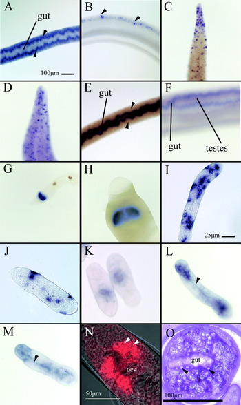
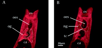
The chorion transcript was selected as a positive control to validate WISH, because of its abundance and known site of expression. Colour developed by 30 min in the posterior two thirds of the female body (Fig. 1A) as 2 irregular parallel lines between the central negative staining gut and surface tegument, corresponding to the position of the vitellaria. Strikingly, colour also developed in all male worms processed in the same sample (Fig. 1B). Staining displayed a distinct, but highly irregular punctate pattern (arrowed) within the dorsal parenchyma, throughout the body, posterior to the ventral sucker. We next sought to establish where the gene encoding the putative tegumental protein Sm29 (of unknown function) was expressed. Coloured product could be observed within 1–2 h of incubation with substrate, as a sharply punctuate pattern over the entire female (Fig. 1C) and male (Fig. 1D) bodies, being particularly dense at the extreme posterior. This distribution indicates that the gene is expressed only in the tegument cell bodies. The protease cathepsin L was investigated as a potential marker of the schistosome gut epithelium. Colour developed within 1–2 h, apparently more intensely in the female (Fig. 1E) than the male (Fig. 1F). In the female worm, there was a reversal of the pattern of the chorion transcript with an intense wavy band of staining down the centre of the body, apart from a faint line representing the gut lumen. The distinctive bifurcation of the male gut around the testes was clearly demarcated (Fig. 1F). The distribution of antigen 10.3 demonstrated the ability of WISH to localize genes to very restricted tissue types. Colour developed within 30 min in both females (Fig. 1G) and males (Fig. 1H) in a bilobed structure, below the oesophagus, immediately anterior to the ventral sucker, but nowhere else in the worm. Staining of adult worms with Langeron's Carmine for analysis of tissues by confocal microscopy revealed the location of both the oesophageal glands and nerve ganglia in the oesophageal region (Fig. 2). The gland envelops the ventral surface of the oesophagus at the extreme posterior, adjoining the gut caecum, as a concave hemisphere (Fig. 2A). The dorsally situated ganglia lie further forward, connected by a transverse commissure (Fig. 2B). The patterns of BM Purple and Fast Red TR staining or antigen 10.3 corresponded precisely with the oeosophageal gland.
Results of whole mount in situ hybridization, (A–H and N) adult worms, (I–M) day 10 in vitro-cultured schistosomula. (O) Toluidine blue-stained thin section of adult female after processing. Distribution of chorion genes expressed in (A) female and (B) male worms. Localization of Sm29 gene expression in (C) female and (D) male worms; extreme posterior only illustrated. Expression of the cathepsin L gene in the female (E) and male worms (F). Antigen 10.3 only present at the anterior of female (G) and male (H) worms. Putative tegumental cell body markers SGTP4 (I) and Gp18-22 (J) localized in differing numbers of foci. Expression of cathepsin L in the embryonic gut (K). SmAg5 (L) and putative protease inhibitor (M) localized in dispersed internal structures at the anterior and posterior. Optical slice showing antigen 10.3 expression localized to the oesophageal gland by Fast Red TR substrate (N).

Sections from a Z-stack of the anterior region of an adult female schistosome between oral (os) and ventral sucker (not visible), stained with Langeron's Carmine showing tissues of interest outlined in white. (A) The ventrally situated oesophageal gland (og) envelops the extreme posterior oesophagus (oes) immediately anterior to the oesophageal-caecal (ca) junction. (B) The dorsally situated nerve ganglia (ng) lie further forward astride the oesophagus, connected by a transverse commissure (tc).

WISH is equally effective in larval tissues
The tegumental glucose transporter SGTP4, with known tissue distribution, was used as a positive control to validate the WISH technique in larval stages. Colour developed within 24 h but the reaction needed 2–3 days to reach its maximum intensity. A very characteristic pattern of staining was observed, comprising approximately 14–20, discrete blue foci, some with a clear centre representing a nucleus, revealing that each aggregate was a single cell (Fig. 1I). A second putative tegumental protein Gp18-22 was also localized to discrete foci of stain within the larval body (Fig. 1J), but on this occasion many fewer were present than for SGTP4 (SGTP4,  =14±s.e. 1·2; Gp18-22, =5±s.e. 0·5, P<0·0001 unpaired t-test, n=12 larvae per probe). As an example of genes likely to be expressed in deeper tissues, the distribution of cathepsin L was examined. A single focus of stain was present in the mid-body region, in the exact position of the embryonic gut diverticula (Fig. 1K). As examples of 2 larva-expressed transcripts with unknown distribution, we localized SmAg5 (Fig. 1L) and the putative protease inhibitor Sm12352 (Fig. 1M). Both produced very similar staining patterns with dual concentrations of colour in internal structures at the anterior and posterior of the larvae. These regions were connected by a fine line of stain (arrowed) corresponding to the position of the rudimentary oesophagus.
Use of fluorescent substrate permits optical dissection of transcript distribution by confocal microscopy
When Fast Red TR is substituted for BM Purple substrate, high resolution distribution of the fluorescent product can be achieved using the Z stack facility of a confocal microscope. It is essential to monitor the development of colour under a fluorescent microscope, due to the sensitivity of detection; high background and non-specific staining ensue if this step is omitted. Judging the colour development under transmitted light results in saturation of the fluorescent signal with loss of definition. Applied to antigen 10.3, the bilobed appearance of the oesophageal glands is evident (Fig. 1N), with the undulating oesophageal lumen lying between. At high magnification (not shown) the red fluorescent product of alkaline phosphatase activity is composed of minute dots which we speculate represent reactions around individual transcripts. The holes left by digested nuclei (arrowed) are also apparent. The loss of nuclear content after proteinase K digestion is readily detected by toluidine blue staining of transverse sections of treated worms (Fig. 1O).
DISCUSSION
With the sequencing, assembly and release of the S. mansoni genome, bolstered by ongoing transcriptome and proteome analyses, schistosome research is now moving into its post-genomic phase. However, compared with the classical model organisms, the post-genomic tools needed to capitalize on the growing body of information are either in their infancy or simply do not exist. Our advocacy of WISH to localize gene expression to specific life-cycle stages and tissues should be seen in this context. It is a practical and essential step in discovering where and how schistosome gene products exert their function. Although high-quality ISH has previously been performed on adult worm sections, and laser microdissection microscopy may permit individual tissues to be assayed for the presence of a specific mRNA (Jones et al. Reference Jones, Randall, McManus and Engwerda2004), neither are suitable for localizing multiple genes in representative numbers of worms. Unless the site of expression is already suspected, highly restricted transcripts may be missed entirely. Two clear examples from this study are antigen 10.3 and the chorion gene. The former was described as a putative adult antigen (Davis et al. Reference Davis, Davis, Carroll, Rajkovic and Rottman1988), which we have now localized solely to the minute oesophageal glands located beneath both the male and female oesophagus (Morris and Threadgold, Reference Morris and Threadgold1968). The chorion gene is abundant in the female vitellaria, but we have also detected discrete foci of expression in the male posterior. Earlier section-based ISH studies (Koster et al. Reference Koster, Dargatz, Schroder, Hirzmann, Haarmann, Symmons and Kunz1988; Osman et al. Reference Osman, Niles and LoVerde2001) did not detect its expression, but our observations accord with ultrastructural studies of male worms that have reported previously the presence of vestigial vitellaria (Shaw and Erasmus, Reference Shaw and Erasmus1982). Analysis of the S. mansoni transcriptome has also revealed this chorion transcript in male worms (see http://bioinfo.iq.usp.br/schisto6/cgi-bin/cap3/cluster_editor/ceditor.pl?show=606889.1.2002-10-01) (Verjovski-Almeida et al. Reference Verjovski-Almeida, DeMarco, Martins, Guimaraes, Ojopi, Paquola, Piazza, Nishiyama, Kitajima, Adamson, Ashton, Bonaldo, Coulson, Dillon, Farias, Gregorio, Ho, Leite, Malaquias, Marques, Miyasato, Nascimento, Ohlweiler, Reis, Ribeiro, Sa, Stukart, Soares, Gargioni, Kawano, Rodrigues, Madeira, Wilson, Menck, Setubal, Leite and Dias-Neto2003). The possession of sexual features associated with the opposite sex is not uncommon in the animal kingdom (e.g. nipples in Homo sapiens and other mammals).
The power of the WISH technique is illustrated by its ability to localize expression of Sm29 solely to the tegument cell bodies. At the worm surface, the syncytial tegument is connected by cytoplasmic processes to underlying cell bodies which contain the nucleus and molecular machinery necessary for protein synthesis and export. Although Sm29 has no homology to molecules outside of the genus Schistosoma, we can infer that the product is tegument specific, which is an added bonus for our independent proteomic analyses of tegument membrane composition (Braschi et al. Reference Braschi, Curwen, Ashton, Verjovski-Almeida and Wilson2005, Reference Braschi and Wilson2006; Braschi and Wilson, Reference Braschi and Wilson2006). Together, these observations imply a role for Sm29 at the tegument surface, possibly in immune evasion. The fact that only the cell bodies are positively stained indicates that this mRNA species, at least, does not pass up the cytoplasmic connections into the tegument syncytium. The results also make Sm29 the ideal tegument cell body marker. Likewise, cathepsin L provides a specific marker for the gut epithelium. It is notable that the level of expression is greater in female compared with male worms; this is probably a reflection of the female's much greater rate of blood ingestion (Lawrence, Reference Lawrence1973). The enzyme cathepsin L was initially localized by immunocytochemistry to the gynecophoric canal of male worms and female reproductive tissues (Michel et al. Reference Michel, Ghoneim, Resto, Klinkert and Kunz1995). However, later studies corrected this error and confirmed gut localization (Bogitsh et al. Reference Bogitsh, Dalton, Brady and Brindley2001). In our opinion, unlike immunocytochemistry, WISH is less susceptible to false positives through non-specific binding or pooling, giving it a significant advantage.
The migrating schistosomulum is a life-cycle stage of particular interest, since it is a validated target of protective immunity in mice (Coulson, Reference Coulson1997). Localizing expression to tissues in this phenotype would thus be particularly useful in the context of finding vaccine candidates. Transcripts for both known tegumental proteins, SGTP4 and Gp18-22, were localized to tegumental cell bodies, although, intriguingly, each probe produced a different staining pattern. The impression gained was that greater numbers of cells were stained using the SGTP4 probe. This may indicate that other tissues, such as muscle, also express SGTP4 or, as hypothesized by Wilson and Barnes (Reference Wilson and Barnes1974), there is more than one kind of tegument cell body. Data from our earlier microarray work (Dillon et al. Reference Dillon, Feltwell, Skelton, Ashton, Coulson, Quail, Nikolaidou-Katsaridou, Wilson and Ivens2006) suggested that the schistosomulum was beginning to express gut cathepsins. WISH confirms the microarray findings by localizing cathepsin L transcripts to the larval gut; this may indicate that the larva is already preparing to digest a bloodmeal or that it is actively digesting serum proteins.
The expression patterns of SmAg5 and the putative protease inhibitor illustrate an aspect of the WISH technique that will repay further refinement. It is difficult to pinpoint the precise cells in which these transcripts are found, apart from the embryonic oesophagus. This situation is partly a consequence of the minute size of schistosome cells, but also the complexity of tissue organization. (Increasing tissue complexity is also a problem for developmental biologists as embryonic tissues differentiate.) Our observations provide a strong argument for the identification of tissue-specific markers, such as Sm29, cathepsin L and antigen 10.3. The adoption of a fluorescent substrate for alkaline phosphatase is a partial solution to tissue complexity, because it allows a 3D image of a worm to be constructed from consecutive optical sections (i.e. Z-stack). As a proof-of-principle, we successfully demonstrated the fine detail of antigen 10.3 localization in the oesophageal gland. Ideally, the combination of a fluorescent alkaline phosphatase substrate for transcript detection with a fluorescent tissue or organelle-specific counterstain, possessing different excitation/emission wave-lengths, would permit simultaneous detection using a confocal microscope. We are actively pursuing this option. We are also investigating the use of pairs of probes with different tags (digoxygenin and fluorescein), one as a tissue-specific marker and the other representing an unknown transcript. The sites of hybridization are then detected by 2 consecutive enzymatic reactions (Kelly et al. Reference Kelly, Nekkalapudi and El Hodiri2007).
We recently reviewed the prospects and limitations for schistosome post-genomics, drawing attention to the technical advances that need to be made before the newly generated genomic data can be fully utilized (Wilson et al. Reference Wilson, Ashton, Braschi, Dillon, Berriman and Ivens2007). Whilst there has been progress in the application of RNA interference to silence genes (Skelly et al. Reference Skelly, Da'dara and Harn2003), both an homologous schistosome cell line and stable transfection into the germ line appear to be distant prospects, although recent reports of cultured schistosome cells (Ming et al. Reference Ming, Dong, Zhong, Grevelding and Jiang2006) and virus-induced transfection (Kines et al. Reference Kines, Mann, Morales, Shelby, Kalinna, Gobert, Chirgwin and Brindley2006) are encouraging developments. Furthermore, the release of the fourth draft of the genome annotation (http://www.genedb.org/genedb/smansoni/) in February 2007 brings a definitive gene list much closer, and with it the feasibility of a genome wide microarray for transcript profiling. The widespread application of WISH to any gene and life-cycle stage should assist in enhancing our understanding of the function of both known and unknown protein products.
This work was funded by the Biotechnology and Biological Sciences Research Council (BBSRC) and National Institutes of Health (NIH) grant AI54711-02. The authors would like to thank Ms M. Stark, York University Technology facility, for resin embedding and tissue sectioning, Dr A. C. Ivens and Ms T. Feltwell, Wellcome Trust Sanger Institute, for their support and access to microarray array clones.