Idiopathic pulmonary hypertension is more frequent at high altitude than at sea level, where hypobaric hypoxia is detrimental in terms of clinical presentation and prognosis. One of the standards of care in treating this population of patients is to move to an area of lower altitude. Reference Díaz Góngora1,Reference Penaloza, Sime and Ruiz2 However, residing in a lower altitude area of Colombia presents the risk for acquiring endemic diseases as Dengue. 3
Dengue is a mosquito-borne viral disease that can present with a wide spectrum of clinical symptoms and complications such as haemorrhage, arrhythmias, and myocardial involvement. Reference Yacoub, Wertheim and Cameron4 Severe cases represent a challenge in the management, when other comorbidities are present. We will describe a patient, previously diagnosed with severe idiopathic pulmonary hypertension, who presented to us with cardiac complications associated with dengue infection.
Case report
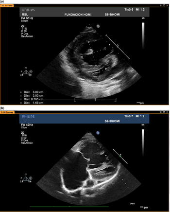
We present a 15-year-old female patient previously diagnosed with severe supra systemic pulmonary hypertension while living in Bogota (2640 m above sea level). Four years before, she arrived at hospital after syncope. She was in functional class 3–4 and brain natriuretic peptide was 2.430 pg/ml. The echocardiogram showed severe pulmonary hypertension (Fig 1). When the patient was stabilised, a catheterism was made: basal pulmonary pressure (PP): 90/43,63 mm Hg; pulmonary resistances (PR): 18.46 WU/M2; with Oxygen: 86/31,51 mm Hg; PR: 14.20 WU/M2; with NO: 78/35,51 mm Hg; PR: 12.73 WU/M2 (negative VRT). As part of treatment, the patient had to be managed by moving from Bogotá to a town located at 400 m above sea level with Sildenafil + Macitentan. Afterward, she stabilised in functional class 3 (3B of Panama Classification). Reference Lammers, Adatia, Del Cerro, Diaz, Freudenthal and Freudenthal5 Two years after treatment a second catheterism was made without changes; the PR were 20.42 WU. Although the patient was stable, taking into account the severe pulmonary hypertension, we recommend to add subcutaneous treprostinil that was refused by the patient, the same as lung transplantation. Selexipag was accepted, but is not approved for children in Colombia.
( a ) Short axis view: this figure shows the huge right ventricle and the collapsed left ventricle in systole, with great alteration of the eccentricity index and the right ventricle/left ventricle ratio. ( b ) Four chamber view: this figure shows the huge right ventricle and right atrium and the collapsed left ventricle. Courtesy of Drs Roy Sanguino and Oscar Arévalo.

She presented to us with a 3-day history of headache, malaise, and joint pain associated with fever. Initial blood tests showed leukopenia, thrombocytopenia, and C-Reactive Protein was 59.9 mg/L. Given the clinical presentation, a serologic test was ordered which was positive for Dengue virus-specific immunoglobulin M. Brain natriuretic peptide was elevated at 840 pg/ml. Alkaline phosphatase and glutamic oxaloacetic transaminase were within the upper limits of normal as well. Echocardiogram showed systolic pulmonary pressure of 100 mmHg and mean pulmonary pressure of 62 mmHg with pericardial effusion. Basal electrocardiogram demonstrated a first-degree atrioventricular block, documented previously.
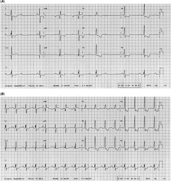
Considering the history of severe pulmonary hypertension, the patient was transferred to the intensive care unit for monitoring. Forty-eight hours past the last febrile episode, on day 7 of symptoms, sinus pauses and bradycardia of 40 beats/minute were documented. Reference Yantie, Gunawijaya, Suradipa and Gustawan6 A new electrocardiogram and Holter showed a second-degree atrioventricular block, Mobitz 1 (Fig 2A). Bradycardia and second-degree atrioventricular block resolved over the next 6 days without requiring additional treatment (Fig 2B). The first-degree atrioventricular block persisted until the time of discharge. The patient continued stabilised and could be followed as an outpatient.
( A ) Date: 03/04/2019. Electrocardiogram that shows the typical second degree atrioventricular block, Mobitz 1 type. It is clear, the progressively prolongation of PR before the not conducted P (3 days after the fever disappeared). ( B ) Date: 15/04/2019. Electrocardiogram 9 days after the fever disappeared: This electrocardiogram is similar to the basal electrocardiogram with first degree atrioventricular block. Very tall R in precordial right leads, indicating severe hypertrofy of the right ventricle due to the severe pulmonary hypertension. Negative ST depresion in several leads by right ventricular overload and abnormal repolarization.

Discussion
There is scarce information on idiopathic pulmonary hypertension in children living at high altitude, although in our experience, given our geographic situation in the Andean region, this pathology can be frequent and severe at early age and the outcome when continuing living in altitude is too bad. As part of our standard of care, we recommend our patients to move to lower altitude cities to improve haemodynamic parameters and decrease the need for oxygen supplementation, impacting positively the survival and quality of life. Nevertheless, in that regions, this fragile population become exposed to endemic and tropical diseases that if acquired, requires transfer to medical centers to receive specialised treatment.
It is interesting that in spite of the severe pulmonary hypertension of our patient, there was not complication of their basal pathology, except the pericardial effusion. In the echocardiogram there were no changes in ventricular function or in the severity of pulmonary hypertension in relation with previous evaluation before dengue. There were no findings that supported the diagnosis of myocarditis which could explain the transient second degree atrioventricular block; however, we can not discard a mild myocarditis not detected by echocardiography. Reference Yantie, Gunawijaya, Suradipa and Gustawan6 The patient did not have deterioration of her pulmonary hypertension maybe by the vasodilators (Sildenafil + Macitentan) and for the effect of living at low altitude. Reference Díaz Góngora1
Dengue is a mosquito-borne viral disease that can cause a wide spectrum of clinical presentation with a significant burden of disease in endemic areas, and significant mortality risk in severe forms of the disease. Close monitoring and early supportive care are pivotal in treating these patients. Among the cardiovascular complications we can find myocarditis, heart failure, arrhythmias, and conduction disorders. Reference Yacoub, Wertheim and Cameron4,Reference Harpreet, Gagan Sukhija and Bala7 These complications can be found in up to 30% of hospitalised patients. Bradyarrhythmias are the most frequent, according to the Singapore study and are thought to be related to the parasympathetic pre-dominance during the defervescence stage. Reference Rajarethinam, Ang and Ong8 We could detect this complication in our patient early in its presentation, due to the close monitoring in the intensive care unit.
Furthermore, few publications on second-degree atrioventricular block Mobitz 1 associated with Dengue are reported. Reference Yacoub, Wertheim and Cameron4,Reference Harpreet, Gagan Sukhija and Bala7 One hypothesis to explain this complication is the “cytokine storm” during the immunologic response to the infection resulting in myocardial inflammation and conduction disorders. Reference Tisoncik, Korth and Simmons9 Bhamarapravati found subendocardial bleeding in 47% of autopsies of patients that died from haemorrhagic dengue. Reference Bhamarapravati, Tuchinda and Boonyapaknavik10 These cardiovascular findings could be underdiagnosed because some are mild and reversible. The Sri Lanka report showed that 62.5% of 120 adults with dengue had electrocardiographic changes, findings that must alert us to the importance of cardiovascular follow up of patients with dengue, mainly in patients with pre-existing cardiovascular disease. Reference Kularatne, Pathirage and Kumarasiri11
Part of the treatment strategies during Dengue infection is supportive care with endovenous crystalloid solution, which in our patient demanded rigorous monitoring due to the severity of her pre-existing condition. We recognised the advantages of having transferred the patient early to the intensive care unit in the disease course. This allowed us to identify the conduction disorder quickly and to infuse crystalloids more accurately, avoiding clinically significant decompensation. It is worth to mention that having a previous first-degree atrioventricular block, increases the likelihood of progression to more complex conduction disorders in the presence of comorbidities. Fortunately, these more complex atrioventricular blocks are usually transitorious and exceptionally need a pacemaker.
Conclusion
Pulmonary hypertension patients living at high altitude represent a fragile population. Relocation to lower altitude, while important in managing their underlying disease process, increases the risk of exposure to endemic diseases. If these endemic diseases are acquired, they still need to be transferred back to high altitude for specialised management when required. Close monitoring is fundamental for early complications detection, which allows better clinical outcomes.
Acknowledgements
To Maria C. Preciado (1), Mary. Vanegas (1), Victor M. Huertas (2), Roy Sanguino R (3) and Oscar Arevalo (3) for the collaboration in preparing the paper: (1) Fellow of Pediatrics Universidad Nacional de Colombia; (2) Catedra Professor, Universidad Nacional de Colombia; (3) Pediatric Cardiologist Hospital La Misericordia, Bogotá, Colombia.
Financial support
Institutional support by Universidad Nacional de Colombia.
Conflicts of interest
None.