Introduction
Attention deficit hyperactivity disorder (ADHD) is defined by age-inappropriate levels of inattention, impulsivity and hyperactivity whereas autism spectrum disorder (ASD) is defined by impairments in communication and social interaction and by restricted, repetitive behaviour (APA, 1994). There is increasing evidence for co-morbidity between the disorders (Rommelse et al. Reference Rommelse, Geurts, Franke, Buitelaar and Hartman2011), which share deficits in executive functions (Willcutt et al. Reference Willcutt, Doyle, Nigg, Faraone and Pennington2005; Corbett et al. Reference Corbett, Constantine, Hendren, Rocke and Ozonoff2009), including working memory (WM), particularly at higher WM loads (Cui et al. Reference Cui, Gao, Chen, Zou and Wang2010; Kasper et al. Reference Kasper, Alderson and Hudec2012). The importance of this clinical and behavioural overlap is highlighted by recent changes to DSM-5, allowing co-diagnosis of ADHD and ASD (www.dsm5.org).
WM refers to the ability to temporarily store and manipulate information to guide and direct behaviour (Baddeley, Reference Baddeley1996). WM can be measured in the N-back task, where subjects identify targets that were shown a few trials back (Baddeley, Reference Baddeley2003). ADHD adolescent studies have shown underactivation during verbal N-back tasks compared to controls in the bilateral dorsolateral prefrontal cortex (DLPFC), parietal lobe and cerebellum, with more pronounced behavioural and functional deficits during higher WM loads (Kobel et al. Reference Kobel, Bechtel, Weber, Specht, Klarhofer, Scheffler, Opwis and Penner2009; Cubillo et al. Reference Cubillo, Smith, Barrett, Giampietro, Brammer, Simmons and Rubia2014). In ASD adults, a functional magnetic resonance imaging (fMRI) study found decreased activation in the left hemispheric DLPFC, inferior frontal cortex (IFC) and inferior parietal lobe during the highest WM load of a verbal N-back task, but this has not been found in children (Koshino et al. Reference Koshino, Carpenter, Minshew, Cherkassky, Keller and Just2005). An important question, however, is whether children with ASD have the same brain dysfunctions as adults with ASD and whether these deficits are shared or are disorder specific relative to ADHD.
Serotonin (5-hydroxytryptamine or 5-HT) mediates verbal WM (Allen et al. Reference Allen, Cleare, Lee, Fusar-Poli, Tunstall, Fu, Brammer and McGuire2006; Rose et al. Reference Rose, Simonotto, Spencer and Ebmeier2006). Pharmacological fMRI studies using the N-back task in healthy adults have shown that selective serotonin reuptake inhibitors (SSRIs), which enhance 5-HT levels, increase left IFC activation (Rose et al. Reference Rose, Simonotto, Spencer and Ebmeier2006) whereas acute tryptophan depletion (ATD), which reduces 5-HT synthesis (Nishizawa et al. Reference Nishizawa, Benkelfat, Young, Leyton, Mzengeza, De Montigny, Blier and Diksic1997), decreases right middle/IFC activation and attenuates default mode network (DMN) deactivation during high WM loads (Allen et al. Reference Allen, Cleare, Lee, Fusar-Poli, Tunstall, Fu, Brammer and McGuire2006).
Few studies have investigated the clinical efficacy of SSRIs in ADHD and ASD. Open-label studies of fluoxetine have shown improvement in ADHD symptoms in co-morbid ADHD children (Barrickman et al. Reference Barrickman, Noyes, Kuperman, Schumacher and Verda1991; Gammon & Brown, Reference Gammon and Brown1993; Quintana et al. Reference Quintana, Butterbaugh, Purnell and Layman2007). Fluoxetine improved ASD symptoms in children (West et al. Reference West, Brunssen and Waldrop2009) and adults with ASD (Williams et al. Reference Williams, Wheeler, Silove and Hazell2010; Hollander et al. Reference Hollander, Soorya, Chaplin, Anagnostou, Taylor, Ferretti, Wasserman, Swanson and Settipani2012), with a meta-analysis showing improvements in repetitive behaviours (Carrasco et al. Reference Carrasco, Volkmar and Bloch2012). However, negative findings with other SSRIs have also been reported (King et al. Reference King, Hollander, Sikich, McCracken, Scahill, Bregman, Donnelly, Anagnostou, Dukes, Sullivan, Hirtz, Wagner, Ritz and Network2009).
Abnormalities in 5-HT have been reported in both disorders. Approximately 30% of ASD patients have enhanced platelet 5-HT levels (Hranilovic et al. Reference Hranilovic, Bujas-Petkovic, Vragovic, Vuk, Hock and Jernej2007) and reduced binding to the 5-HT reuptake transporter and 5-HT2A receptors in the frontal lobe and PCC, which have been linked to poor social communication (Murphy et al. Reference Murphy, Daly, Schmitz, Toal, Murphy, Curran, Erlandsson, Eersels, Kerwin, Ell and Travis2006; Nakamura et al. Reference Nakamura, Sekine, Ouchi, Tsujii, Yoshikawa, Futatsubashi, Tsuchiya, Sugihara, Iwata, Suzuki, Matsuzaki, Suda, Sugiyama, Takei and Mori2010). ADHD patients have lower platelet 5-HT levels relative to controls (Spivak et al. Reference Spivak, Vered, Yoran-Hegesh, Averbuch, Mester, Graf and Weizman1999), along with genetic (Gizer et al. Reference Gizer, Ficks and Waldman2009) and biochemical 5-HT abnormalities (Oades, Reference Oades2007). Furthermore, both animal and human studies have shown an association between 5-HT and impulsiveness (Dalley & Roiser, Reference Dalley and Roiser2012) and between 5-HT and stimulant medication response (Gainetdinov et al. Reference Gainetdinov, Wetsel, Jones, Levin, Jaber and Caron1999; McGough et al. Reference McGough, McCracken, Loo, Manganiello, Leung, Tietjens, Trinh, Baweja, Suddath, Smalley, Hellemann and Sugar2009).
Neurotransmitter abnormalities underlie abnormal neurocognitive functioning. An important question that may help to elucidate potential disorder-specific neurotransmitter underpinnings of neurocognitive abnormalities is whether 5-HT agonists modulate verbal WM networks in both disorders in a shared or disorder-specific manner. An investigation of acute effects can reveal true drug effects, not confounded by indirect effects on symptom improvement after chronic administration. A better understanding of the shared and disorder-specific neurofunctional and neurotransmitter abnormalities in ADHD and ASD is of clinical importance as it may ultimately aid future differential diagnosis and improve disorder-specific treatment.
The aim of this fMRI study was therefore to investigate (1) shared and disorder-specific brain dysfunctions in children with ADHD and children with ASD during a verbal N-back task relative to controls and each other, and (2) shared and disorder-specific neurofunctional effects of an acute dose of fluoxetine on these relative to placebo.
Based on previous findings, we hypothesized that, under placebo, relative to controls, both disorders would show reduced left DLPFC and parietal activation (Koshino et al. Reference Koshino, Carpenter, Minshew, Cherkassky, Keller and Just2005; Kobel et al. Reference Kobel, Bechtel, Weber, Specht, Klarhofer, Scheffler, Opwis and Penner2009; Cubillo et al. Reference Cubillo, Smith, Barrett, Giampietro, Brammer, Simmons and Rubia2014), with ADHD patients exhibiting additional right DLPFC and cerebellar abnormalities (Koshino et al. Reference Koshino, Carpenter, Minshew, Cherkassky, Keller and Just2005; Kobel et al. Reference Kobel, Bechtel, Weber, Specht, Klarhofer, Scheffler, Opwis and Penner2009; Cubillo et al. Reference Cubillo, Smith, Barrett, Giampietro, Brammer, Simmons and Rubia2014). Based on evidence for decreased serotonin levels in ADHD (Barrickman et al. Reference Barrickman, Noyes, Kuperman, Schumacher and Verda1991; Gammon & Brown, Reference Gammon and Brown1993; Quintana et al. Reference Quintana, Butterbaugh, Purnell and Layman2007), we hypothesized that increased serotonin levels under fluoxetine would increase frontal brain activation in ADHD, as observed previously in pharmaco-fMRI studies of verbal WM and serotonin agonists in healthy adults (Allen et al. Reference Allen, Cleare, Lee, Fusar-Poli, Tunstall, Fu, Brammer and McGuire2006; Rose et al. Reference Rose, Simonotto, Spencer and Ebmeier2006). Although there is some evidence for higher platelet levels of serotonin in ASD (Hranilovic et al. Reference Hranilovic, Bujas-Petkovic, Vragovic, Vuk, Hock and Jernej2007), the poor binding of serotonin to 5-HT receptors in the frontal lobe suggests that this may be ineffective at eliciting frontal activation (Murphy et al. Reference Murphy, Daly, Schmitz, Toal, Murphy, Curran, Erlandsson, Eersels, Kerwin, Ell and Travis2006; Nakamura et al. Reference Nakamura, Sekine, Ouchi, Tsujii, Yoshikawa, Futatsubashi, Tsuchiya, Sugihara, Iwata, Suzuki, Matsuzaki, Suda, Sugiyama, Takei and Mori2010). Therefore, we hypothesized that a fluoxetine-mediated increase in serotonin would also enhance frontal activation in ASD.
Method
Participants
Fifty-six right-handed boys (Edinburgh Handedness Inventory; Oldfield, Reference Oldfield1971) aged 10–17 years (22 controls, 17 with ADHD and 17 with ASD), with IQ > 70 [Wechsler Abbreviated Scale of Intelligence – Revised (WASI-R; Wechsler, Reference Wechsler1999], participated in this study.
ADHD boys had a clinical DSM-IV diagnosis of non-co-morbid ADHD, inattentive/hyperactive-impulsive combined subtype, assessed using the standardized Maudsley diagnostic interview (Goldberg & Murray, Reference Goldberg and Murray2002). They scored above the clinical threshold for ADHD symptoms on the Strengths and Difficulties Questionnaire (SDQ; Goodman & Scott, Reference Goodman and Scott1999) and the revised Conners’ Parent Rating Scale (CPRS-R; Conners et al. Reference Conners, Sitarenios, Parker and Epstein1998); one boy was below the cut-off on the SDQ but had diagnostic confirmation from a child psychiatrist. Three ADHD boys were medication naïve, one had stopped methylphenidate for 3 months and 13 had received chronic stimulants but had a 48-h medication wash-out prior to scanning. To ensure there was no co-morbidity with ASD, ADHD boys had to score below 15 on the Social Communication Questionnaire (SCQ; Rutter et al. Reference Rutter, Bailey and Lord2003).
ASD diagnosis was made using ICD-10 (WHO, 1994) diagnostic criteria and confirmed by the Autism Diagnostic Interview – Revised (ADI-R; Lord et al. Reference Lord, Rutter and Couteur1994) and the Autism Diagnostic Observation Schedule (ADOS; Lord et al. Reference Lord, Risi, Lambrecht, Cook, Leventhal, DiLavore, Pickles and Rutter2000). All ASD subjects were medication naïve except one, who took melatonin, but underwent a 2-week medication wash-out. To ensure no co-morbidity with ADHD, the ASD boys had to score below 7 on the Hyperactivity/Inattention subscale of the SDQ.
Exclusion criteria were co-morbidity with other psychiatric or neurological disorders, and drug/alcohol dependency, as assessed by the standardized Maudsley diagnostic interview. Patients were recruited from local clinics and support groups.
Patients were scanned twice in a double-blind, counterbalanced randomized, placebo-controlled design. Because of the half-life of fluoxetine (1–3 days) and its metabolite (5–16 days) (Wong et al. Reference Wong, Bymaster and Engleman1995), scans were 3–4 weeks apart. To ensure that fluoxetine had reached its peak plasma level, after 5–8 h (Wong et al. Reference Wong, Bymaster and Engleman1995), patients were scanned 5 h after administration. During this 5-h time period, patients practised the N-back task once each time, and underwent IQ and clinical assessments (the first time), had lunch and rested. Liquid fluoxetine was titrated to age and weight: boys aged between 10 and 13 years and weighing < 30 kg received 8 mg, those weighing > 30 kg received 10 mg. Boys aged between 14 and 17 years and weighing < 30 kg received 10 mg, those > 30 kg received 15 mg. Placebo was equivalent amounts of peppermint water with a similar taste to fluoxetine.
Twenty-two healthy, right-handed and age-matched boys were recruited by advertisement and scored below clinical thresholds on the SDQ, SCQ and CPRS-R.
Written informed consent/assent was obtained from all participants. The study was approved by the local ethics committee. Participants were paid £50 for each scan.
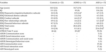
For recruitment, demographic and clinical details of participants, see Table 1 and the online Supplementary material.
Sample characteristics for healthy control boys and boys with ADHD and ASD

ADHD, Attention deficit hyperactivity disorder; ASD, autism spectrum disorder; SDQ, Strength and Difficulties Questionnaire; SCQ, Social Communication Questionnaire; CPRS-R, revised Conners’ Parent Rating Scale; ADOS, Autism Diagnostic Observation Schedule; ADI, Autism Diagnostic Interview.
a and b indicate a significant difference between groups. aRefers to a significantly enhanced value in ADHD relative to controls and ASD patients (p < 0.05). bRefers to a significantly higher level in ASD relative to controls and ADHD patients (p < 0.05).
Values are given as mean (standard deviation).
fMRI paradigm
Subjects practised the 6-min block design WM task once before scanning (Ginestet & Simmons, Reference Ginestet and Simmons2011; Cubillo et al. Reference Cubillo, Smith, Barrett, Giampietro, Brammer, Simmons and Rubia2014). During the 1-back, 2-back and 3-back conditions, subjects are presented with series of letters (A–Z) (1 s duration; intertrial interval 2 s) and must respond with a button press (using their right thumb) whenever the letter presented is the same as one, two or three before it respectively (e.g. 2-back: B/J/A/J). This requires both storage and continuous updating of stimuli being held in WM. In the baseline vigilance 0-back condition, subjects must respond to each X that appears on the screen. The task consists of 12 randomized blocks. Before each block, written instructions (3 s) indicate which condition is next (i.e. 0-back, 1-back, etc.). In each of the WM blocks of 30 s duration, only one WM condition is presented (i.e. 2-back), and contains 15 stimuli: three targets and 12 non-targets. Each condition is presented three times. Performance data were recorded during scanning. The dependent variable is accuracy (percentage of correctly identified targets) and commission errors (false alarms).
Performance data analysis
A repeated-measures ANOVA within patients was conducted on the main performance measures of omission and commission errors, with drug condition (placebo, fluoxetine) and WM load (1-back, 2-back, 3-back) as within-subject factors and group as the between-subjects factor. For case–control comparisons, two repeated-measures ANOVAs (controls versus ADHD and ASD under placebo/under fluoxetine) were conducted with WM load as the within-subjects factor and group as the between-subjects factor.
fMRI image acquisition
Gradient-echo echo planar imaging (EPI) MR data were acquired on a GE Signa 3T Horizon HDx system (GE Healthcare, UK) at the Centre for Neuroimaging Sciences, Institute of Psychiatry, King's College London, UK. A semi-automated quality control procedure ensured consistent image quality. A quadrature birdcage headcoil was used for RF transmission and reception. In each of the 39 non-contiguous planes parallel to the anterior–posterior commissure, 186 T2*-weighted MR images depicting blood oxygen level-dependent (BOLD) contrast covering the whole brain were acquired, with echo time (TE) = 30 ms, repetition time (TR) = 2 s, flip angle = 75°, in-plane voxel size = 3 mm, slice thickness = 3.5 mm, slice skip = 0.5 mm. A whole-brain high-resolution structural scan (inversion recovery gradient echo planar image) on which to superimpose the individual activation maps was also acquired in the intercommissural plane with TE = 30 ms, TR = 3 s, flip angle = 90°, 43 slices, slice thickness = 3.0 mm, slice skip = 0.3 mm, in-plane voxel size = 1.875 mm. This EPI dataset provided complete brain coverage.
fMRI image analysis
Blocked designed fMRI data were acquired in randomized block presentation and analysed using the XBAM software package (www.brainmap.co.uk; Brammer et al. Reference Brammer, Bullmore, Simmons, Williams, Grasby, Howard, Woodruff and Rabe-Hesketh1997), which makes no normality assumptions (often violated in fMRI data), but instead uses median statistics to control outlier effects and permutation rather than normal theory-based inference (Thirion et al. Reference Thirion, Pinel, Meriaux, Roche, Dehaene and Poline2007). The fMRI analyses methods for individual and group analyses are described in detail elsewhere (Brammer et al. Reference Brammer, Bullmore, Simmons, Williams, Grasby, Howard, Woodruff and Rabe-Hesketh1997; |Bullmore et al. Reference Bullmore, Brammer, Rabe-Hesketh, Curtis, Morris, Williams, Sharma and McGuire1999a , Reference Bullmore, Suckling, Overmeyer, Rabe-Hesketh, Taylor and Brammer b , Reference Bullmore, Long, Suckling, Fadili, Calvert, Zelaya, Carpenter and Brammer2001; Cubillo et al. Reference Cubillo, Smith, Barrett, Giampietro, Brammer, Simmons and Rubia2014) and in the online Supplementary material.
Individual- and group-level analyses for each of the three contrasts (i.e. 1-back/2-back/3-back versus 0-back) are described in more detail elsewhere (Brammer et al. Reference Brammer, Bullmore, Simmons, Williams, Grasby, Howard, Woodruff and Rabe-Hesketh1997; Bullmore et al. Reference Bullmore, Brammer, Rabe-Hesketh, Curtis, Morris, Williams, Sharma and McGuire1999 a, Reference Bullmore, Suckling, Overmeyer, Rabe-Hesketh, Taylor and Brammer b , Reference Bullmore, Long, Suckling, Fadili, Calvert, Zelaya, Carpenter and Brammer2001; Cubillo et al. Reference Cubillo, Smith, Barrett, Giampietro, Brammer, Simmons and Rubia2014) and in the online Supplementary material. In brief, the fMRI data were realigned to minimize motion-related artefacts and smoothed using a 7.2-mm full-width at half-maximum (FWHM) Gaussian filter (Bullmore et al. Reference Bullmore, Brammer, Rabe-Hesketh, Curtis, Morris, Williams, Sharma and McGuire1999a ). Time-series analysis of individual subject activation was performed with a wavelet-based resampling method described previously (Bullmore et al. Reference Bullmore, Long, Suckling, Fadili, Calvert, Zelaya, Carpenter and Brammer2001). We convolved the task epoch of the contrasts of interest (i.e. 3-back–0-back; 2-back–0-back; 1-back–0-back) with two Poisson model functions (delays of 4 and 8 s). Individual activation maps were recalculated by testing the goodness of fit of this convolution with the BOLD time series; the goodness-of-fit calculation used the ratio of the sum of squares (SSQ) of deviations from the mean intensity value due to the model (fitted time series) divided by the sum of squares due to the residuals (original time series minus model time series). This statistic, the SSQ ratio, was used in further analyses. Using rigid body and affine transformation, the individual maps were registered into Talairach standard space (Talairach & Tournoux, Reference Talairach and Tournoux1988). A group brain activation map was then produced for each medication and each experimental condition (see online Supplementary material).
Investigation of group by WM load interaction
To test for group by WM load interaction effects on brain activation, we conducted a repeated-measures ANCOVA with motion measures as covariates (i.e. rotational and translation movement in Euclidian 3D space), group as the between-subject variable and WM load as the within-subject variable. We conducted randomization-based tests for voxel- or cluster-wise differences as described in detail elsewhere (Brammer et al. Reference Brammer, Bullmore, Simmons, Williams, Grasby, Howard, Woodruff and Rabe-Hesketh1997; Bullmore et al. Reference Bullmore, Brammer, Rabe-Hesketh, Curtis, Morris, Williams, Sharma and McGuire1999a ,Reference Bullmore, Suckling, Overmeyer, Rabe-Hesketh, Taylor and Brammer b , Reference Bullmore, Long, Suckling, Fadili, Calvert, Zelaya, Carpenter and Brammer2001; Cubillo et al. Reference Cubillo, Smith, Barrett, Giampietro, Brammer, Simmons and Rubia2014) and in the Supplementary material. A significant WM load effect was shown in 12 clusters in the right DLPFC, left DLPFC/IFC, anterior cingulate cortex (ACC)/supplementary motor area (SMA), right IFC/basal ganglia/thalamus, left basal ganglia/thalamus, thalamus/midbrain, bilateral precentral/postcentral gyri, precuneus/cuneus and bilateral PCC. The BOLD response was extracted for each region and each group. This showed that, for all three groups, activation in the bilateral ACC/SMA, DLPFC, IFC, basal ganglia, thalamus, midbrain and precuneus/cuneus increased progressively with increasing WM load, whereas activation in the precentral/postcentral gyri and PCC was progressively more deactivated with increasing WM load (online Supplementary Fig. S1). However, no group by WM load interaction effects were observed. Consequently, the 3-back–0-back contrast was used in all subsequent analyses, as this contrast was the most challenging WM condition with the highest WM load and elicited the strongest brain activation for all groups.
ANCOVA between-group difference analyses
For between-group comparisons between controls and patients under either placebo or fluoxetine for the 3-back condition, one-way ANCOVAs with group as factor and motion as covariate were conducted using randomization-based tests for voxel- or cluster-wise differences as described in detail previously (Bullmore et al. Reference Bullmore, Suckling, Overmeyer, Rabe-Hesketh, Taylor and Brammer1999b , Reference Bullmore, Long, Suckling, Fadili, Calvert, Zelaya, Carpenter and Brammer2001). For these between-group comparisons, less than one false-positive activated cluster was expected at p < 0.05 for voxel and p < 0.01 for cluster comparisons. Standardized BOLD response values (SSQ ratios) for each participant were then extracted for the significant clusters of the ANCOVAs, and post-hoc t tests [correcting for multiple comparisons using least significant difference (LSD)] were conducted to identify the direction of the group differences.
ANCOVA within-patients interaction effects
A 2 × 2 ANCOVA (two medication conditions, two groups) with motion parameters as covariate was conducted using randomized-based testing for voxel- or cluster-wise differences as described previously (Bullmore et al. Reference Bullmore, Long, Suckling, Fadili, Calvert, Zelaya, Carpenter and Brammer2001). Less than one false-positive activation cluster was expected at p < 0.05 at the voxel level and at p < 0.01 at the cluster level. Statistical measures of the BOLD response for each participant were then extracted in each of the significant clusters and post-hoc t tests (correcting for multiple comparisons with LSD) were conducted to identify the direction of the interaction effects.
Normalization effects
To test for the statistical significance of any apparent normalization effects of fluoxetine on case–control activation differences observed under placebo, repeated-measures t tests were used on the extracted BOLD responses during each medication condition for each of the significant difference clusters in the comparison between controls and patients during placebo. This test was only conducted within patients, given that controls were only tested once, and hence was constant across comparisons.
Correlations with clinical behaviour, performance and IQ
To test whether clusters that showed group or group by drug interaction effects were related to IQ, performance or clinical behaviour, the BOLD response in these clusters was extracted for each subject and Pearson correlations were performed with IQ and performance (omission and commission errors) in each group, with SDQ Hyperactive/Inattentive and CPRS-R scores in the ADHD group, and with the ADOS Social and Communication and ADOS Stereotyped and Repetitive Behaviours subscales in the ASD group.
Results
Participant characteristics
ANOVAs showed no significant group differences in age, but significant group differences were seen in IQ (F 2,55 = 15, p < 0.0001), which was significantly lower in ADHD relative to control and ASD boys (p < 0.0001), who did not differ from each other (see Table 1). ADHD children have typically lower IQ than their healthy peers (Bridgett & Walker, Reference Bridgett and Walker2006). Therefore, IQ was not covaried because covarying for a measure that is associated with the condition, and hence differs between groups that were not randomly selected, would violate ANCOVA assumptions (Dennis et al. Reference Dennis, Francis, Cirino, Schachar, Barnes and Fletcher2009). Furthermore, WM is included in the Wechsler Intelligence Scale for Children, 4th edition (WISC-IV; Wechsler, Reference Wechsler2004). Covarying for IQ would therefore also covary for the function of interest (Conway et al. Reference Conway, Kane and Engle2003). However, to assess potential effects of IQ on group difference findings, these were correlated with IQ.
Performance data
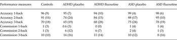
Across all subjects there was a significant linear WM load effect on accuracy (F 2,53 = 49, p < 0.0001) and commission errors (F 2,53 = 104, p < 0.0001) under both placebo and fluoxetine (accuracy: F 2,53 = 35, p < 0.0001; commission errors: F 2,53 = 20, p < 0.0001). Accuracy and commission errors were lowest and highest in the more difficult conditions respectively. No group or drug by group interaction effects were observed. Within patients, repeated-measures ANOVA showed a trend for a drug effect for accuracy (F 1,32 = 3, p < 0.1) because both patient groups showed more accuracy under fluoxetine than under placebo (p < 0.08). No other effects were significant (Table 2).
Performance measures for the N-back WM task for healthy controls, ADHD and ASD boys

WM, Working memory; ADHD, attention deficit hyperactivity disorder; ASD, autism spectrum disorder.
Values are given as mean (standard deviation).
fMRI data
Movement
Repeated-measures ANOVAs using group as the independent factor and maximum x, y, z rotation and translation as repeated measures showed that there were no significant group by movement interaction effects in rotation (F 4,106 = 1, p = n.s.) or translation (F 4,106 = 2, p = n.s.). Nevertheless, to eliminate potential effects of even small, non-significant motion variance, the two motion parameters of rotation and translation in 3D Euclidean space were used as a covariate in the fMRI analyses.
Group brain activation maps
Findings are reported in the online Supplementary material and Supplementary Figs S2 and S3.
Between-group differences between controls and patients under placebo for 3-back
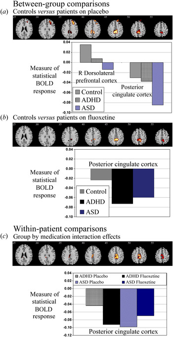
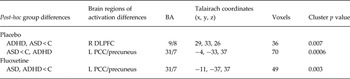
ANCOVA between controls and patients on placebo for the 3-back condition showed a significant group effect in the right DLPFC and the PCC (Fig. 1a , Table 3). Post-hoc analyses showed that the right DLPFC was significantly decreased in activation in the ASD (p < 0.0001) and ADHD (p < 0.05) groups relative to controls, and trend-wise in ASD compared to ADHD boys (p < 0.08). In the PCC, the ASD group had significantly increased deactivation compared to both controls (p < 0.0001) and ADHD participants (p < 0.005), both of which also deactivated this cluster but did not differ from each other (Fig. 1a , Table 3). Right DLPFC activation in the ASD group (which was reduced relative to controls) was negatively correlated with commission errors (r = −0.6, p < 0.05). No other significant correlations were observed.
Between-group and within-patient comparisons. Axial sections show the between-group ANCOVA findings between controls and patients under (a) placebo and (b) fluoxetine, and (c) the group by medication interaction effects within the two patient groups. Also shown are the statistical measures of the blood oxygen level-dependent (BOLD) response for (a, b) each of the three groups for each of the brain regions that showed a significant group effect and (c) each of the brain regions that showed a significant group by medication interaction effect within patients. ADHD, Attention deficit hyperactivity disorder; ASD, autism spectrum disorder; R, right. Talairach z coordinates are indicated for slice distance (in mm) from the intercommissural line. The right side of the image corresponds to the right side of the brain.

Brain activation differences between controls and patients on placebo or fluoxetine

BA, Brodmann area; R, right; L, left, DLPFC, dorsolateral prefrontal cortex; PCC, posterior cingulate cortex.
Between-group differences between controls and patients under fluoxetine for 3-back
ANCOVA between controls and patients on fluoxetine showed a significant group effect in the PCC, as observed under placebo, but not in the DLPFC (Fig. 1b , Table 3). Post-hoc analyses revealed that both ADHD (p < 0.0001) and ASD boys (p < 0.008) deactivated the PCC more than controls.
Repeated-measures t tests to test for significant normalization showed that fluoxetine relative to placebo significantly increased activation in right DLPFC in the ASD group only (p < 0.007), whereas in the ADHD group this was not significant. However, fluoxetine relative to placebo significantly increased the deactivation of the PCC in ADHD (p < 0.05) whereas PCC activation changes were not significant for ASD.
Correlations with behaviour showed that there was a significant positive correlation between PCC activation and scores on the Hyperactive/Inattentive subscale of the SDQ in the ADHD group (r = 0.6, p < 0.05) and PCC activation and scores on the Social and Communication subscale of the ADOS in the ASD group (r = 0.6, p < 0.05), suggesting that less deactivation of the PCC is associated with more severe ADHD and ASD symptoms. The findings suggest that fluoxetine is more efficacious in the deactivation of the PCC, an area of the DMN, in boys who show less severe forms of ADHD and ASD. No significant correlations between brain activation and IQ were observed.
Within-patients group by medication interaction effects
ANCOVA showed a significant group by medication interaction effect in the PCC [74 voxels, peak Talairach coordinates (x, y, z): 11, − 30, 37; Brodmann area (BA): 31/7] due to fluoxetine increasing deactivation of this area in the ADHD group and attenuating its deactivation in the ASD group (p < 0.005) (Fig. 1c ). No significant correlations between brain activation and behaviour or IQ were observed.
Discussion
Under placebo, while performing a WM task, ADHD and ASD boys shared underactivation relative to controls in the right DLPFC, a key area for WM (Wager & Smith, Reference Wager and Smith2003), which at trend level was more severe in ASD relative to ADHD. Furthermore, ASD boys showed disorder-specific increased deactivation of the PCC compared to controls and ADHD boys. Fluoxetine, at a trend level, improved performance in both disorders relative to placebo, but had a disorder-dissociated effect on task-positive and task-negative activations. Fluoxetine significantly normalized the right DLPFC underactivation in ASD whereas it only non-significantly ameliorated it in ADHD. However, fluoxetine did significantly increase the deactivation of the PCC DMN region in ADHD boys while decreasing it in ASD. Thus, we show shared deficits in both disorders (DLPFC underactivation), albeit more pronounced in ASD, and also disorder-specific differences (deactivation of the PCC in ASD). Furthermore, we show disorder-dissociated drug effects on task-positive and task-negative activations, with fluoxetine normalizing task-positive right DLPFC underactivation in ASD but enhancing task-negative PCC deactivation in ADHD, both of which may have contributed to the trend for improving WM performance.
The shared underactivation relative to controls in the right DLPFC, a key area of manipulation of information during WM (Wager & Smith, Reference Wager and Smith2003), during a verbal N-back task replicates previous findings of right DLPFC underactivation in ADHD children during a verbal N-back task (Cubillo et al. Reference Cubillo, Smith, Barrett, Giampietro, Brammer, Simmons and Rubia2014) and in an fMRI meta-analysis of attention tasks (Hart et al. Reference Hart, Radua, Nakao, Mataix-Cols and Rubia2013). The finding shows for the first time that DLPFC underactivation in adult ASD during a verbal N-back task (Koshino et al. Reference Koshino, Carpenter, Minshew, Cherkassky, Keller and Just2005) is also observed in children with ASD and, furthermore, that this is shared with ADHD and is trend-wise even more impaired. The finding of shared right DLPFC deficits in ADHD and ASD extends prior evidence for shared reduction in left DLPFC activation during a parametric sustained attention task (Christakou et al. Reference Christakou, Murphy, Chantiluke, Cubillo, Smith, Giampietro, Daly, Ecker, Robertson, Murphy and Rubia2013) to the right-hemispheric homologous area, suggesting that DLPFC dysfunction during attention tasks may be shared between the two disorders and mediate the cognitive overlap of attention and WM problems (Rommelse et al. Reference Rommelse, Geurts, Franke, Buitelaar and Hartman2011).
The disorder-specific increased deactivation of the PCC, a key region of the DMN, in ASD boys compared to control and ADHD boys may have been a compensation for their DLPFC underactivation, given that deactivation of the DMN is typically anti-correlated with task-positive activation in the DLPFC (Christakou et al. Reference Christakou, Murphy, Chantiluke, Cubillo, Smith, Giampietro, Daly, Ecker, Robertson, Murphy and Rubia2013) and associated with better task performance (Northoff et al. Reference Northoff, Qin and Nakao2010).
Under fluoxetine, right DLPFC underactivation appeared normalized in both groups, but rigorous normalization testing showed significance only for ASD. However, fluoxetine led to an increased deactivation of the PCC in ADHD that, while not different from controls under placebo, was now significantly more pronounced relative to controls under fluoxetine. This disorder-differential effect of fluoxetine on a task-positive area in ASD (the DLPFC) and a task-negative DMN area in ADHD (the PCC) suggests that fluoxetine had a positive effect on performance and brain activation in both disorders but through different underlying mechanisms, by decreasing task-unrelated self-referential thinking processes mediated by the DMN in ADHD, which has been shown to interfere with task-positive activation and lead to poor inattention in the disorder (Christakou et al. Reference Christakou, Murphy, Chantiluke, Cubillo, Smith, Giampietro, Daly, Ecker, Robertson, Murphy and Rubia2013), and by upregulating and normalizing a key task-relevant prefrontal area of WM in ASD.
The disorder-specific normalization effects on DLPFC activation and the inverse upregulation/downregulation effects on the PCC in ADHD/ASD respectively may be due to differences in the underlying biochemical abnormalities in the two disorders. Approximately 30% of individuals with ASD have enhanced platelet 5-HT levels (Hranilovic et al. Reference Hranilovic, Bujas-Petkovic, Vragovic, Vuk, Hock and Jernej2007) and reduced binding to the serotonin reuptake transporter and 5-HT2A transporter in the frontal lobe and PCC, which have been linked to poor social communication (Murphy et al. Reference Murphy, Daly, Schmitz, Toal, Murphy, Curran, Erlandsson, Eersels, Kerwin, Ell and Travis2006; Nakamura et al. Reference Nakamura, Sekine, Ouchi, Tsujii, Yoshikawa, Futatsubashi, Tsuchiya, Sugihara, Iwata, Suzuki, Matsuzaki, Suda, Sugiyama, Takei and Mori2010). Hyperserotonaemia may hence be an adaptation to counteract poor 5-HT receptor binding. The increase in 5-HT with fluoxetine may have increased ligand–receptor binding sufficiently to enhance activation in areas where 5-HT receptor density is typically low (Murphy et al. Reference Murphy, Daly, Schmitz, Toal, Murphy, Curran, Erlandsson, Eersels, Kerwin, Ell and Travis2006; Nakamura et al. Reference Nakamura, Sekine, Ouchi, Tsujii, Yoshikawa, Futatsubashi, Tsuchiya, Sugihara, Iwata, Suzuki, Matsuzaki, Suda, Sugiyama, Takei and Mori2010). This extends prior evidence that chronic fluoxetine can increase metabolic activity in prefrontal areas in a small group of adults with ASD, which was associated with an improvement in ASD behaviour (Buchsbaum et al. Reference Buchsbaum, Hollander, Haznedar, Tang, Spiegel-Cohen, Wei, Solimando, Buchsbaum, Robins, Bienstock, Cartwright and Mosovich2001).
Conversely, in ADHD there is evidence for lower platelet 5-HT levels compared to controls (Spivak et al. Reference Spivak, Vered, Yoran-Hegesh, Averbuch, Mester, Graf and Weizman1999) and biochemical serotonergic abnormalities (Oades, Reference Oades2007, Reference Oades2008, Reference Oades2010). Furthermore, serotonergic genotypes are implicated in ADHD and in responsiveness to stimulant medication (Gizer et al. Reference Gizer, Ficks and Waldman2009; Sonuga-Barke et al. Reference Sonuga-Barke, Kumsta, Schlotz, Lasky-Su, Marco, Miranda, Mulas, Oades, Banaschewski and Mueller2011). Therefore, the increase in 5-HT induced by fluoxetine may not have been sufficient to normalize their DLPFC deficit relative to controls. The increased deactivation of the PCC in ADHD under fluoxetine may be due to the regulatory role of 5-HT as 5-HT1A autoreceptors and heteroreceptors on gamma-aminobutyric acid (GABA)/glutamate neurons have been shown to modulate PCC deactivation in healthy controls during resting-state fMRI, with lower levels of 5-HT1A autoreceptor binding/more serotonin release being associated with increased deactivation of the PCC (Hahn et al. Reference Hahn, Wadsak, Windischberger, Baldinger, Höflich, Losak, Nics, Philippe, Kranz, Kraus, Mitterhauser, Karanikas, Kasper and Lanzenberger2012).
Despite significant normalization effects on brain activation, fluoxetine only improved WM performance at a trend level in patients relative to placebo. Practice effects in patients may have overshadowed performance effects, as controls were only tested once. In addition, brain function is more sensitive to pharmacological manipulations than WM performance (Allen et al. Reference Allen, Cleare, Lee, Fusar-Poli, Tunstall, Fu, Brammer and McGuire2006; Rose et al. Reference Rose, Simonotto, Spencer and Ebmeier2006; Cubillo et al. Reference Cubillo, Smith, Barrett, Giampietro, Brammer, Simmons and Rubia2014), which could be due to insufficient statistical power for neuropsychological but not fMRI effects (Thirion et al. Reference Thirion, Pinel, Meriaux, Roche, Dehaene and Poline2007) in a sample of 17 subjects.
The strengths of this study lie in the carefully selected and non-co-morbid patient groups with ADHD and with ASD who were free of psychiatric co-morbidities and, in the case of the ASD group, were medication naïve. A limitation is that, for ethical reasons, the control group was only scanned once whereas patients were scanned twice. The significantly lower IQ in the ADHD group is another limitation. However, low IQ is intrinsically associated with ADHD (Bridgett & Walker, Reference Bridgett and Walker2006) and it is therefore difficult to separate IQ and diagnosis effects. Moreover, brain activation did not correlate with IQ, suggesting that IQ did not to play a key role. The relatively high IQ of our ASD sample also reduces the generalizability of the findings. In this study we were interested in acute effects of fluoxetine to control for chronic effects on behavioural improvement. However, future studies of chronic fluoxetine administration in ADHD and ASD are essential to assess the clinical relevance of these findings.
To summarize, ADHD and ASD patients shared underactivation in the right DLPFC, a key area for WM, with ASD patients showing more pronounced deactivation of the PCC relative to both groups. Fluoxetine had a disorder-dissociated region-specific effect of significantly normalizing the task-positive DLPFC dysfunction in ASD and enhancing task-negative deactivation in the PCC in ADHD, both of which were concomitant with a trend-level improved WM task performance relative to placebo. The region-specific disorder-dissociated effects of fluoxetine may be due to differing biochemical abnormalities underlying the two disorders.
Supplementary material
For supplementary material accompanying this paper visit http://dx.doi.org/10.1017/S0033291714002232.
Acknowledgements
This study was supported by the UK Department of Health through the National Institute for Health Research (NIHR) Biomedical Research Centre (BRC) for Mental Health at the South London and the Maudsley National Health Service (NHS) Foundation Trust and the Institute of Psychiatry, King's College London.
Declaration of Interest
K.R. has received funding from Lilly for another project and consultancy fees from Lilly, Shire and Novartis. The other authors have no conflicts of interest to declare.