Salmonella are Gram-negative bacteria that are capable of infecting a wide range of host species from mammals to birds and reptiles. Infection with Salmonella usually occurs as a result of consuming contaminated food or water. There are two main clinical syndromes associated with Salmonella infection: non-typhoidal salmonellosis – a gastrointestinal disease also known as enteritis; and the systemic disease typhoid fever. The nature and severity of disease depends on the Salmonella serotype and the host species involved. There are over 2500 serotypes of Salmonella, and in many cases infection usually results in a mild and self-limiting disease with symptoms that may include nausea, vomiting, diarrhoea and abdominal cramps. However, in some immunocompromised individuals a severe bacterial infection can result, including the development of life-threatening septicaemia. The more severe typhoid fever is caused by Salmonella enterica serovar Typhi (S. Typhi), which is a predominantly human pathogen. Mortality rates in untreated typhoid fever infections can be 10–15%. In some instances patients recover but remain carriers of the bacteria for many years.
Salmonella infections in humans and food-producing animals cause substantial economic and welfare problems worldwide each year. In England and Wales alone, there were 11 447 recorded cases of non-typhoidal salmonellosis in 2005 (http://www.hpa.org.uk/infections/topics_az/Salmonella/data_human.htm). However, this is likely to be a massive underestimate of the actual number of Salmonella infections because the figure represents only those individuals who tested positive following a visit to a doctor. A large proportion of people will have been ill with enteritis, but as the disease is of a relatively short duration, they will have recovered without seeking medical attention.
During the infection process, Salmonella survive the acidic conditions of the stomach and pass into the intestine where they interact with epithelial cells lining the intestinal mucosa. It is through interactions with these cells that Salmonella initiates the disease process. This review focuses on how Salmonella causes enteritis at the molecular level and how understanding the nature of the Salmonella–host interactions can impact on the development of therapeutics.
Model systems for studying Salmonella-induced enteritis
There are many in vivo and in vitro model systems that together have provided a wealth of data on the pathogenesis of enteritis (enteropathogenesis) induced by Salmonella. The bovine ligated intestinal loop model system (bovine ligated loop model) has been used for studying early events in Salmonella enteropathogenesis for more than a decade. In this model, the mid-ileum of a terminally anaesthetised calf is ligated into up to 36 individual loops. Salmonella strains are then injected in triplicate and enteropathogenic responses such as recruitment of neutrophils and fluid secretion into the discrete loops can then be measured. This system allows a large number of strains or conditions, including internal controls, to be studied in triplicate in a single animal, thereby reducing inter-animal variation. This model showed for the first time the pivotal importance of genes within the chromosomal region Salmonella pathogenicity island 1 (SPI-1) in intestinal invasion in vivo (Ref. Reference Watson1).
Recently a modified bovine ligated loop model has been developed to permit longer (5-day) studies of the interaction between Salmonella and host intestines (Ref. Reference Coombes2). As yet, there are limited published data available from this model, but they do suggest distinct temporal roles for different Salmonella virulence factors in vivo (Ref. Reference Coombes2) and they support data obtained from the streptomycin-treated mouse model (described below).
Traditionally, mice were not used for enteropathogenesis studies because infection of mice with Salmonella enterica serovar Typhimurium (S. Typhimurium) – a serotype that causes enteritis in humans and ruminants – results in a systemic disease similar to typhoid fever. However, it has been known for almost half a century that pre-treatment of mice with the antibiotic streptomycin enhances susceptibility of the mice to Salmonella infection (Refs Reference Bohnhoff3, Reference Bohnhoff4) and recently it was found that the streptomycin-pretreated mice developed acute intestinal inflammation following oral inoculation with S. Typhimurium (Ref. Reference Barthel5). The streptomycin pre-treatment disrupts the commensal gut microflora, allowing the pathogen to colonise the intestine. Using this model, it has been shown that two regions of the Salmonella chromosome associated with virulence – SPI-1 and SPI-2 – play a role in Salmonella-induced enteritis in mice (Refs Reference Coburn6, Reference Hapfelmeier7). There are many immunological ‘tools’ and reagents available for mouse experiments compared with a somewhat limited number of bovine reagents. Coupled with the fact that technology for developing knockout animals is much more advanced and cost-effective for mice than cattle, the streptomycin mouse model allows additional studies to be performed – particularly on host-cell responses occurring during Salmonella-induced enteritis. For example, MyD88-knockout mice, which lack an adaptor molecule required for Toll-like-receptor signalling in the innate immune response, have been used to characterise two different mechanisms used by Salmonella to trigger enteritis (Ref. Reference Hapfelmeier8).
Oral challenge models using S. Typhimurium in calves are another in vivo system for studying Salmonella-induced enteritis and provide relevant data because they reflect the natural infection process occurring with this serotype in humans. The role of various genes in Salmonella virulence can be assessed by using strains containing defined mutations and comparing clinical outcomes. invH is one gene that was mutated and assessed in this system and was found to reduce the severity of Salmonella-induced enteritis (Ref. Reference Watson9).
In vitro systems have also yielded informative results. One particularly useful in vitro system is the polarised epithelial cell model, where intestinal epithelial cell lines (e.g. T84 or CACO-2) are cultured in a monolayer on a permeable support. The polarised monolayer mimics the situation in vivo where epithelial cells express certain receptors only at one pole of the cell. This system has been used to identify the host and bacterial factors driving recruitment of neutrophils to the site of Salmonella infection (Refs Reference Criss10, Reference Lee11, Reference McCormick12, Reference McCormick13). The cell monolayers are infected apically with Salmonella and neutrophils isolated from donor blood are added basolaterally. Movement of the neutrophils across the epithelial layer can then be assessed. The polarised epithelial cell system has also recently been used to identify bacterial proteins required for the disruption of tight junctions between the epithelial cells (Ref. Reference Boyle, Brown and Finlay14) – a process thought to lead to diarrhoea during Salmonella-induced enteritis. Nonpolarised cultured cells have also provided information about Salmonella–host-cell interactions. For example, many of the initial studies on cytoskeletal changes accompanying Salmonella invasion were performed on COS cells (Refs Reference Hardt15, Reference Stender16), presumably because these cells express genes from eukaryotic expression vectors at high levels and can be readily attained, labelled and visualised.
Molecular basis of Salmonella-induced enteritis
The use of animal and cell culture models has greatly improved the understanding of the molecular mechanisms of Salmonella-induced enteritis. The initial interaction of enteritis-inducing Salmonella serotypes with the host occurs within the intestine where, following ingestion, Salmonella uses a highly specialised machinery termed the type III secretion system 1 (T3SS-1) – often likened to a ‘molecular syringe’ – to interact with intestinal epithelial cells. T3SSs are found in many pathogenic Gram-negative bacteria and are used to secrete and translocate a range of proteins, termed effectors, from the bacteria directly into the host cell cytosol (reviewed in Ref. Reference Galan and Wolf-Watz17). Salmonella itself possesses two T3SSs encoded by the large chromosomal DNA SPIs. To date, 14 SPIs have been identified (reviewed in Ref. Reference Morgan, Rhen, Maskel, Mastroeni and Threlfall18). T3SS-1 encoded by SPI-1 is responsible for invasion of host cells and inflammatory responses occurring in the initial stages of infection, whereas the SPI-2-encoded T3SS-2 is used later in the infection process, promoting intracellular survival and replication within the host.
T3SSs are multiprotein complexes, requiring a number of different sub-classes of proteins: structural proteins, regulatory proteins, and secreted proteins and their chaperones. SPI-1 encodes more than 30 different proteins. Regulatory proteins control the expression and secretion of various T3SS-1 proteins. The secretion organelle, termed the needle complex, spans the inner and outer bacterial membranes and comprises at least five structural proteins. Needle complexes have been purified in vitro (Ref. Reference Kubori19) and the narrow diameter of the needle suggests that proteins delivered by the system may be in a partially unfolded conformation. T3SS chaperone proteins regulate the secretion of proteins delivered via the needle complex (reviewed in Ref. Reference Page and Parsot20). Chaperones are believed to interact with, stabilise and maintain their substrate protein in a secretion-competent state, and prevent premature association of the substrate with interacting proteins. Some chaperones are associated with only one substrate, whereas others are promiscuous and have multiple substrate proteins. For example, SicP is the chaperone for only one protein (Ref. Reference Fu and Galán21), whereas InvB chaperones at least four (Refs Reference Ehrbar22, Reference Ho Lee and Galan23, Reference Bronstein, Miao and Miller24, Reference Ehrbar25). Chaperone release and unfolding of the cognate substrate protein immediately before secretion via the needle complex is induced by InvC, the ATPase associated with T3SS-1 (Ref. Reference Akeda and Galan26).
Proteins secreted via the assembled T3SS-1 can be divided into two subsets: translocators and effectors. The three translocator proteins – the Salmonella invasion proteins (Sips) B, C and D –localise at the tip of the needle complex and are believed to form a pore in the host cell membrane, facilitating the delivery of effector proteins into the host cell (Ref. Reference Scherer, Cooper and Miller27). Translocators can also have an effector function; for example, SipC modulates the actin cytoskeleton (Ref. Reference Hayward and Koronakis28). The class of proteins known as effectors are secreted into the host cell where they modulate host-cell pathways and functions for the benefit of the pathogen. To date, ten T3SS-1 effectors have been identified; some belong to a family of molecules known as Salmonella outer proteins (Sops), whereas others are named according to their structural or functional features. For example, AvrA is named because it shares sequence homology with an avirulence determinant of a plant pathogen (Ref. Reference Hardt and Galán29) and SptP is Salmonella protein tyrosine phosphatase (Ref. Reference Kaniga30). Some T3SS-1-secreted effector molecules, such as SptP, are encoded within SPI-1, whereas others are located elsewhere on the chromosome. For example, SopE is encoded within a temperate bacteriophage (Ref. Reference Hardt, Urlaub and Galán31) and SopB is encoded within SPI-5 (Ref. Reference Wood32).
T3SS effectors and enteritis
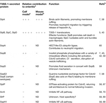
Several T3SS-1 effectors have been extensively studied, and the function of these proteins and their relative contribution to Salmonella-induced enteritis are summarised in Table 1 and reviewed below.
Proteins secreted by Salmonella type III secretion system 1 and their relative contribution to enteritis

a++++, major role; +++, moderate role; ++, intermediate role; +, slight role; –, no role; ND, not determined.
bReferences refer to articles where effect of gene mutation on enteritis in vivo is tested.
cAssessed in calves:ligated loops or oral challenge.
cAssessed in streptomycin-treated mouse model.
SopE and SopE2
Invasion of epithelial cells is an early step in the infection process. Salmonella induces its own uptake into non-phagocytic cells through the activity of the effector proteins SopE and SopE2. SopE, the first identified substrate of T3SS-1, was isolated from the supernatants of S. Dublin and S. Typhimurium cultures and found to be required for efficient entry of Salmonella into host cells (Refs Reference Hardt, Urlaub and Galán31, Reference Wood33). Subsequent work found that SopE induces cellular changes, such as membrane ruffling and actin cytoskeletal rearrangements, within the host cells in a manner dependent on members of the Rho family of GTPases – particularly Cdc42 and Rac1 (Ref. Reference Hardt15). This family of G proteins cycles between GTP-bound (active) and GDP-bound (inactive) conformations, regulated by the activity of guanine-nucleotide-exchange factors (GEFs), GTPase-activating proteins (GAPs) and guanine-nucleotide-dissociation inhibitors (GDIs). SopE, found in some but not all Salmonella serotypes (Refs Reference Hardt, Urlaub and Galán31, Reference Mirold34), and the highly homologous and more widely distributed SopE2, function as GEFs (Refs Reference Stender16, Reference Bakshi35). Both SopE and SopE2 activate Cdc42, and SopE additionally acts on Rac1, demonstrating the specificity of individual effectors (Ref. Reference Friebel36). SopE is associated with some epidemic strains of Salmonella, which may suggest a specific role for this effector in the emergence of epidemic strains (Ref. Reference Mirold34).
SptP
The membrane ruffling and cytoskeletal rearrangements stimulated by SopE and SopE2 are reversible, with infected cells regaining their architecture shortly after bacterial internalisation through the antagonistic effect of SptP. This T3SS-1 effector has two distinct enzymatic activities at its N- and C-termini. The N-terminus of SptP shares homology with two other bacterial toxins: ExoS from Pseudomonas aeruginosa and YopE from Yersinia (Ref. Reference Kaniga30). The SptP C-terminus is homologous to the tyrosine phosphatase catalytic domain of Yersinia YopH – hence the name of this Salmonella effector (Ref. Reference Kaniga30). In vitro, cells infected with an sptP mutant continued to show membrane ruffling even after bacterial internalisation (Ref. Reference Fu and Galán37), and microinjection of SptP alone disrupted the actin cytoskeleton (Ref. Reference Fu and Galán38). Co-injection of SptP and SopE did not cause any cytoskeletal changes within cells (Ref. Reference Fu and Galán37), thus demonstrating the contrasting and antagonistic effects of these two effector molecules (Ref. Reference Fu and Galán37). The SptP-induced actin cytoskeletal changes were independent of the tyrosine phosphatase activity of SptP (Ref. Reference Fu and Galán37), but were dependent on its ability to directly bind the GTP-bound (active) form of the Rho GTPase Rac1 (Ref. Reference Fu and Galán37). SptP therefore acts as a GAP for Cdc42 and Rac1, stimulating the conversion of GTP to GDP and downregulating signalling through the Rho proteins. X-ray crystallography studies of SptP bound to Rac1 revealed that SptP mimics eukaryotic GAP proteins in the host cell (Ref. Reference Stebbins and Galán39). Despite the distinctive function in invasion, SptP does not play a role in the induction of enteropathogenic responses in vivo.
SopB
An extensively studied and well-characterised T3SS-1 effector protein that does contribute to Salmonella-induced enteritis is SopB, also known as SigD (Refs Reference Galyov40, Reference Hong and Miller41). A sopB mutant induces less fluid secretion and inflammatory responses (neutrophil influx) in the bovine ligated loop model compared with wild-type bacteria (Ref. Reference Galyov40); however, enteropathogenesis is not totally abolished as it is with a T3SS-1 mutant such as sipB (Ref. Reference Galyov40). SopB is an inositol phosphate phosphatase (Ref. Reference Norris42) capable of hydrolysing phosphate groups on various inositol phosphate substrates (Ref. Reference Norris42). There has been much work, with some conflicting results, on the biological significance of the inositol phosphate phosphatase function of SopB in host cells.
SopB has been shown to activate Akt, a serine/threonine kinase (Ref. Reference Steele-Mortimer43), and a region at the C-terminus of SopB, which is homologous to the inositol 5-phosphatase synaptojanin, is required for Akt activation (Ref. Reference Marcus44). Akt is also known as protein kinase B (PKB) and plays an important role in cell-signalling pathways, regulating several cellular functions including proliferation, survival and apoptosis. Akt possesses a pleckstrin homology (PH) domain at the N-terminus, which interacts with the second messenger molecule phosphatidylinositol (Reference Bohnhoff3,Reference Bohnhoff4,Reference Barthel5)-trisphosphate [PtdIns(Reference Bohnhoff3,Reference Bohnhoff4,Reference Barthel5)P 3], generated by phosphoinositide 3-kinase (PI 3-kinase). PtdIns(Reference Bohnhoff3,Reference Bohnhoff4,Reference Barthel5)P 3 itself does not directly activate Akt, but recruits Akt from the cytoplasm to the plasma membrane and induces a conformational change that allows phosphorylation and activation of Akt by phosphoinositide-dependent kinase 1 and 2 (PDK1 and PDK2). In the case of Salmonella infection, it is hypothesised that Akt is recruited to membrane ruffles that have a high concentration of PtdIns(Reference Bohnhoff3,Reference Bohnhoff4,Reference Barthel5)P 3 in a SopB-independent manner (Ref. Reference Marcus44). SopB dephosphorylates PtdIns(Reference Bohnhoff3,Reference Bohnhoff4,Reference Barthel5)P 3 to phosphatidylinositol (Reference Bohnhoff3,Reference Bohnhoff4)-bisphosphate [PtdIns (Reference Bohnhoff3,Reference Bohnhoff4,)P 2] – a lipid that can also recruit Akt, resulting in conformational change and full activation of Akt (Ref. Reference Marcus44). Since Akt is involved in cellular survival, it is hypothesised that activation of Akt would allow Salmonella more time within the cell in which to replicate (Ref. Reference Steele-Mortimer43).
Through its phosphatase activity, SopB is also able to induce membrane ruffling and actin cytoskeletal changes via Cdc42 (Ref. Reference Zhou45). However, additional studies indicate that the action of SopB on actin is dependent on the N-terminus of SopB and independent of its phosphatase activity (Ref. Reference Alemán46), suggesting a bifunctional role for SopB. Most recently, experimental evidence suggests that SopB, through its inositol phosphate phosphatase activity, disrupts vesicular trafficking in the endocytic pathway in host cells by preventing accumulation of PtdIns(Reference Bohnhoff3,Reference Barthel5)P 2 (Refs Reference Dukes47, Reference Hernandez48) – an inositol phosphate required for multiple membrane-trafficking pathways. Given the array of cellular signalling pathways involving inositol phosphates, it is not surprising that a number of functions are attributed to SopB. The challenge remains to determine the true significance of SopB in the context of Salmonella infection in vivo.
SipA
The multifunctionality of T3SS effector proteins is further illustrated by the case of SipA, which was originally identified more than a decade ago to be encoded within SPI-1, along with SipB, SipC and SipD (Refs Reference Hueck49, Reference Kaniga, Trollinger and Galán50). SipA was not originally thought to be required for Salmonella invasion of host cells (Refs Reference Hueck49, Reference Kaniga, Trollinger and Galán50), but later studies indicated a role in invasion soon after infection (Ref. Reference Jepson, Kenny and Leard51). SipA was subsequently found to modulate the host cell cytoskeleton by: (Reference Watson1) binding F-actin, thereby preventing depolymerisation of actin filaments (Refs Reference McGhie, Hayward and Koronakis52, Reference Zhou, Mooseker and Galán53); (Reference Coombes2) decreasing the critical concentration of G-actin required for polymerisation of actin filaments (Ref. Reference Zhou, Mooseker and Galán53); and (Reference Bohnhoff3) binding fimbrin (also known as T-plastin), which increased the actin-bundling (crosslinking) activity of this protein (Ref. Reference Zhou, Mooseker and Galán54). X-ray crystallography and electron microscopy studies revealed that SipA acts like a ‘molecular staple’, binding actin filaments via two non-globular protein arms (Ref. Reference Lilic55). By binding actin filaments, SipA displaces and inhibits the proteins cofilin and ADF (Ref. Reference McGhie, Hayward and Koronakis56), which themselves bind actin filaments to promote depolymerisation. Another actin-binding protein, gelsolin, which severs and caps actin filaments, is also inhibited by SipA (Ref. Reference McGhie, Hayward and Koronakis56). Through these actin-modulation activities, SipA contributes to the membrane-ruffling process and enhances the activities of SipC (Ref. Reference McGhie, Hayward and Koronakis52).
In addition to the role of SipA in modulating the actin cytoskeleton, this effector has also been shown to be involved in neutrophil transmigration across epithelial monolayers in a protein kinase C (PKC)-dependent manner (Ref. Reference Lee11). SipA is believed to activate a signal transduction pathway involving ADP-ribosylation factor 6 (ARF6), resulting in PKC activation and culminating in the release of pathogen-elicited epithelial chemoattractant (PEEC) (subsequently identified as hepoxilin A3) (Ref. Reference Mrsny57) from the apical side of epithelial cells (Ref. Reference Criss10). PEEC/hepoxilin A3 coupled with the cytokine IL-8 drives neutrophil movement across the intestinal epithelium (Ref. Reference McCormick13). In vivo, SipA elicits secretory and inflammatory responses in the bovine ligated loop model (Ref. Reference Zhang58) and inflammatory responses in the streptomycin-treated mouse model (Ref. Reference Hapfelmeier7).
SopD
A number of other T3SS-1 effector proteins have been identified and shown to play a role in Salmonella-induced enteritis, but their mechanisms of action remains unclear. SopD is not required for invasion of host cells, but like sopB mutants, sopD mutants induce less fluid secretion and neutrophil influx compared with the wild type in the bovine ligated loop model (Ref. Reference Jones59). Interestingly, a double sopB–sopD mutant induced significantly less fluid secretion and neutrophil influx compared with the single mutants, indicating that the effector proteins act in concert (Ref. Reference Jones59).
SopA
SopA is also involved in the induction of enteritis in the bovine ligated loop model (Ref. Reference Wood60), and was found to induce neutrophil transmigration across polarised T84 epithelial cell monolayers in vitro (Ref. Reference Wood60). Recent data suggest that SopA is ubiquitylated and degraded by a host cell pathway and this activity may be involved in the escape of Salmonella from vacuoles in the host cell (Ref. Reference Zhang61). Amino acid residues at the N-terminus of SopA have been shown to be required for SopA targeting to the mitochondria of host cells (Ref. Reference Layton, Brown and Galyov62); the biological significance of this targeting is unknown. Additionally, SopA itself is a HECT-like E3 ubiquitin ligase (Ref. Reference Zhang63). This activity is important for neutrophil transepithelial migration because a catalytically inactive SopA induced less neutrophil movement than wild-type Salmonella in vitro (Ref. Reference Zhang63). Taken together these data suggest that SopA demonstrates molecular mimicry of a host cell protein and is an important T3SS effector in the induction of inflammatory responses during Salmonella infection.
SipB and SipC
SipB and SipC are T3SS-1 translocators required for the delivery of effector proteins into host cells, but they also display effector functions themselves. SipC contributes to cytoskeletal rearrangements by nucleating actin and bundling actin filaments (Ref. Reference Hayward and Koronakis28), which is enhanced by the activity of SipA (Ref. Reference McGhie, Hayward and Koronakis52). The translocation and effector protein functions of SipC are attributed to distinct regions of the protein (Ref. Reference Chang, Chen and Zhou64). SipB, in its role as an effector molecule, induces two distinct types of programmed cell death in macrophages. The first is a rapid, necrotic-like cell death, dependent on the protease caspase-1 (Ref. Reference Hersh65). In the second type of macrophage cell death, which is independent of caspase-1, SipB induces autophagy by disrupting mitochondria in the macrophages (Ref. Reference Hernandez66). In animal models, SipB abolishes the induction of enteritis as a result of its essential role as a translocator (Refs Reference Galyov40, Reference Tsolis67).
Other T3SS-1 effectors
Other T3SS-1 effectors appear to play no role in enteropathogenesis because mutants behave like the wild-type strain in terms of diarrhoeal symptoms and mortality in bovine oral infection models (Refs Reference Tsolis67, Reference Miao68, Reference Tsolis69) and do not induce secretory or inflammatory responses in the bovine ligated loop model (Refs Reference Zhang58, Reference Schesser70). This collection of proteins includes: SptP, which is described in detail above; AvrA, which inhibits the pro-inflammatory NF-κB pathway in vitro (Refs Reference Hardt and Galán29, Reference Collier-Hyams71); SspH1, which can be secreted by both T3SS-1 and T3SS-2, is localised to the host cell nucleus and inhibits NF-κB-dependent gene expression by interacting with protein kinase N1 (Refs Reference Haraga and Miller72, Reference Haraga and Miller73); and SlrP, whose function is unknown, but is suggested to be involved in host adaptation because an slrP mutant was found to have a colonisation defect in mice but not calves (Ref. Reference Tsolis69).
Other bacterial factors
T3SS-1 and its associated effector proteins are not the only bacterial factors contributing to enteritis. Other bacterial factors involved include lipopolysaccharide (LPS) and flagellin. Both of these factors, found on the bacterial surface, are recognised by the host immune system and trigger cell-signalling pathways and responses that contribute to Salmonella-induced enteritis. The mechanisms by which flagellin and LPS contribute to enteritis are discussed in more detail below. Interestingly, flagellin engages in a cooperative interaction with the T3SS effector SopE2 whereby flagellin facilitates translocation of SopE2 into host cells and SopE2 enhances signalling pathways activated by flagellin (Ref. Reference Huang74).
Host factors contributing to enteritis
Induction of enteritis by Salmonella is a complex process involving not only bacterial factors, but also host-cell molecules that are directly or indirectly activated by Salmonella. Such host-cell molecules include: sensors, such as Toll-like receptors; signals, such as cytokines; and cells of the immune system, such as neutrophils.
Toll-like receptors
Toll-like receptors (TLRs) (reviewed in Ref. Reference Janssens and Beyaert75) recognise specific ligands termed PAMPs (pathogen-associated molecular patterns) in pathogenic microorganisms. To date, 13 different mammalian TLRs have been identified and they recognise distinct PAMPs, including CpG DNA (TLR9), LPS (TLR4) and flagellin (TLR5). Activation of a TLR via interaction with its respective ligand triggers an intracellular signalling cascade (reviewed in Ref. Reference Akira and Takeda76), which ultimately results in expression of host defence products and generation of an innate immune response.
The flagellin–TLR5 interaction has been well studied in Salmonella. Many enteric bacteria possess flagella – organelles that rotate and provide the bacteria with a means of motility. The filament portion of a flagellum comprises polymerised flagellin subunits and the structure of the protein flagellin is highly conserved amongst diverse bacterial species. Flagellin secreted by Salmonella is a potent stimulator of pro-inflammatory responses such as NF-κB activation and iNOS expression (Ref. Reference Eaves-Pyles77), and was found to be translocated across intestinal epithelial cells in a manner dependent upon SPI-2 genes involved in vesicle trafficking (Refs Reference Gewirtz78, Reference Lyons79). The cellular receptor for flagellin, TLR5 (Ref. Reference Hayashi80), is found only on the basolateral side of polarised epithelial cells (Ref. Reference Gewirtz81). However, there is some controversy surrounding this observation because TLR5 expression on apical surfaces has been detected in mice (Ref. Reference Bambou108). It is suggested that polarised expression of TLR5 may prevent induction of pro-inflammatory gene expression by commensal bacteria because only bacterial pathogens able to transcytose flagellin would activate the TLR5 pathway. Flagellin and TLR5 signalling activates the NF-κB pathway, which drives proinflammatory responses such as cytokine expression. Recent data suggest that TLR5 signalling via NF-κB can also activate anti-apoptotic pathways in epithelial cells in vitro and in vivo (Refs Reference Feuillet82, Reference Vijay-Kumar83). The contribution of flagellin and TLR5 to enteric infections is discussed in a recent review (Ref. Reference Steiner84).
Cytokines
Host defence products generated through TLR activation and signalling include cytokines, which attract cells such as neutrophils and macrophages to the infection site. Cytokines induced during Salmonella infection have been extensively studied (reviewed in Ref. Reference Eckmann and Kagnoff85). Studies have mainly involved cultured intestinal epithelial cell lines from various species, including humans. The cells are often polarised to investigate the direction of cytokine release in response to Salmonella infection. IL-8 is one such cytokine that has been well studied in terms of Salmonella enteropathogenesis. IL-8 is a member of the CXC chemokine family and is produced by macrophages and epithelial cells – hence its importance in intestinal infections – and also attracts neutrophils. To reach the intestinal epithelium, which is the site of Salmonella infection, neutrophils have to leave the vasculature and traverse an endothelial layer and the lamina propria. IL-8, which is produced basolaterally by intestinal epithelial cells in response to Salmonella infection (Refs Reference McCormick12, Reference Eckmann, Kagnoff and Fierer86), drives the initial stage of the neutrophil migration to the subepithelial space by imprinting the subepithelial matrices with a chemotactic gradient (Ref. Reference McCormick87). IL-8 was unable to direct neutrophil migration in the final step across the intestinal epithelium. Instead, this stage of the process is driven by PEEC/hepoxilin A3, which, as discussed above, is released apically from intestinal epithelial cells (Ref. Reference McCormick13).
During the migratory process, damage can occur to the intestinal epithelium as neutrophils breach the tight junctions between epithelial cells. It is suggested that this damage can contribute to Salmonella enteropathogenesis. There is some controversy surrounding neutrophil migration during Salmonella infection as to whether the neutrophils are actively driven by Salmonella via T3SS-mediated effects, or alternatively whether the influx is driven by the host recognising PAMPs on the bacterial surface and responding accordingly. The arguments favouring both hypotheses are extensively discussed in a recent review by Tükel and colleagues (Ref. Reference Tükel88).
Fluid and ion secretion
Fluid secretion accompanies neutrophil influx into the Salmonella-infected intestine and is a host response that is characteristic of enteric infections. Secretory responses are believed to aid rapid elimination of bacteria from the intestine. Fluid secretion is regulated by ion secretion from intestinal epithelial cells – in particular chloride ion fluxes. Cl− secretion itself is controlled by stimulatory and inhibitory factors acting on intestinal epithelial cells. For example, epidermal growth factor (EGF) inhibits Cl− secretion via a PI3K-dependent pathway (Refs Reference Uribe89, Reference Uribe90). Salmonella infection causes an increase in intracellular inositol (Reference Watson1,Reference Bohnhoff4,Reference Barthel5,Reference Coburn6)-tetrakisphosphate, which indirectly increases Cl− secretion by antagonising the inhibitory effect of EGF (Ref. Reference Eckmann91). The T3SS-1 effector SopB plays a role in controlling the Cl− secretory responses, probably via its inositol phosphate phosphatase activity (Ref. Reference Bertelsen92). Through the action of SopB on secretory pathways, it appears that Salmonella is once again utilising a host pathway for its own benefit.
The secretory or diarrhoeal response is thought to be exacerbated by disruption of tight junctions in the intestinal epithelium. Tight junctions, which are located between adjoining enterocytes, are protein complexes that restrict paracellular movement of harmful materials and control movement of fluid and ions. Infection of epithelial monolayers with Salmonella disrupts tight junctions (Ref. Reference Jepson93) in a manner dependent on protein geranylgeranylation (Ref. Reference Tafazoli, Magnusson and Zheng94). Recently, the T3SS-1 effectors SopB, SopE, SopE2 and SipA were found to be responsible for S. Typhimurium disruption of tight junctions (Ref. Reference Boyle, Brown and Finlay14), demonstrating that the damage to the epithelial monolayer is a process actively induced by Salmonella. Although increased fluid secretion and Cl− secretion will undoubtedly result in removal of Salmonella from the intestine, it may be a mechanism used by the bacteria to provide it with access to the environment, allowing it to disseminate throughout the population.
Clinical implications and applications
Taken together these experimental data show that Salmonella has evolved successful mechanisms to cause enteric disease, with the T3SSs playing a major role (summarised in Fig. 1). The characterisation of effector proteins has led to the idea that Salmonella utilises the T3SS and its associated secreted proteins to hijack host-cell pathways and mimic host-cell proteins. The phenomenon of molecular mimicry by bacterial proteins is a relatively recent discovery with SptP being the first Salmonella T3SS effector to demonstrate such activity (Ref. Reference Stebbins and Galán39). Since this discovery, other Salmonella T3SS virulence factors have been shown to demonstrate molecular mimicry (reviewed in Ref. Reference Stebbins and Galan95).
Molecular events contributing to Salmonella-induced enteritis. (a) Salmonella injects effector proteins into intestinal epithelial cells via T3SS-1. (b) SopE2, a guanine-nucleotide-exchange factor, activates Cdc42, leading to actin cytoskeletal changes and membrane ruffling. SptP has an antagonistic effect, mimicking eukaryotic GTPase-activating proteins to return cell architecture to normal after bacterial internalisation. (c) SipA contributes to membrane ruffling by binding actin filaments, preventing depolymerisation. SipC nucleates actin and bundles actin filaments, enhanced by SipA. (d) Akt is recruited to PtdIns(3,4,5)P 3-rich membrane ruffles. SopB dephosphorylates PtdIns(3,4,5)P 3 to PtdIns(3,4)P 2, allowing activation of Akt, which is involved in various cell survival and signalling pathways. (e) SopB also contributes to membrane ruffling and chloride ion secretion. (f) SopD acts in concert with SopB to promote fluid secretion. Intracellular targets of SopD are unknown. (g) SipA recruits an ARF6 GEF such as ARNO to the apical plasma membrane. This results in activation of ARF6, triggering a signalling pathway that culminates in PKC recruitment, leading to the apical release of the chemoattractant hepoxilin A3. (h) Via a pathway dependent on SPI-2 genes, flagellin is translocated across epithelial cells to encounter basolaterally expressed TLR5. TLR5 activation stimulates NF-κB pathways leading to production and basolateral release of the chemokine IL-8. Together IL-8 and hepoxilin A3 drive neutrophil migration from the vasculature into the intestinal lumen. (i) SopA contributes to neutrophil migration via a mechanism dependent on its HECT-like E3 ubiquitin ligase activity. (j) SipA, SopB and SopE2 disrupt tight junctions between epithelial cells. Transepithelial migration of neutrophils also contributes to tight junction disruption. (k) SopB disrupts vesicle trafficking in epithelial cells by preventing accumulation of PtdIns(3,5)P 2. ARF6, ADP-ribosylation factor 6; ARNO, ADP-ribosylation factor nucleotide-binding site opener; DAG, diacylglycerol; IL-8, interleukin-8; PA, phosphatidic acid; PC, phosphatidylcholine; PKC, protein kinase C; PLD, phospholipase D; TLR5, Toll-like receptor 5.

Host-cell pathways hijacked by T3SS proteins include those involved in cytoskeleton rearrangements and various signal transduction pathways resulting in promotion of cell survival and activation of pro-inflammatory molecules. All these activities are believed to aid the success of Salmonella as a virulent pathogen, promoting invasion and survival within host cells. Interestingly, a small class of effectors, including AvrA and SspH1 described above, appear to have an opposing effect, targeting and inhibiting the NF-κB pathway, which presumably prevents activation of various pro-inflammatory cytokines and enables Salmonella to evade the host immune response and ‘hide’ within the host. Although some of these activities appear to be conflicting, it should be noted that secretion of effector proteins by T3SS is temporally regulated (Ref. Reference Kubori and Galan96), such that the effectors elicit their effects in a carefully coordinated manner.
Clinical treatment for Salmonella infection traditionally involved the use of antibiotics. However, the emergence and increase in antibiotic resistance in bacteria, often driven by the misuse of antibiotics, is a major concern to animal and human welfare. Conventional antibiotics target metabolic processes in the bacteria essential for survival, such as DNA or protein synthesis. However, commensal bacteria are also affected by such antibiotic treatments and disrupting the commensal microflora of the intestine can make it easier for pathogenic microorganisms to establish an infection resulting from a lack of competition. The plasticity of bacterial genomes also permits the easy transfer of resistance genes between bacterial strains, exacerbating the problem of antibiotic resistance. In addition, there are suggestions that antibiotic treatment for non-typhoidal salmonellosis can actually lead to prolonged excretion of Salmonella (Ref. Reference Aserkoff and Bennett97), although this has been disputed in other studies (Refs Reference van Duijkeren and Houwers98, Reference Barbara99).
As a result of all these factors, antibiotics are no longer recommended for treating non-typhoidal Salmonella infection. Instead, the medical profession recommends exclusion from work or school until the patient has been free of diarrhoea and/or vomiting for 48 h, and increased fluid intake (oral rehydration therapy) to replace fluids lost through diarrhoea (www.cks.library.nhs.uk/gastroenteritis/scenario). The illness usually lasts 4–7 days and most individuals will recover without the need for drug treatments. Exemptions to these guidelines are in cases where the individual may develop a more severe illness comprising fever and bloody diarrhoea. Certain groups of the population such as the elderly, the young and immunocompromised individuals are more at risk of this. In these cases, fluoroquinolones such as ciprofloxacin are the antibiotics of choice for adults, whereas third-generation cephalosporins tend to be used for children because fluoroquiolones are not usually recommended for this age group (http://www.who.int/mediacentre/factsheets/fs139/en/). Ampicillin, chloramphenicol, amoxicillin and trimethoprim-sulfamethoxazole can be used as alternatives.
Vaccination against Salmonella infections has been researched for many years. A vaccination is available for the severe systemic disease in humans – typhoid fever – caused by S. Typhi (reviewed in Ref. Reference Guzman100). As with the enteritis-inducing Salmonella serotypes, infection with S. Typhi occurs via contaminated food or water; thus developing countries with poor sanitation are classed as high-risk areas. People travelling to countries in Asia, Africa, Central and South America are immunised usually at least two weeks before travelling to allow immunity to develop. There are currently two types of vaccine licensed for use in preventing typhoid fever: a subunit vaccine and a live attenuated vaccine. The subunit vaccine is based on the Vi capsular polysaccharide of S. Typhi. This vaccine is administered via intramuscular or subcutaneous injection and a single dose protects for up to three years, whereas a combined Vi-subunit and hepatitis A vaccination will protect for up to one year. A major drawback of the subunit vaccine is that it is not effective in very young children, and because T cells are not involved in the immune response to polysaccharides, immunological memory is not established so there is no booster effect from revaccination. The alternative vaccine is a live attenuated mutant strain of S. Typhi Ty2 (Ty21a). This strain, made by chemical mutagenesis, lacks an essential metabolic enzyme and contains mutations in a number of other genes, including the gene encoding the Vi capsular polysaccharide. The vaccine, which is administered orally via capsules, is tolerated well and induces a strong protective response. The one limitation is the requirement to administer multiple (three or four) oral doses to achieve protection. As a result, a number of companies are investing an effort in developing single-dose oral typhoid vaccines.
A number of Salmonella vaccines are available for food-producing animals. In particular, the poultry industry has been a major focus for vaccine development because contaminated products (eggs and meat) from this industry are a leading source of human Salmonella infection. A regulation on zoonoses (2160/2003/EC) issued by the European Commission requires EU member states to monitor for Salmonella in poultry and pigs, and use control measures to reduce the risk of transmission. Vaccination can form part of these control measures. In the UK, 85% of eggs carry the Lion mark (http://www.britegg.co.uk), indicating that they come from flocks compulsorily vaccinated against S. Enteritidis –the predominant serotype associated with human infections from poultry – as detailed in the Lion Quality code of practice issued by The British Egg Industry Council.
The two major types of vaccination strategy employed by the poultry industry are live attenuated vaccines and inactivated vaccines. Live attenuated vaccines can be given orally to a flock via the water supply, making vaccine administration an easy process, but they induce only a localised gut immune response. By contrast, inactive vaccines can be administered via injection, and although this is more time consuming and labour intensive, it does result in the generation of antibodies. The limitation of poultry vaccination is the dependence on a mature immune system and the time it takes to mount a specific antibody response. In young chicks, these factors mean the birds remain highly susceptible to Salmonella infection. An alternative approach is that of competitive exclusion (reviewed in Ref. Reference Van Immerseel101) where a normal gut flora preparation can be administered orally to newly hatched chicks and prevents colonisation by pathogenic bacteria such as Salmonella.
Vaccines are also available for other food-producing animals such as cattle and pigs, but presently there is no vaccine for humans against serotypes causing enteritis. Coupled with the worrying prospect of bacterial infections that are untreatable with antibiotics in the future, there is clearly a need to develop new agents as alternatives to antibiotics. One emerging alternative is the development of chemotherapeutic agents that target bacterial virulence factors (reviewed in Ref. Reference Patel, Rossanese and Galan102). The targets could include the T3SS apparatus or specifically the individual effectors, which would permit a highly targeted approach to preventing enteropathogenesis. Additionally, because virulence factors are not found in non-pathogenic bacteria, such therapies would not be expected to disrupt commensal bacteria.
Research in progress and outstanding research questions
Recent work has identified a set of compounds capable of inhibiting T3SSs in Yersinia (Refs Reference Kauppi103, Reference Nordfelth104), enteropathogenic E. coli (Ref. Reference Gauthier105) and Chlamydia trachomatis (Refs Reference Muschiol106, Reference Wolf107) and therefore could be a viable alternative to conventional antibiotics. This class of compounds, which are acylated hydrazones of salicylic aldehydes, appear to have a generic effect on T3SSs, probably because the structure of the T3SS apparatus is highly conserved across bacterial species. As yet, the mechanisms by which the compounds inhibit type III secretion is unknown and further work is needed to elucidate this. Work is also needed to assess whether these compounds are effective in Salmonella.
There is still much work to be done on characterising T3SSs. For example, what is the signal that switches T3SS on and off in Salmonella? How is it sensing the host? Is there a hierarchy for delivery of effector proteins? What is the true biological function of each effector in vivo? Can T3SS mutants ever be good vaccine candidates? Understanding the exact mechanisms of action of the individual T3SS effectors and how they work in concert to induce enteropathogenesis will greatly help the development of new treatments and therapeutics for Salmonella-induced enteritis.
Acknowledgements and funding
Work on Salmonella in the Galyov laboratory is funded by the Biotechnology and Biological Sciences Research Council (BBSRC), including grant BB/D010632/1. We thank Dr Jo Stevens for helpful comments on the manuscript and the anonymous referees for their advice. We apologise to the authors of the many interesting and important papers that we were not able to cite in this review.