Introduction
Systemic lupus erythematosus (SLE) is a complex, multisystem autoimmune disease that involves several pathogenic mechanisms, including autoantibody production, formation of immune complexes and cytokine dysregulation (Refs [Reference Fava and Petri1–Reference Rees3]). Clinical manifestations of SLE include fatigue, photosensitive rash, arthralgia, renal disease and seizures. The treatment of SLE generally consists of immunomodulators such as hydroxychloroquine and immunosuppressives such as cyclophosphamide, as well as targeted therapy (Ref. Reference Fava and Petri1). Although the aetiology of SLE remains unclear, it is fairly accepted to be a result of an interplay between genetic and environmental factors (Ref. Reference Fava and Petri1). Nevertheless, the mechanisms by which they interact to develop a clinically evident disease are unknown. For this reason, epigenetic dysregulation is suggested to be the bridge between susceptibility and clinical manifestation (Ref. Reference Hedrich4).
MicroRNAs (miRNAs) are single-stranded, small non-coding RNAs that are approximately 22 nucleotides in length (Ref. Reference Friedman5). miRNAs are primarily involved in post-transcriptional regulation of gene expression. The role of microRNA-155 (miR-155) in immune cells is well-recognised and encompasses the regulation of immune cell development as well as establishing an immune response (Refs Reference Rodriguez6, Reference Mehta and Baltimore7). Owing to its pivotal role in regulating the immune system, the dysregulation of miR-155 has been implicated in SLE pathogenesis. Of note, miR-155 is involved in autoantibody production and cytokine release in SLE and other autoimmune diseases (Ref. Reference Su8). However, with its history of contradictions and inconsistent accounts, it is unclear whether miR-155 should be labelled as a friend or foe of SLE. Here, we review literature that describes miR-155 as a miRNA of clashing functions and varying expression levels and suggest possible reasons for these paradoxical observations.
Is miR-155 an ally of SLE?
The role of miR-155 in autoantibody production
One of the main pathophysiological mechanisms of SLE involves the production of antibodies against self-antigens by autoreactive B cells. In fact, anti-dsDNA antibodies are one of the most recognised antibodies in SLE diagnosis (Ref. Reference Pisetsky, Bossuyt and Meroni9). Although the mechanism of their induction still remains largely unknown, a recent study by Wen et al. revealed that miR-155 is upregulated in peripheral blood mononuclear cells (PBMCs), and it is involved in the production of anti-dsDNA autoantibodies. miR-155 was found to be regulated by high-mobility group box 1 protein in a MyD88-dependent manner. miR-155 increases autoantibody production by downregulating a transcription factor essential in the inhibition of plasma cell differentiation and antibody production, Ets-1 (Fig. 1). Moreover, the levels of miR-155 were reported to be upregulated in PBMCs and positively correlated with serological levels of anti-dsDNA Ab (Ref. Reference Wen10).
Summary of miR-155's role in SLE pathogenesis in PBMCs in humans. miR-155 is a key player in immune cell development and therefore important in autoimmune diseases. Studies have revealed miR-155 as a miRNA of contradicting functions in different scenarios. miR-155 in PBMCs was found to induce autoantibody production through upregulating Blimp-1 via Ets-1 repression. miR-155 also increases IL-21 release through increasing STAT3 phosphorylation via repressing SOCS1, all of which exacerbate SLE. On the contrary, other studies report miR-155 to be an inhibitor of the processes CSR and SMH that are crucial in autoantibody production through AID. In addition, miR-155 is seen as enemy of SLE as well in that it indirectly reduces the production of the inflammatory cytokine TNF-α via PU.1 and increases expression levels of the immune response regulator IL-2 via repressing PP2Ac indirectly through CREB. AICDA (AID), activation induced cytidine deaminase; BAFF, B cell-activating factor; Blimp-1, B lymphocyte-induced maturation protein-1; CD19, cluster of differentiation 19; CSR, class-switch recombination; HMGB1, high mobility group box 1 protein; IL-2, interleukin 2; IL-21, interleukin 21; IL-21R, interleukin 21 receptor; JAK, Janus kinase; MAPK, mitogen-activated protein kinase; MyD88, myeloid differentiation primary response 88; miR-155, microRNA-155; PBMC, peripheral blood mononuclear cell; pCREB, phosphorylated cyclic AMP response element binding; PP2Ac, catalytic subunit of protein phosphatase 2A; SMH, somatic hypermutation; SOCS1, suppressor of cytokine signalling 1; STAT3, signal transducer and activator of transcription 3; TGF-β1, transforming growth factor β 1; TNF-α, tumour necrosis factor alpha.

Recent observations describe conditions in which increased interleukin (IL)-21 levels result in a subsequent increase of immunoglobulin G levels in SLE (Ref. Reference Ozaki11). Rasmussen et al. studied the relationship between IL-21 release and miR-155. Interestingly, signal transducer and activator of transcription 3 (STAT3) signalling is attenuated in SLE patients despite increased IL-21 levels. This is explained by the upregulation of suppressor of cytokine signalling 1 (SOCS1), a known inhibitor of STAT3 and a target of miR-155, as a result of miR-155 downregulation. Overexpressing miR-155, therefore, repressed SOCS1, which increased STAT3 phosphorylation and thus, resulted in an unfavourable increase in IL-21 release, which may lead to an increase in antibody production owing to the role of IL-21 in autoimmune diseases (Fig. 1) (Ref. Reference Rasmussen12).
The antagonistic role of miR-155
miR-155 inhibits autoantibody production
Fascinatingly, contradictory findings in the control of autoantibody production by miR-155 were reported by White et al. miR-155 was seen to act as an inhibitor of crucial processes involved in autoantibody production, class-switch recombination (CSR), and somatic hypermutation (SMH), via activation-induced cytidine deaminase. Moreover, miR-155 was shown to be down-regulated in human B cells, and that subsequent upregulation of miR-155 using histone deacetylase inhibitors (HDIs) suppressed CSR and SMH. The same study reported a reduction in autoantibody production, including anti-dsDNA Ab and others, in lupus-prone mice following HDI treatment. However, the study lacks the involvement of SLE patients (Ref. Reference White13).
miR-155 manipulates cytokine levels
Tumour necrosis factor-alpha (TNF-α) is a well-known pro-inflammatory and immunoregulatory cytokine found to be upregulated in various autoimmune diseases (Ref. Reference Chatzantoni and Mouzaki14). Previous studies have reported that TNF-α favours inflammation, tissue injury and organ destruction in SLE (Refs Reference Aringer and Smolen15, Reference Jacob16). In line with these findings, our group has previously revealed the role of miR-155 in TNF-α regulation. miR-155 indirectly repressed TNF-α via PU.1, a transcription factor involved in lymphoid and myeloid development. Ultimately, TNF-α increased the B-cell marker CD19 through BAFF (B cell-activating factor). Ectopic expression of miR-155, therefore, suppressed the TNF-α/BAFF/CD19 signalling pathway in PBMCs (Fig. 1) (Ref. Reference Aboelenein17).
Another cytokine miR-155 is involved with is IL-2. The central role of IL-2 in the regulation of the immune system has been appreciated since the 1990s when manifestations of a rapidly progressive autoimmune disease were observed in IL-2-deficient mice (Ref. Reference Gutierrez-Ramos18). Accordingly, some studies revealed downregulation of IL-2 levels in autoimmune diseases, including SLE in humans (Refs Reference Linker-Israeli19, Reference Melamed20). In line with these results, our group reported findings of miR-155 downregulation in PBMCs that positively correlated with IL-2 levels. This relationship was confirmed by overexpressing miR-155 in PBMCs, which significantly increased IL-2 release via repressing protein phosphatase 2A (PP2Ac) mRNA expression. PP2Ac is a phosphatase enzyme involved in multiple cellular processes, including cytokine regulation owing to the diversity of its subunits, and is therefore implicated in several diseases' pathogeneses. The repression of PP2Ac relieves IL-2 inhibition, possibly through the repression of cyclic adenosine monophosphate regulatory element-binding protein (Fig. 1) (Ref. Reference Lashine21).
miR-155 and lupus nephritis
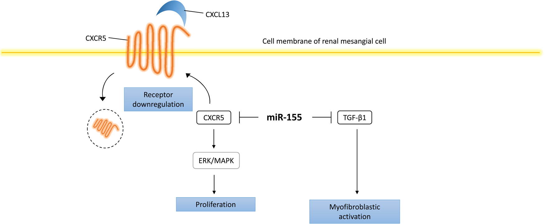
Lupus nephritis (LN) is one of the major causes of death in SLE patients. Histologically, LN is characterised by abnormal mesangial cell proliferation, leucocytic infiltration and overproduction of extracellular matrix, all of which can lead to renal sclerosis and end-stage renal failure (Ref. Reference Lewis and Schwartz22). The role of miRNAs in LN hinges on the regulation of leucocytic function and pro-inflammatory cytokines (Ref. Reference Dai and Ahmed23). An example of this would be the fact that miR-155 regulates the extracellular signal-regulated kinase-mitogen-activated protein kinase (ERK-MAPK) inflammatory pathway of T cells through C-X-C motif chemokine receptor 5 (CXCR5). Interestingly, ERK-MAPK was also found to be involved in the regulation of human renal mesangial cells (HRMCs) through miR-155 as well. miR-155 mimics inhibit HRMC proliferation via inhibiting the phosphorylation of ERK through downregulating CXCR5. Moreover, miR-155 transfection abrogates the increased levels of the cytokine transforming growth factor β1 (TGF-β1) in HRMCs, an inducer of myofibroblastic activation in vitro (Fig. 2) (Ref. Reference Kong24).
Summary of miR-155's role in SLE pathogenesis in HRMCs in humans. miR-155 exhibits anti-proliferative effects on renal mesangial via inhibiting the phosphorylation of ERK through the repression of CXCR5. miR-155 transfection also abrogates the increased levels of the cytokine transforming growth factor β1 (TGF-β1) in HRMCs, which reduces myofibroblastic activation in vitro. CXCL13, C-X-C motif chemokine ligand 13; CXCR5, C-X-C motif chemokine receptor 5; ERK, extracellular-signal-regulated kinase.

Is miR-155 a good potential biomarker in SLE?
Several studies have attempted to evaluate the diagnostic and prognostic value of miR-155 in SLE in different body fluids and compartments. The heterogeneity between the studies and inconsistent findings have made it unclear whether miR-155 is suitable to be used as a biomarker in SLE progression and diagnosis. For example, miR-155 was found to be both upregulated and downregulated in PBMCs of SLE patients in different studies (Refs Reference Lashine21, Reference Kaga25–Reference Stagakis27). Furthermore, differential expression of miR-155 in PBMCs has been reported to exist between different subgroups of SLE patients (Ref. Reference Chauhan28). Studying a more stable population of miRNAs such as cell-free circulating miRNAs may provide more accurate, reliable findings.
Similarly, the expression of miR-155 in plasma was found to be reduced in SLE patients in one study (Ref. Reference Wang29), whereas another study reported no overall statistically significant results (Ref. Reference Carlsen30). Moreover, miR-155 was also reported to be upregulated in plasma samples of LN patients (Ref. Reference Vahed31). Such inconsistent data may be a result of the pitfalls related to miRNA research, including methodological inconsistencies and cohort heterogeneity.
Moreover, a meta-analysis was conducted to identify the diagnostic capability of serum miRNAs in SLE. Interestingly, miR-155 was revealed to be upregulated and possessed the highest specificity in diagnosing SLE patients amongst single miRNAs (Ref. Reference Dai32). However, miR-155 was found to be downregulated in serum in another study (Ref. Reference Wang33).
Finally, the use of miRNAs as biomarkers of LN activity has intrigued the interest of many researchers owing to their high stability in tissues and fluids. Consequently, Abulaban et al. studied the use of urinary cell-free miRNAs (including miR-155) as biomarkers for LN activity and compared it with the LN-Panel in 15 paediatric SLE patients (Ref. Reference Abulaban34). However, it was concluded that miRNAs were not superior to the LN-Panel as biomarkers. Similarly, Wang et al. reported that cell-free urinary levels of miR-155 were similar to the levels found in healthy controls and not statistically significant (Ref. Reference Wang33). However, they reported significant results in another study when they used urinary sediment instead (Ref. Reference Wang35).
In conclusion, the heterogeneity across studies implies the need to conduct further research that addresses these inconsistencies to obtain a better understanding of SLE, which is discussed later in this review. Table 1 summarises the findings of articles that have evaluated the role of miR-155 as a biomarker in SLE in different body fluids and compartments.
Summary of the role of miR-155 as a biomarker in SLE in different body fluids

C3, complement component 3; C4, complement component 4; CVD, cardiovascular disease; PBMCs, peripheral blood mononuclear cells; PBL, peripheral blood leucocytes; SLEDAI, systemic lupus erythematosus disease activity index.
↑, increased level of miR-155; ↓, decreased level of miR-155.
ns, not statistically significant (P > 0.05).
What do lupus mouse models tell us about miR-155?
Lupus mouse models have helped us understand the pathogenesis of the disease because of their high resemblance to human lupus. Several methods have been used to induce lupus-like phenotypes in mice, such as crossbreeding of specific mouse strains, drug induction or gene knockout (Ref. Reference Richard and Gilkeson36). It is important to note that despite the existence of a large number of SLE models, no model completely resembles human lupus. In fact, these animal models differ in the age at which they manifest the disease, antibody profiles and symptoms coverage and severity (Ref. Reference Richard and Gilkeson36). Nevertheless, murine models do not fail to provide us with a clear insight into the different aspects of the disease, including genetic and epigenetic factors contributing to SLE development. In this section, the role of miR-155 in murine SLE pathogenesis is elucidated in an attempt to understand the antagonistic roles of miR-155.
miR-155 in murine lupus pathogenesis
The expression profile of miR-155 has been studied in several SLE models. For example, Dai et al. aimed to identify a set of miRNAs that are commonly dysregulated in three different lupus-prone mice models. Interestingly, they found an upregulation in miR-155 levels in whole splenocytes and both splenic T cells and B cells that is consistent in all models. miR-155 was also found to be upregulated in lipopolysaccharide-activated splenocytes isolated from young mice as well as lupus-expressing 3–4-month-old MRL/MpJ-Faslpr/J (MRL-lpr) mice compared with 1-month old MRL-lpr mice, confirming that miR-155 is involved in lupus development and progression (Ref. Reference Dai37).
Strikingly, Zarate et al. developed a different mouse model of an autoimmune disease that resembled SLE, which on the contrary exhibited miR-155 downregulation. Furthermore, they constructed a target network for miR-155 that may be responsible for its function (Ref. Reference Zarate-Neira38). The findings of both studies highlight the difficulty in interpreting the role of miR-155 in SLE because of differences in experimental contexts and the use of different animal models with different disease backgrounds.
The effects of miR-155 deficiency in mice
One particular advantage of using animal models is the ability to manipulate the mouse genetic background and delete a specific gene of interest to observe its loss of function effects. In this case, the effects of miR-155 loss were observed in miR155−/−-Faslpr mice. miR155−/−-Faslpr mice showed reduced spleen size, smaller peripheral lymph nodes, less kidney inflammatory infiltration and a decrease in serum antibody levels via regulating src homology 2 domain-containing inositol phosphatase isoform 1 and sphingosine-1-phosphate receptor 1 levels (Refs Reference Thai39, Reference Xin40).
In a pristine-induced miR155-deficient mouse model with no genetic susceptibility to SLE, Leiss et al. observed a reduction in lupus-associated autoantibodies and decreased symptomatic lung and spleen involvement along with no marked increase in interferon-signature genes (Ref. Reference Leiss41). This comes in agreement with the findings of Zhou et al., who also concluded that miR-155 promotes pristine-induced lung inflammation through the activation of nuclear factor-κB signalling pathways via direct targeting of one of its negative regulators peroxisome proliferator-activated receptor a (Ref. Reference Zhou42). Therefore, miR-155 deficiency in mice appears to decrease autoantibody production and organ involvement.
miR-155 regulates B and T cells in mice
B cells, along with other antigen-presenting cells, express CD1d through which it interacts and activates invariant natural killer T (iNKT) cells. This interaction is associated with the tolerance of NKT cells and may partially inhibit autoreactive B cells. Previous studies have reported that inflammation causes a decrease in CD1d expression levels, which exacerbates SLE in mouse models. The underlying mechanisms by which this occurs involve the transcription of miR-155 upon TLR9 activation, which in return increased miR-155 target interaction with the 3′ UTR of CD1d mRNAs, lowering its levels and impairing CD1d-mediated antigen presentation (Ref. Reference Liu43).
The role of miR-155 extends to regulatory T (Treg) cell regulation in SLE as well. In MRL-Faslpr/lpr the number of CD4+CD25+Foxp3+ Treg cells is increased; however, these Tregs are phenotypically different (CD62L–CD69+) compared with normal mice (Ref. Reference Divekar44). This decrease in CD62L and increase CD69 expression is associated with decreased suppressive capacity and contributes to the pathogenesis of SLE. Interestingly, Divekar et al. demonstrated that miR-155 is overexpressed in these Treg cells and targets CD62L, lowering its levels and contributing to this altered phenotype even though the expression of Dicer, an enzyme required for miRNA biogenesis and maturation, was reduced. In its absence, it is expected that all miRNAs will have a reduced expression. However, miR-155, along with few other miRNAs, seem to oppose this rule, hinting towards the existence of other miRNA processing pathways (Refs Reference Divekar44, Reference Cifuentes45). The increase in miR-155 expression in Treg cells comes in agreement with other reports that found that Pbx1-d overexpression in transgenic mice induces the expression of miR-155 and may regulate Treg/TFH homoeostasis (Ref. Reference Choi46), highlighting that miR-155 may play important roles in immune cell regulation.
An attempt to solve the conflict
One miRNA, different net results
In the literature we have reviewed, miR-155 was seen to play a role both as a friend and as a foe of SLE. This duplicity can be explained by the fact that miRNAs heavily rely on sequence complementarity as their targeting mechanism, which consequently results in that a single miRNA may have hundreds of different mRNA targets in a single cell. These targets can have opposing effects on disease progression, and hence the balance between these targets as well as the factors that regulate miRNAs affinity to their target determine the type of net effect exerted by the miRNA (Ref. Reference Svoronos, Engelman and Slack47). In the context of SLE, it would be the balance between inflammatory mRNAs and protective mRNAs that determine whether miR-155 exhibits a net protective or a net inflammatory effect. For such opposing actions to be targeted by the same miRNA, they must be subjected to other regulatory mechanisms for one to dominate over the other (Ref. Reference Zan and Casali48). Therefore, studies exploring integrated regulatory networks of the non-coding transcriptome may help in reaching a better understanding of SLE.
Methodological inadequacies
Undoubtedly, methodological inadequacies are of extreme importance when discussing heterogeneity across the literature. For example, the average sample sizes in most studies involving humans are quite small, consequently resulting in skewed results (Refs Reference Vahed31, Reference Shumnalieva49). Second, the sample patients used in each study were of single ethnic groups, and therefore of the same genetic background. Different ethnic groups are also exposed to different environmental factors that may affect miRNA expression. To further elaborate, it was previously mentioned that dietary factors, exposure to infections and lifestyles affect miRNA expression (Refs Reference Chung50, Reference Palmer51). Moreover, previous studies describe differential miRNA expression profiles between adult and paediatric patients (Refs Reference Cortes-Marquez52, Reference Shah and Singh53); therefore, more studies conducted on paediatric SLE patients are needed. In addition, different methodologies used for RNA extraction may affect expression studies as well. Expression profiles may also be changed with time and are influenced by different treatment regimens (Ref. Reference Chafin54). This influence can be because of the effect of treatments on miRNA expression or the effect of miRNA expression on treatment response. It is also noteworthy to mention that an increase in miRNA expression following therapy does not necessarily suggest that the miRNA is anti-inflammatory. Therefore, studying changes in miRNA expression levels with therapy may produce misleading results. Finally, the physiological differences between animal models and humans make it difficult to interpret the precise and complex pathogenic mechanisms and prognoses of SLE, so not all findings can be extrapolated across species.
Addressing the inconsistencies
Conducting larger, longitudinal, multi-racial studies and dividing patients into subgroups according to autoantibody profiles may help us develop a more comprehensive understanding of SLE. Furthermore, the discrepancies found between whole peripheral blood, PBMCs and subtypes of immune cells suggest different regulatory mechanisms in different body fluids. Studying subtypes of cells rather than PBMCs as a whole may provide a deeper understanding of SLE pathogenesis. Finally, we reviewed that miR-155 possesses antagonistic functions; therefore, investigating its shared targets with other non-coding RNAs may provide a more holistic insight into the epigenetic regulation of SLE. Unravelling the signalling pathways miR-155 is involved in will also help further elucidate the role of miR-155 in immune cells.
Conclusion
miR-155 preserves the integrity and normal responses exerted by both the innate and adaptive immune systems; hence it is of no surprise that its dysregulation has been reported in various autoimmune diseases. Similar to other pathologies, the miR-155 functional axis is disrupted in SLE as well. Its irregular pattern of expression has been reported in both immune and non-immune cells, highlighting its contribution to hallmark events occurring in the course of SLE development, such as autoantibody production and cytokine release. As evident from the studies we reviewed, miR-155 exerts several actions that at times appear to have overall contradicting effects. The heterogenous mechanistic actions of miRNAs also extend to other clinical applications, such as their use as prognostic and diagnostic markers. Thus far, miR-155 displayed marked inconsistencies regarding its use as a biomarker in SLE, even though this can be partly attributed to population and methodological differences between the reported studies. This phenomenon of contradicting miRNA functions and expression has been reported many times before in various pathological entities, and it rather highlights the functional diversity of miRNAs as biological molecules in different biological contexts.
Author contributions
S.A. Ibrahim and A.Y. Afify collected the data and wrote the manuscript. A.I. Abdelaziz conceived the idea. I.O. Fawzy, N. El-Ekiaby and A.I. Abdelaziz revised and critically appraised the manuscript.
Conflict of interest
The authors declare no conflict of interest.