



Introduction
Lichenicolous fungi inhabit lichens as parasites, saprobes or commensals (Diederich et al. Reference Diederich, Lawrey and Ertz2018a). Of these fungi, c. 9% are basidiomycetes but they are taxonomically diverse, belonging to ten orders and five classes (Diederich et al. Reference Diederich, Millanes, Wedin and Lawrey2022a).
The Corticiales are an ecologically diverse order of entirely resupinate fungi in the class Agaricomycetes, comprising four families: Corticiaceae, Dendrominiaceae, Punctulariaceae and Vuilleminiaceae (Ghobad-Nejhad et al. Reference Ghobad-Nejhad, Nilsson and Hallenberg2010, Reference Ghobad-Nejhad, Langer, Nakasone, Diederich, Nilsson, Rajchenberg and Ginns2021; Diederich et al. Reference Diederich, Lawrey, Sikaroodi and Gillevet2011; He et al. Reference He, Zhao, Hyde, Begerow, Kemler, Yurkov, McKenzie, Raspé, Kakishima and Sánchez-Ramírez2019; Wijayawardene et al. Reference Wijayawardene, Hyde, Dai, Sánchez-García, Goto, Saxena, Erdoğdu, Selçuk, Rajeshkumar and Aptroot2022). Species within the order are mostly saprotrophic, mainly wood-rotting, but several are also parasitic on plants or lichens, or are endolichenic (Lawrey et al. Reference Lawrey, Diederich, Sikaroodi and Gillevet2008; Lücking & Moncada Reference Lücking and Moncada2017). The lichenicolous Corticiales are characterized by the production of pink to orange or red basidiomata or bulbils (structures also called sclerotia). They all belong to the family Corticiaceae within four genera: Corticium, Erythricium, Laetisaria and Marchandiomyces (Diederich et al. Reference Diederich, Zimmermann, Sikaroodi, Ghobad-Nejhad and Lawrey2018b, Reference Diederich, Ghobad-Nejhad, Lawrey, Diederich, Millanes, Wedin and Lawrey2022b). Seven endolichenic species were also included in Lawreymyces (Lücking & Moncada Reference Lücking and Moncada2017), a genus that is invalidly described since all these species are known only from DNA sequences obtained from lichen thalli. Identification keys to the genera and to the species of Corticiaceae are provided by Ghobad-Nejhad et al. (Reference Ghobad-Nejhad, Langer, Nakasone, Diederich, Nilsson, Rajchenberg and Ginns2021), except for the endolichenic Lawreymyces.
Ancestral character state reconstruction showed that saprotrophy is the ancestral nutritional mode in the Corticiaceae, while multiple transitions have occurred to various other nutritional modes, such as the lichenicolous and parasitic lifestyles, within this family (Ghobad-Nejhad et al. Reference Ghobad-Nejhad, Langer, Nakasone, Diederich, Nilsson, Rajchenberg and Ginns2021). There is a high tendency to form asexual structures within the family and some lichenicolous Corticiaceae are known only from the asexual stage. Their bulbils are regarded as resting or dispersal structures remaining viable for extended periods of time under unfavourable conditions (Lawrey et al. Reference Lawrey, Diederich, Sikaroodi and Gillevet2008).
In the framework of lichen surveys in the Massif Central (France) in May 2024, an unknown lichenicolous fungus was collected on Physcia growing in Juniperus thickets at the summit of the Puy de Manson near Saint-Genès-Champanelle. The species was reminiscent of Laetisaria lichenicola Diederich et al., but appeared different due to the vivid pink colour present mainly on the lower host thallus surface (vs upper surface in L. lichenicola). A more detailed microscopical examination confirmed that it was a distinct species.
The aims of this study were 1) to describe the new species, 2) to determine its phylogenetic position and 3) to describe the habitat where the new species was discovered.
Materials and Methods
Voucher specimens are deposited in the herbaria BR and GZU. The macroscopic characteristics were studied and measured using a Leica MZ7.5 dissecting microscope. Macroscopic images were captured using a Canon 6D camera with a Nikon BD Plan 5 objective, and StackShot (Cognisys) with Helicon Focus (HeliconSoft) for increasing the depth of field. Hand-cut sections and squash preparations of the basidiomata were mounted in water, 5% KOH, phloxine B, Congo red, and Lugol’s iodine solution, and studied under a Leica DM 750 compound microscope with a Leica ICC50 W ×1000 camera. The size of the basidia and basidiospores was measured in water. For the basidiospores, the average (x̄) and standard deviation (SD) were calculated. These measurements are given as x̄ ± SD, surrounded by the extreme values (between parentheses), followed by the number of measurements (n).
Molecular techniques
A well-preserved and freshly collected specimen (Pinault 101; five weeks old) was used for DNA isolation. Hand-cut sections of the basidiomata were used for direct PCR as described in Ertz et al. (Reference Ertz, Tehler, Irestedt, Frisch, Thor and van den Boom2015). The material was then added to a tube containing the PCR reaction mixture and amplified directly. Amplification reactions were prepared for a 50 μl final volume containing 5 μl 10× DreamTaq Buffer (Fermentas), 1.25 μl of each of the 20 μM primers, 5 μl of 2.5 mg ml−1 bovine serum albumin (Fermentas #B14), 4 μl of 2.5 mM each dNTPs (Fermentas), 1.25 U DreamTaq DNA polymerase (Fermentas) and tiny fragments of the lichenicolous fungus. The nuITS rDNA (ITS1 + 5.8 S + ITS2) was amplified using the primers ITS1F and ITS4 (White et al. Reference White, Bruns, Lee, Taylor, Innis, Gelfand, Sninsky and White1990). The PCR cycling conditions for the nuITS consisted of the following steps: 1) 10 min at 95 °C; 2) 35 cycles of 45 s at 95 °C, 1 min at 52 °C, 75 s at 72 °C; 3) 10 min of final elongation at 72 °C. Both strands were sequenced by Macrogen® using the amplification primers. Sequence fragments were assembled with Sequencher v. 5.4.6 (Gene Codes Corporation, Ann Arbor, Michigan).
Taxon selection and phylogenetic analyses
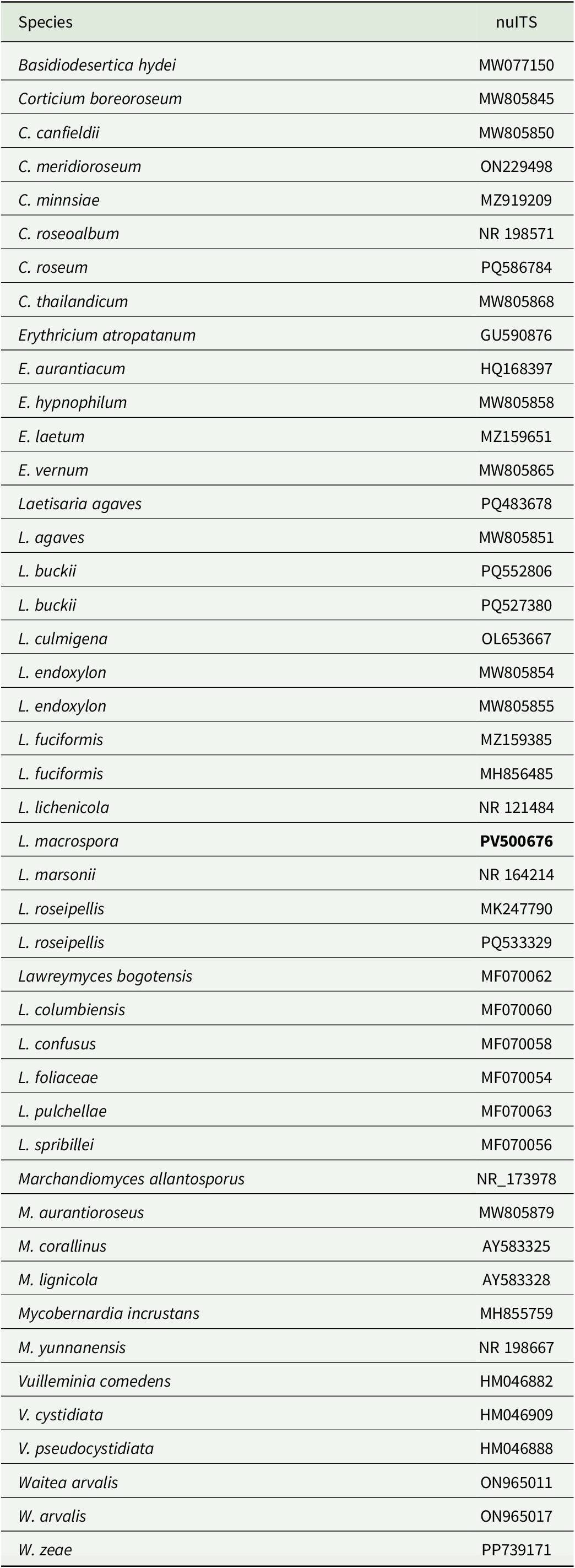
The newly generated sequence was subjected to NCBI BLAST searches (https://www.ncbi.nlm.nih.gov) in order to obtain a first approximation of phylogenetic affiliation. A dataset of nuITS sequences was assembled for placing the newly sequenced taxon in a phylogeny of the Corticiaceae. Sequences published mainly in DePriest et al. (Reference DePriest, Sikaroodi, Lawrey and Diederich2005), Lawrey et al. (Reference Lawrey, Diederich, Sikaroodi and Gillevet2008), Diederich et al. (Reference Diederich, Lawrey, Sikaroodi and Gillevet2011), Ghobad-Nejhad & Hallenberg (Reference Ghobad-Nejhad and Hallenberg2011), Lücking & Moncada (Reference Lücking and Moncada2017), Vu et al. (Reference Vu, Groenewald, de Vries, Gehrmann, Stielow, Eberhardt, Al-Hatmi, Groenewald, Cardinali and Houbraken2019), Ghobad-Nejhad et al. (Reference Ghobad-Nejhad, Langer, Nakasone, Diederich, Nilsson, Rajchenberg and Ginns2021), Maharachchikumbura et al. (Reference Maharachchikumbura, Wanasinghe, Cheewangkoon and Al-Sadi2021), Dong et al. (Reference Dong, Li, Yuan, Luo, Zhang, Dai, Zhou, Liu, Deng and Zhou2024) and Li et al. (Reference Li, Luo and Zhao2024) were retrieved from GenBank. The sequences (Table 1) were aligned using MAFFT v. 7.505 (Katoh & Standley Reference Katoh and Standley2013) on the CIPRES Web Portal (Miller et al. Reference Miller, Pfeiffer and Schwartz2010) and manually edited using Mesquite v. 3.04 (Maddison & Maddison Reference Maddison and Maddison2015). Terminal ends of sequences and ambiguously aligned regions were delimited manually and excluded from the datasets. The resulting matrix of Corticiaceae consisted of 45 terminals and 537 unambiguously aligned sites. Three species of Vuilleminiaceae, viz. Vuilleminia comedens (Nees) Maire, V. cystidiata Parmasto and V. pseudocystidiata Boidin et al., were selected as the rooting taxa from Diederich et al. (Reference Diederich, Millanes, Wedin and Lawrey2022a).
GenBank Accession numbers of nuITS sequences used in this study. The newly generated sequence is marked in bold.
The best-fit evolutionary model was estimated using the Akaike Information Criterion (AIC) as implemented in jModelTest v. 2.1.6 (Darriba et al. Reference Darriba, Taboada, Doallo and Posada2012). The GTR + I + G model was selected.
Bayesian analyses were carried out on the nuITS dataset under the selected model using the Metropolis-coupled Markov chain Monte Carlo method (MCMCMC) in MrBayes v. 3.2.7a (Huelsenbeck & Ronquist Reference Huelsenbeck and Ronquist2001; Ronquist & Huelsenbeck Reference Ronquist and Huelsenbeck2003) on the CIPRES Web Portal (Miller et al. Reference Miller, Pfeiffer and Schwartz2010). Two parallel MCMCMC runs were performed, each using four independent chains and 20 million generations, sampling trees every 1000th generation. Posterior probabilities (PP) were determined by calculating a majority-rule consensus tree generated from the 30 002 post burn-in trees of the 40 002 trees sampled by the two MCMCMC runs using the sumt option of MrBayes. Tracer v. 1.7.1 (Rambaut et al. Reference Rambaut, Drummond, Xie, Baele and Suchard2018) was used to ensure that stationarity was reached by plotting the log-likelihood values of the sample points against generation time, making sure that the ESS values were much higher than 200. Convergence between runs was also verified using the PSRF (Potential Scale Reduction Factor), where values were all equal or close to 1.000.
In addition, a maximum likelihood (ML) analysis was performed using RAxML v. 8.2.12 (Stamatakis Reference Stamatakis2014) on the CIPRES Web Portal (Miller et al. Reference Miller, Pfeiffer and Schwartz2010) with 1000 ML bootstrap iterations (ML-BS) and the GTRGAMMA model.
The ML tree did not contradict the Bayesian tree topology for the strongly supported branches. Therefore, only the ML tree is shown with the ML-BS values added above or near the internal branches. Internodes with ML-BS ≥ 70 and PP ≥ 0.95 are represented by thicker lines (Fig. 1). Phylogenetic trees were visualized using FigTree v. 1.4.2 (Rambaut Reference Rambaut2012).
Phylogeny of Corticiaceae (with three species of Vuilleminiaceae as outgroup) based on a dataset of nuITS sequences that resulted from the RAxML analysis. Maximum likelihood bootstrap values are shown above or near internal branches. Internodes considered strongly supported by the RAxML and Bayesian analyses (ML-BS ≥ 70 and PP ≥ 0.95) are represented by thicker lines. The newly sequenced sample is in bold. The lineage corresponding to the genus Laetisaria is highlighted. Lichenicolous species of Laetisaria are marked with an asterisk. In colour online.

Results
Phylogenetic analyses
One new sequence was obtained for this study and 44 additional nuITS sequences were retrieved from GenBank representing a total of 39 species (Table 1). The RAxML tree obtained from this Corticiaceae dataset is shown in Fig. 1. The monophyly of the genera within Corticiaceae is generally well supported, with a small number of exceptions (Erythricium and Waitea); however, the relationships among the genera are unresolved, including the sister relationship of Laetisaria with Mycobernardia. The main well-supported lineages are in accordance with the results obtained by Diederich et al. (Reference Diederich, Millanes, Wedin and Lawrey2022a). All genera are recovered as monophyletic except Erythricium, which is paraphyletic because of the placement of E. atropatanum Ghob.-Nejh. & Hallenb. as sister to the genus Lawreymyces. This was also the case in the phylogeny of Diederich et al. (Reference Diederich, Millanes, Wedin and Lawrey2022a). The new species is nested in the Laetisaria lineage which is fully supported by both analyses, but its relationship to the other species is unresolved.
Taxonomy
Laetisaria macrospora P. Pinault, Ertz & Diederich sp. nov.
MycoBank No.: MB 858766
Differs from Laetisaria lichenicola by the larger and 4-sterigmate (vs 2-sterigmate) basidia, 40–110 × 9–27 μm (vs 25–40 × 7.5–12 μm), the much larger, ovoid to reniform (vs pyriform to lacriform) basidiospores, 23–34 × 11–18 μm (vs 14.5–18.5(–20.0) × (8–)10.5–12.5 μm), the occasionally clamped subhymenial hyphae (vs clampless), and the basidiomatal development mainly on the lower (vs upper) host thallus surface.
Type: France, Puy-de-Dôme, Saint-Genès-Champanelle, sommet du Puy de Manson, 45.7496°N, 3.0070°E, 980 m, sur Physcia adscendens et P. stellaris sur Juniperus communis et sur vieux Genista, 3 May 2024, P. Pinault 101 (BR—holotype; GZU—isotype).
(Fig. 2)
Laetisaria macrospora, holotype. A, infected thallus of Physcia adscendens, with pink to coral coloured infections mainly on the lower thallus surface. B–D, young basidia. E, mature basidium with one mature basidiospore attached. F, sterigmata of mature basidia with young basidiospores. G & H, hyphae with clamp connections. I–M, mature basidiospores. B–D, G & K in ammoniacal Congo red; E, H & M in phloxine; F, I, J & L in water. Scales: A = 1 mm; B–M = 10 μm. In colour online.
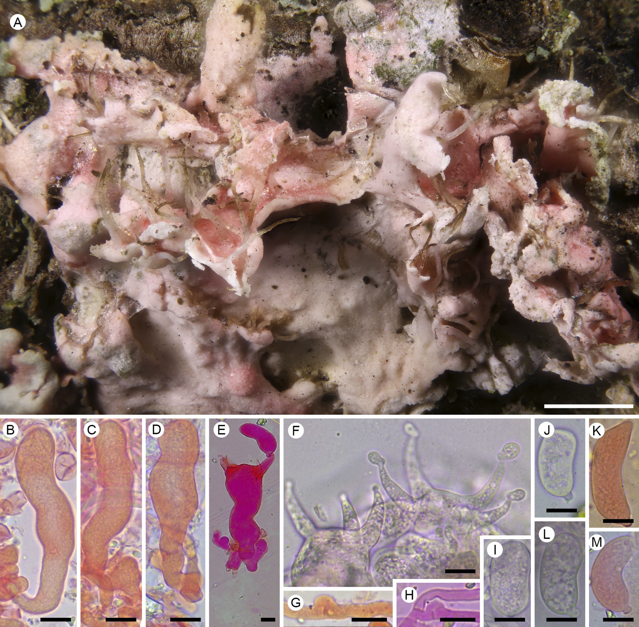
Basidiomata light pink to coral colour, effuse, thin, adnate, granulose to floccose, margin indeterminate, reaching 2 mm diam., usually on the lower surface of the host thallus. Basal hyphae hyaline, straight or occasionally contorted, lacking clamps, smooth, 2.5–3 μm wide, wall c. 0.5 μm. Subhymenial hyphae hyaline, thin-walled, without or with clamps, septate, smooth, 4–5 μm wide. Hymenium comprising one layer of basidia on vertically branching, thin-walled hyphae. Hyphidia, cystidia and other sterile hymenial elements not observed. Basidia initially elongate cylindrical, basally occasionally narrower, without basal, lateral probasidial bladder; when mature, generally becoming clavate to suburniform, aseptate or rarely with one transverse septum, 40–110 × 9–27 μm (n = 15), distinctly wider than the supporting hyphae; wall 0.5–1.0 μm; basal clamps not observed; sterigmata 4 per basidium, 18–24 μm long and 6–10 μm wide at the base, curved. Basidiospores hyaline, smooth, aseptate, nonamyloid, ovoid to reniform, one side frequently flattened or slightly concave, with a prominent truncate apiculus 3–4 μm diam., (23–)25–31(–34) × (11–)12–15(–18) μm (n = 45); wall c. 1 μm.
Conidial and bulbilliferous morphs unknown.
Etymology
The epithet refers to the large basidiospores, compared to closely related species.
Distribution and ecology
The new species is currently known only from its type locality at the summit of the Puy de Manson. It inhabits thalli of Physcia adscendens H. Olivier and P. stellaris (L.) Nyl. growing on the branches of Juniperus communis, more rarely on dead branches of Genista. The Puy de Manson is predominantly covered by sheep pastures, except for the northern edges, where Juniperus thickets are present and from which the holotype specimen was collected. Located at the eastern edge of the Plateau des Dômes, the site is characterized by Hercynian metamorphic plutonic rocks which are extensively faulted. The climate is cool sub-oceanic, with annual rainfall c. 500 mm and annual temperatures ranging from 0 °C to 25 °C on average. Situated 4.5 km south-east of the Puy de Dôme, the area is somewhat sheltered from the dominant westerly winds and rainfall, benefiting from a slight foehn effect.
Notes
The new species strongly resembles Laetisaria lichenicola, which also has a predilection for corticolous Physcia thalli. While basidiomata of L. lichenicola mainly develop on the upper thallus surface that becomes strongly pink or coral coloured, basidiomata of the new L. macrospora are mainly situated on the lower thallus surface, with the upper surface only pale pinkish. Microscopically, the new species is distinguished by the 4- (vs 2-) sterigmate basidia, by the occasional presence of clamps on the subhymenial hyphae, by the ovoid to reniform basidiospores (vs pyriform to lacriform), but mainly by the overall much larger basidia and basidiospores. Basidia of L. lichenicola are just 25–40 × 7.5–12 μm, sterigmata are 6.5–11.5 μm long and 3–3.5 μm wide at the base, and basidiospores are 14.5–18.5(–20.0) × (8–)10.5–12.5 μm (Diederich et al. Reference Diederich, Lawrey, Sikaroodi and Gillevet2011).
Although using multiple collections is preferable when describing a new species, this is not always possible, as is the case with rare taxa. It is then recommended to support the species description with multiple lines of evidence (e.g. Aime et al. Reference Aime, Miller, Aoki, Bensch, Cai, Crous, Hawksworth, Hyde, Kirk and Lücking2021). In the case of Laetisaria macrospora, the evidence supporting its recognition as a new species is compelling. Morphologically, it is distinguished by key characteristics such as the number of sterigmata per basidium and the spore size. Genetically, it is supported by a nuITS sequence that is highly divergent from all previously sequenced Laetisaria species, including all known lichenicolous members of the genus.
Discussion
Laetisaria is a small genus described by H. H. Burdsall in 1979 for L. fuciformis (Berk.) Burds., a widespread fungal pathogen occurring on grasses, forming delicate, pink basidiocarps or clavarioid sclerotia. A second species was included in the genus three years later, viz. L. agaves Burds. & Gilb., which grows on Agave leaves but with an uncertain nutritional habit (Burdsall & Gilbertson Reference Burdsall and Gilbertson1982). Diederich et al. (Reference Diederich, Lawrey, Sikaroodi and Gillevet2011) added the first lichenicolous fungus to the genus, viz. L. lichenicola, with phylogenetic analyses showing a close relationship with fungal pathogens of plants. With the advent of molecular data, phylogenetic analyses were used to revise the circumscription of the genus Laetisaria and its relatives. Two species of Limonomyces and three of Marchandiomyces were transferred to Laetisaria by Diederich et al. (Reference Diederich, Zimmermann, Sikaroodi, Ghobad-Nejhad and Lawrey2018b), while two species of Corticium were combined in Laetisaria by Ghobad-Nejhad et al. (Reference Ghobad-Nejhad, Langer, Nakasone, Diederich, Nilsson, Rajchenberg and Ginns2021). Among these species, Laetisaria buckii (Diederich & Lawrey) Diederich et al. was the second lichenicolous species added to the genus, in addition to plant pathogens and saprotrophs. It is known from North America and reported as a parasite on the lichens Bacidia heterochroa (Müll. Arg.) Zahlbr., Huneckia pollinii (A. Massal.) S. Y. Kondr. et al. and Pertusaria hypothamnolica Dibben, but it has also been confirmed as lignicolous on Juniperus (Diederich et al. Reference Diederich, Millanes, Wedin and Lawrey2022a). With the discovery of L. macrospora, the genus currently includes 11 species, mostly parasitic on leaves of monocotyledons (Poaceae, Agave, Pandanus) and on lichen thalli, rarely lignicolous (Lawrey et al. Reference Lawrey, Diederich, Sikaroodi and Gillevet2008; Ghobad-Nejhad et al. Reference Ghobad-Nejhad, Langer, Nakasone, Diederich, Nilsson, Rajchenberg and Ginns2021; Diederich et al. Reference Diederich, Millanes, Wedin and Lawrey2022a). Our phylogenetic analyses recover the lichenicolous species as a polyphyletic group within the genus Laetisaria, suggesting several transitions to diverse nutritional modes within the genus.
The diversity of lichenicolous species of Corticiaceae that inhabit lichens of Xanthorion parietinae communities (James et al. Reference James, Hawksworth, Rose and Seaward1977) is striking. The species Erythricium aurantiacum (Lasch) D. Hawksw. & A. Henrici, Laetisaria lichenicola and L. macrospora are predominantly or exclusively found on Physcia species growing on nutrient-enriched bark, making them ecologically similar. This habitat has obviously played a significant role in the speciation of these lichenicolous Corticiaceae, which contribute to the dynamics of this lichen vegetation by killing their hosts. Erythricium aurantiacum, in particular, is often abundant and can sometimes cause the mortality of entire populations of corticolous lichens. Further research should explore other nitrophilous habitats in different regions of the world to find additional lichenicolous Corticiaceae species.
The host specificity of lichenicolous fungi is usually an important factor in delimiting species. However, the host range of lichenicolous basidiomycetes that kill their hosts rapidly appears broader than that of commensals. For example, the virulent parasites Athelia arachnoidea (Berk.) Jülich and Marchandiomyces corallinus (Roberge) Diederich & D. Hawksw. have a wide host spectrum, occurring on many lichen genera. These virulent parasites are often easily isolated in pure cultures, unlike highly host-specific lichenicolous species, probably due to their lower nutrient specificity. Similarly, two species of lichenicolous Laetisaria (L. buckii and L. lichenicola), which are also virulent parasites that kill their host thalli, were isolated in culture for sequencing (Diederich & Lawrey Reference Diederich and Lawrey2007; Lawrey et al. Reference Lawrey, Binder, Diederich, Molina, Sikaroodi and Ertz2007; Diederich et al. Reference Diederich, Lawrey, Sikaroodi and Gillevet2011), while a culture of the new species was not needed and thus not attempted (see Methods). However, in contrast to Laetisaria buckii that is not host specific, L. lichenicola shows a strong preference for species of Physcia based on the data currently available. Therefore, the host specificity, the seasonability and the distribution of L. macrospora warrant further study because the new species is currently known only from the type material. Additionally, the material of L. lichenicola should be revised, since some specimens may have been misidentified and may represent the new species, which is morphologically and ecologically similar.
Acknowledgements
We wish to warmly thank Arne Mertens and Wim Baert (Meise Botanic Garden) for their help with the molecular work.
Author ORCIDs
Damien Ertz, 0000-0001-8746-3187; Paul Diederich, 0000-0003-0357-7414; Patrick Pinault, 0009-0006-6751-5908.
Competing Interests
The authors declare none.



