Significant outcomes
-
∙ Patients with obsessive–compulsive disorder (OCD) showed the brain metabolic abnormalities of glutamate/glutamine (Glx) and myo-inositol (mI) in the right dorsolateral prefrontal cortex (DLPFC). This outcome is valuable for an understanding of the pathophysiologic mechanism for the progress of OCD.
Limitations
-
∙ The participants consisted of two relatively small groups of 14 patients and 14 healthy individuals.
-
∙ There was no segmentation of the white matter and the grey matter in the single-voxel localisation on the right DLPFC in magnetic resonance spectroscopy (MRS).
-
∙ There was no control region showing the metabolic specificity, which was comparable with that of the right DLPFC in MRS.
Introduction
OCD is a psychiatric disorder characterised by obsessive thinking and compulsive actions that are often uncontrollable (1). The lifetime prevalence rate of OCD in the general population is ~2–3% (Reference Karno, Golding and Sorenson2). The major causes of OCD stem from a number of psychological and biological factors, which can be worsened by precarious life styles in a rapidly changing modern society. Effective prevention and treatment for OCD is essential.
Treatment for OCD involves behavioural therapy and/or medication with selective serotonin reuptake inhibitors (SSRIs). Psychiatric treatment is generally performed to improve psychologically rooted behaviours. Knowledge of the benefits of SSRIs on OCD (Reference Zohar, Chopra and Sasson3) has spawned OCD therapy linked with abnormalities of the neurotransmitter, serotonin. However, this approach is not successful in about 30% of those with OCD (Reference Greist, Jefferson and Kobak4).
Recent studies on the neurochemical mechanisms associated with OCD have utilised MRS. Russell et al. (Reference Russell, Cortese and Lorch5) reported that OCD patients showed higher intensity of N-acetylaspartate (NAA) in the left DLPFC. Ebert et al. (Reference Ebert, Speck and König6) described that OCD patients showed decreased intensity of NAA in the right striatum and anterior cingulate cortex (ACC). In addition, there was a negative correlation between the OCD symptom and the intensity of NAA in the right ACC. Bartha et al. (Reference Bartha, Stein and Williamson7) reported a similar outcome, in which OCD patients had decreased intensity of NAA in the left striatum. Recently, Simpson et al. (Reference Simpson, Shungu and Bender8,Reference Simpson, Kegeles and Hunter9) demonstrated patients with OCD showed a decreased γ-aminobutyric acid (GABA) in the medial prefrontal cortex, and more recently, they revealed that there was no significant difference in glutamate in the striatal subregions: dorsal caudate, dorsal putamen, and ventral striatum. Rosenberg et al. (Reference Rosenberg, MacMaster and Keshavan10) reported that paediatric/adolescent (8–17 years of age) OCD patients showed an increased intensity of Glx in the caudate nucleus. On the contrary, Ohara et al. (Reference Ohara, Isoda and Suzuki11) reported no different intensity of NAA in the lenticular nuclei in OCD patients. Despite the inconsistency of the results mentioned above, it is considered that metabolic changes are associated with the neural dysfunction and abnormality in conjunction with OCD symptoms. These symptoms are closely related with the cortico-striatal-thalamo-cortical (CSTC) circuit, which forms a neural network interconnected with the orbitofrontal cortex (OFC), ACC, DLPFC, striatum, and thalamus (Reference den Braber, van ‘t Ent and Cath12). The DLPFC is an important area of cognitive functions including attention, executive function, and memory that are damaged in individuals with OCD (Reference Togao, Yoshiura and Nakao13).
The right DLPFC is considered more sensitive to OCD symptoms than left. Clinical treatment with use of repetitive transcranial magnetic stimulation targeting the DLPFC has revealed that stimulation of the right DLPFC is more effective in lessening OCD symptoms than left DLPFC stimulation (Reference Sachdev, McBride and Loo14). A voxel-based morphometry study reported a positive correlation between grey matter volumes within the right DLPFC and OCD symptoms in the ‘sexual/religious’ dimension (Reference Alvarenga, do Rosário and Batistuzzo15).
Based on the hypothesis that OCD symptom is associated with abnormalities in the biochemical metabolism of the DLPFC, the present study examined the right DLPFC as the region of interest (ROI) to compare brain metabolic changes between OCD patients and healthy controls using proton magnetic resonance spectroscopy (1H-MRS) at 3-Tesla (3T).
Materials and methods
Participants
The participants were 14 patients with OCD and 14 gender-, age-, and education-matched healthy individuals with no history of neurological or psychiatric illness who were all right-handed. In total, 14 patients with OCD were diagnosed by diagnostic and statistical manual of mental disorders 4th edition text revision (DSM-IV-TR) and the severity of OCD symptoms was evaluated by Yale–Brown Obsessive Compulsive Scale (Y-BOCS) (Reference Goodman, Price and Rasmussen16). The mean ages of the OCD patients and controls were 28.9±7.2 years and 32.6±7.1 years, respectively. The duration of OCD exceeded 6 years and the educational level exceeded 14 years. All the participants underwent three types of clinical interviews including Y-BOCS, Hamilton Depression Scale 17 (HAM-D 17) and Hamilton Anxiety Scale (HAM-A).
The Y-BOCS is a valid and comprehensive clinician interview for OCD symptoms to give a total score of 0 (no symptoms) to 40 (extreme symptoms): 0–7 (subclinical), 8–15 (mild), 16–23 (moderate), 24–31 (severe), and 32–40 (extreme). The HAM-D 17 measures the symptom of depression, yielding 0–7 (normal range), 8–13 (mild), 14–18 (moderate), 19–22 (severe), and over 23 (very severe). The HAM-A measures the symptoms of anxiety, yielding a total score of 0–56: 0–13 (normal range), 14–17 (mild), 18–24 (moderate), and over 25 (severe).
The OCD patients received prescriptions for multiple psychiatric medications including antidepressants (escitalopram, n=12; fluvoxamine, n=2), atypical antipsychotic drugs (amisulpride, n=3; paliperidone, n=1), anxioytics (clonazepam, n=2; alprazolam, n=3) and an anticonvulsant (Divalproex Na, n=1). All participants recruited from Chonbuk National University Hospital (CNUH) were right-handed and had no history of neurophysiological disorder. All subjects provided informed written consent before their participation. The study protocol was approved by the Institutional Review Board of CNUH.
Magnetic resonance imaging
Subjects were examined using a 3T Magnetom Verio MR Scanner (Siemens Medical Solutions, Erlangen, Germany) with a birdcage head coil. High-resolution T1-weighted images (T1WI) were obtained with a repetition time/echo time (TR/TE) of 2000/2.35 ms, field-of-view of 22×22 cm2, matrix of 256×256, number of excitations of 1 and slice thickness of 1 mm. The total acquisition time was 193 s.
1H-MRS
After T1WI had been obtained, the single-voxel 1H-MRS measurements were performed using a point-resolved spectroscopy sequence with TR/TE=2000/30 ms, 96 acquisitions (scanning time 3 min, 20 s), 1200 Hz spectral width, 1024 data points, and 8 cm3 (20×20×20 mm) voxel size. Before the MRS experiment, manual shimming of the B0 magnetic field and optimisation of the transmitter pulse power were performed. To ensure the reproducibility of spectral quality, a full-width at half-maximum (FWHM) of the water peak at 4.7 ppm was measured in this study. The FWHM was in the range of 12–19 Hz. Water suppression was accomplished with three 50 Hz bandwidth chemical shift selective saturation pulses, followed by spoiling gradients at the beginning of data acquisition. 1H-MRS was performed on the right DLPFC.
Data processing and statistical analysis
For the analysis of MRS, seven major brain metabolites were assigned as follows: NAA (2.02 ppm), creatine (Cr, 3.02 ppm), choline (Cho, 3.20 ppm), mI (3.56 ppm), lactate (Lac, 1.33 ppm), lipid (Lip, 0.91 ppm) and Glx (2.05–2.50, 3.65–3.80 ppm). The MR spectra were post-processed and analysed by using a MRS data analysis package (Siemens Medical Solutions) as previously described (Reference Reyngoudt, Claeys and Vlerick17). The free induction decay data were multiplied by Gaussian broadening function with 2.5 Hz, zero-filled (two times giving 2048 data points) and Fourier transformed to frequency domain. The spectral baseline was corrected with eight polynomial fitting, followed by phase correction with zero and first order. The total Glx concentration was measured from six resonance peak groups: 2.09–2.17 ppm (centred at 2.12) with a maximal bandwidth (MBW) of 1–7 Hz; 2.17–2.23 ppm (centred at 2.20) with a MBW of 1–7 Hz; 2.25–2.35 ppm (centred at 2.28) with a MBW of 1–7 Hz; and 2.31–2.40 ppm (centred at 2.35) with a MBW of 1–7 Hz; 3.70–3.80 ppm (centred at 3.72) with a MBW of 1–10 Hz; and 3.76–3.86 ppm (centred at 3.79) with a MBW of 1–10 Hz. For relative quantification in vivo, the Cr peak was used as an internal reference of the resonance peak groups of other metabolites. All the metabolite ratios relative to Cr were expressed as mean±standard deviation for considering the variability within sample size.
Results
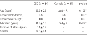
Demographic data of the OCD and control groups are described in Table 1. There were no differences between the two groups in gender distribution, age and in length of education. The clinical interviews of the patients with OCD gave severe scores on the Y-BOCS (27.3±4.4), but normal range for both the HAM-A (10.93±7.78) and HAM-D (7.07±4.63). The control group showed the normal ranges of all the Y-BOCS (0.0±0.0), HAM-A (0.5±1.2), and HAM-D (0.4±0.5).
Demographic information of obsessive–compulsive disorder (OCD) patients and control individuals

Y-BOCS, Yale–Brown Obsessive Compulsive Scale.
* Independent t-test.
† χ2 test.
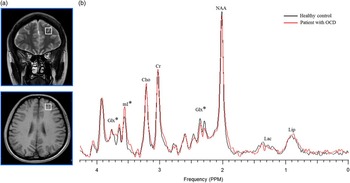
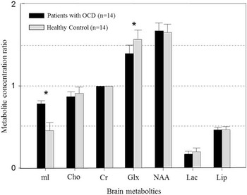
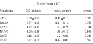
Figs 1 and 2, and Table 2 provide comparative data of the metabolite between two groups in the right DLPFC detected. MRS was performed with a short TE (30 ms) to detect metabolites with short T2 relaxation time, such as Glx, Lip and mI. Although Glx is one of the most abundant neurotransmitters in the normal brain, it is not easy to assign and quantify because of its signal pattern with multiples on the spectrum. Typical Glx peaks overlapped with each other in the chemical shift ranges of 2.20–2.40 ppm for β·γ-Glx and 3.60–3.80 ppm for α-Glx. Nevertheless, these Glx peak groups were combined overall with consideration for contamination with other metabolites such as GABA in β·γ-Glx. The metabolite concentration ratio of Glx/Cr and mI/Cr was significantly different between the OCD patients and healthy controls. However, no significant difference was observed in NAA/Cr, Cho/Cr, Lac/Cr and Lip/Cr. The concentration ratios of Glx/Cr and mI/Cr in OCD patients were significantly decreased by 11% and increased by 52%, respectively, compared with healthy controls (p<0.05).
1H-magnetic resonance localizer image showing placement of the volume of interest in the right DLPFC (a) and representative spectra (b) acquired from a patient with OCD and a healthy control. *Indicates a significant difference of the metabolite concentrations (Mann-Whitney U-test, p < 0.05).

Comparison of the brain metabolites in the right DLPFC between patients with OCD and healthy controls. *Indicates a significant difference between the metabolite concentrations of patients with OCD and healthy controls (Mann-Whitney U-test, p < 0.05).

Variation of the metabolites concentration in the right dorsolateral prefrontal cortex between patients with obsessive–compulsive disorder (OCD) and healthy controls

Cho, choline; Cr, creatine; Glx, glutamine/glutamate; Lac, lactate; Lip, lipid; mI, myo-inositol; NAA, N-acetylaspartate.
* The statistical analysis was performed with the independent t-test.
Discussion
In the right DLPFC the concentration ratios of Glx/Cr of OCD patients decreased by 11%, whereas the concentration ratio of mI/Cr of the OCD patients increased by 52%, as compared with that of the healthy controls (p<0.05). Glx is one of the excitatory neurochemicals released from axon terminals, which bind to and interact with proteins of the neurotransmitter receptors in the postsynaptic membranes. Glx is transformed into glutamines in the receptors, which are sent back to the presynaptic neurons. Furthermore, there is a neurological significance in the change of Glx levels in the DLPFC, which is a subregion of the CSTC circuit. The CSTC circuit can be subdivided into ACC (the affective circuit), DLPFC (the dorsal cognitive circuit) and OFC (the ventral cognitive circuit). Each of these subdivisions is associated with an emotional and compensatory reaction, work remembrance/execution function or suppressive response (Reference Milad and Rauch18). The mechanism of neurochemicals in the normal CSTC circuit involves signals of glutamate released from the prefrontal cortex, which are transmitted to the striatum. Signals of GABA in the activated striatum are sent to the globus pallidus pars interna/substantia nigra pars reticulate (GPi/SNr) through direct and indirect neurologic pathways. Neurons are directly connected to the GPi/SNr in the direct pathway. In the indirect pathway, neurons are connected to the GPi/SNr through the glubus pallidus pars externalis and subthalamic nucleus. In the direct pathway, disinhibited signals are generated to promote activation, whereas an indirect pathway suppresses activation by producing suppressive signals. The amount of GABA signal transmitted from the GPi/SNr to the thalamus via these two pathways adjusts the final amount of glutamate signal at the thalamus that is sent to the prefrontal cortex (Reference Ting and Feng19). Homoeostasis with respect to emotion and activities of cognitive function can be maintained by the feedback mechanism of neurotransmission. OCD is considered to be closely associated with functional abnormalities of the CSTC circuit. An MRS study (Reference Whiteside, Port and Deacon20) revealed that the levels of Glx and NAA in the OCD patients with CSTC circuitry abnormality tend to increase in the OFC, whereas the mI levels are likely to decrease in the head of the caudate nucleus. Moreover, OCD patients have decreased Glx levels in the ACC (Reference Rosenberg, Mirza and Russell21) and an increase of Glx levels in the caudate (Reference Rosenberg, MacMaster and Keshavan10), as compared with healthy individuals. The results were correlated with those of previous studies that had reported an inversely proportional relationship with dimensional changes of the ACC and basal ganglia (Reference Rosenberg and Keshavan22,Reference Szeszko, MacMillan and McMeniman23). This can explain the tonic-phase Glx system in the CSTC circuit (Reference Keshavan24). Activation of tonic Glx in the cerebral cortex plays the function of suppressing the release of phasic glutamate in the striatum. On this basis, a reduction of tonic Glx in the ACC of the cerebral cortex can explain the over-activation of phasic Glx in the striatum. Among the subregions of the CSTC circuit, the OFC and the ACC are primary models for OCD symptoms in most MRS studies. It is assumed that abnormal metabolic processes are also associated with the DLPFC, in addition to either the OFC or the ACC, on the basis of prior reports of functional injury like cognitive injury and structural injury (Reference Togao, Yoshiura and Nakao13) in the DLPFC of OCD patients. In this study, the observation of a reduced level of Glx/Cr in OCD patients suggests that there are metabolic abnormalities of Glx in the ACC and the OFC, which have been reported before, as well as the DLPFC.
Among brain metabolites, mI is associated with the pathophysiology and treatment of mental disorders. mI is a brain metabolite related to important functions of phosphatidylinositol second messenger system (PI-cycle) (Reference Kim, McGrath and Silverstone25). Particularly, mental disorders such as OCD, generalised anxiety disorder or schizophrenia are associated with the PI-cycle, which is a pathway of neurotransmission (Reference Kim, McGrath and Silverstone25,Reference Vaden, Ding and Peterson26). mI transformed by various receptors attached to the PI-cycle, mI concentration or function change of the PI-cycle affects specific neurons (Reference Vaden, Ding and Peterson26). Such an abnormal metabolism of mI likely has extensive effects on neurologic pathways and changes PI-cycle activation, which may induce OCD. Nevertheless, most MRS studies of OCD patients have not found a significant difference in mI levels between OCD patients with or without a drug treatment and the healthy controls (Reference Simpson, Shungu and Bender8,Reference Rosenberg, MacMaster and Keshavan10,Reference Rosenberg and Keshavan22). Therefore, further systematic studies are necessary on the relevance of OCD symptoms and PI-cycle. mI is also a marker of gliocytes. The presumption is that an increase in the level of mI is due to changes in metabolic processes that act to heal the damaged neurons by gliocytes when the neurons are damaged (Reference Chang, Ernst and Grob27). An increased level of mI can be explained by proliferation and increased activation of the gliocytes. Accordingly, with respect to increased mI/Cr levels in this study, OCD-induced abnormality in the CSTC circuit damages DLPFC circuitry, with the level of mI increasing as a healing response to the neuronal damage.
There are several limitations of this study. First, a normality test (Kolmogorov-Smirnov test) was performed for the participants to ensure a normal distribution of data. However, the study included only 14 OCD patients and 14 healthy individuals, which could compromise statistical reliability. Second, the OCD patients received SSRI (escitalopram or fluxovamine), and the effect of these pharmacologic agents could not be avoided. However, this study recruited and enrolled only OCD patients with the Y-BOCS score of 24 or greater. Third, in MRS, the possibility of the white matter contamination on the single voxel which localised in the right DLPFC was not excluded. Accurate segmentation of the white and grey matters in the voxel of interest is needed. Fourth, there was no control region demonstrating the metabolic specificity comparable with that of the right DLPFC. In this current study, the ROI was determined by reference studies (Reference Sachdev, McBride and Loo14,Reference Alvarenga, do Rosário and Batistuzzo15).
In conclusion, this study demonstrates that the OCD patients have differential mI and glutamatergic metabolite concentrations in the DLPFC. These findings will be helpful to understand DLPFC-related glutamatergic and metabolic abnormalities within CSTC circuits associated with OCD.
Acknowledgements
This work was supported by the National Research Foundation of Korea (NRF) grant funded by the Korea government (MSIP) (2015R1A2A2A01007827) and Chonnam National University Hospital Biomedical Research Institute (CRI 15011-1). Authors’ Contribution: S.-E.P. and G.-W.J. have designed the studies and acquired the data. S.-E.P. and N.-G.C. contributed to the analysis and interpretation of the data. S.-E.P. wrote the first draft of the manuscript under the supervision of G.-W.J., and then G.-W.J. approved the final manuscript. In addition, all authors are in agreement with the content of the manuscript. All authors met the following authorship criteria: (1) substantial contributions to the conception and design of this study, as well as acquisition of data and analysis and interpretation, (2) drafting and revising the article critically for important intellectual content, and (3) final approval of the version to be published.
Conflicts of Interest
None.
Ethical Standards
The authors assert that all procedures contributing to this work comply with the ethical standards of the relevant national and institutional committees on human experimentation and with the Helsinki Declaration of 1975, as revised in 2008.