The population of adults with CHD is growing and ageing.Reference Marelli, Mackie, Ionescu-Ittu, Rahme and Pilote 1 This development has been driven mainly by improvement in diagnosis, surgical techniques, and survival over the last five decades.Reference Botto, Correa and Erickson 2 – Reference Moller, Taubert, Allen, Clark and Lauer 4 Ventricular septal defect is one of the most common CHDs. Patients with ventricular septal defect have traditionally been thought to have an unaltered post-surgical prognosis with low complication rates both in the short and the long termReference Bol-Raap, Weerheim, Kappetein, Witsenburg and Bogers 5 – Reference Perrault, Drblik and Montigny 10 ; however, with increasing longevity and cardiac surgery follow-up comes a growing number of physiological and cardiac abnormalities.Reference Möller, Brun and Fredriksen 11 – Reference Heiberg, Schmidt, Redington and Hjortdal 16
Menting et al recently demonstrated increasing late biventricular dysfunction and occurrence of aortic regurgitation in patients with ventricular septal defects.Reference Menting, Cuypers and Opi 15 Our group reported a substantially disrupted right ventricular force–frequency relationship during exercise, indicating that patients’ contractile function was impaired almost 20 years after surgical ventricular septal defect closure.Reference Heiberg, Schmidt, Redington and Hjortdal 16 This finding was directly correlated with a considerable reduction in peak oxygen uptake.Reference Möller, Brun and Fredriksen 11 , Reference Heiberg, Laustsen, Petersen and Hjortdal 12 A number of clinically important right ventricular abnormalities and their underlying mechanisms therefore have yet to be fully described. Such mechanisms may include both myocardial and pulmonary vascular components.
Cardiac magnetic resonance has become the gold standard for non-invasive measurement of cardiac output, and can be performed non-invasively and during exercise. The aim of this prospective, cardiac magnetic resonance study was therefore to determine any differences in cardiac output and its determinants between ventricular septal defect patients and age- and sex-matched controls more than two decades after the patients underwent surgery.
Materials and methods
Design
All consecutive children who underwent surgical closure of a congenital ventricular septal defect at our institution in the years between 1990 and 1995 formed the original study cohort of 182 patients. Review of charts led to exclusion of 117 patients according to the following predefined criteria: coexistence of other CHDs (n=89), operation by ventriculotomy (n=7), associated syndromes such as Down syndrome (n=14), and documented arrhythmia requiring a pacemaker (n=1). Among the remaining 65 patients, who were all operated through a right atrial approach, 25 were randomly selected and invited to participate. From May 2014 to September 2014, 20 patients and 20 control subjects were examined. These patients presented with a homogenous collection of perimembranous (n=14) and muscular (n=6) ventricular septal defects. The same group of patients had previously undergone cardiopulmonary exercise testing and echocardiography as reported elsewhere.Reference Heiberg, Laustsen, Petersen and Hjortdal 12 , Reference Heiberg, Schmidt, Redington and Hjortdal 16 The control subjects were healthy, young adults, matched for age and sex, recruited by local flyers and internet advertisements.
Bicycle ergometry
A MRI-compatible ergometer bicycle (MRI cardiac ergometer, Lode, Groningen, the Netherlands) mounted on the MRI table was used for a supine exercise set-up. Heart rate was monitored using a standard electrocardiography-monitoring system and by pulse oximetry (Nonin 7500FO pulse oximeter with a fibre optic sensor cable 8000FC-30, Nonin Medical, Inc. Plymouth, MN, USA). Each participant was positioned with his or her feet strapped to the ergometer pedals, with the shoulders in a restraining system to minimise movement during exercise, and with maximum knee joint extension standardised to 30°. A standardised workload protocol with automatically adjustable electronic resistance was used. Workloads commenced at 25 W for two minutes and 15 seconds and gradually increased by 25 W every 75th second. The participant was instructed to maintain a cycling speed of 60–70 rounds/minute, and to keep pedalling until he/she was told to stop or was unable to continue. Exercise was continued until the patient reached the submaximal exercise level, which was defined as 75% of maximal heart rate calculated as 220 beats/minute subtracted by age. Once the patient reached this level, the exercise was continued until completion. Exercise intensities were divided into four intervals defined by the percentage of each participant’s obtained submaximal workload: very low (>12.5% ⩽37.5%), low (>37.5% ⩽62.5%), moderate (>62.5% ⩽87.5%), and submaximal (>87.5%).
MRI
MRI during rest and exercise was performed using a 1.5-T Philips Achieva dStream whole-body scanner and an ergometer bicycle as previously described.Reference Hjortdal, Emmertsen and Stenbøg 17 – Reference Pedersen, Pedersen and Pedersen 19 The MRI scanner was equipped with 33 mT/m gradients with a slew rate of 180 mT/m/ms and software release R517 using an 18-cm surface coil and spine coil arrays. Electrocardiography was performed using an integrated four-electrode electrocardiography system. The respiratory rate was monitored using an air-filled belt mounted on the abdomen. Standard scout images of the heart and the large vessels were acquired in three orthogonal planes. Left and right ventricular outflow tract views were located using clinical cardiac MRI guidelines and an interactive MRI platform. On left ventricular outflow tract views, an orthogonal plane was placed at the upper edge of the aortic bulb to obtain transversal views of the ascending aorta. On right ventricular outflow tract views, an orthogonal plane was placed just above the valve to obtain transversal views of the pulmonary trunk. Before commencement of the exercise test, an electrocardiography-triggered, multi-frame, phase-contrast MRI scan of one cardiac cycle of the ascending aorta and pulmonary trunk was recorded at rest. The multi-frame, phase-contrast MRI scans consisted of 50 heart phases obtained during free breathing. The field-of-view was 300×300 mm with an acquisition voxel size of 2.34×3.13 mm and a slice thickness of 7 mm. For the exercise test, a real-time, phase-contrast MRI sequence was used to measure blood flow in the ascending aorta and pulmonary trunk. Measurements were recorded at rest and during continuous exercise with free breathing. The real-time, phase-contrast MRI scans consisted of 170 consecutive, phase-contrast flow acquisitions without electrocardiography triggering, each lasting 81 ms, yielding a frame rate of 12.2 frames/second. The sequence and parameters were as follows: segmented gradient-echo phase-contrast echo planar imaging, field of view of 320×305mm, matrix 88×68 (pixel size, 3.64×4.49 mm2, reconstructed to 1.43×1.44 mm2), 10-mm slice thickness, flip angle 10°, echo planar imaging factor 17, half-scan factor 0.6, echo time 3.48 ms, and repetition time of 10 ms. Velocity encoding varied from 170 to 290 cm/second depending on the exercise level. Real-time MRI scans were obtained of the ascending aorta and the pulmonary trunk in the resting state and at increasing exercise workloads. The order of scans was randomised – first the aorta and second the pulmonary trunk or first the pulmonary trunk and second the aorta – before testing was commenced. At all stages, images were acquired during free breathing and continuous exercise.
Body composition
Bioelectrical impedance analysis (ImpediMed Imp SFB7 bioimpedance analysis device) was performed before initiating the exercise test to assess body composition, defined as the amount of fat mass and fat-free mass of total body weight.
Questionnaire
Each participant was asked to fill out the questionnaire International Physical Activity Questionnaire to assess the weekly physical activity levels.
Data analysis
Blinded, post-hoc MRI analyses were performed by a primary observer who used an in-house developed software (Siswin version 0.9© 2008). Vessel wall contours of the ascending aorta and the pulmonary trunk were traced manually using a four-point adjustable, elliptical region-of-interest tool. From each flow scan, the cardiac output, heart rate, and stroke volume were calculated as mean values. Cardiac output was defined as the antegrade aortic flow subtracted by the retrograde flow. Cardiac output and retrograde flow measures were adjusted for body surface area using the Du Bois method.Reference Du Bois and Du Bois 20 The cross-sectional areas of the ascending aorta and the pulmonary trunk were assessed from an electrocardiography-triggered, multi-frame, phase-contrast MRI scan of one cardiac cycle at resting state and calculated using the traced vessel-wall region of interest. Hereof, the vessel diameter was calculated as for a simple circle. The inter-observer variability was assessed by two blinded observers by measuring 41 real-time MRI scans from six randomly selected participants. Intra-observer variability was assessed by the primary observer with two repeated measurements of 37 real-time MRI scans from five randomly selected participants.
Statistical analysis
Normally distributed continuous data are reported as mean±standard deviations, and are reported as medians with either total ranges or 95% confidence intervals otherwise. Continuous data were compared using unpaired Student’s t-tests, and non-normally distributed data were compared using the Mann–Whitney–Wilcoxon rank-sum test. Correlations were checked using simple regression analyses. Cardiac index measures in relation to percentage of achieved submaximal workloads were plotted with 95% confidence intervals and connecting lines. Statistical significance was defined as a p-value <0.05. Inter- and intra-observer agreements were tested using the intra-class correlation coefficient calculated with a two-way mixed model for absolute difference between measures, as described by Shrout and Fleiss.Reference Shrout and Fleiss 21 All analyses were performed using Stata/IC 12.1 for Mac (StataCorp, Texas, United States of America), and figures were made using Prism 6 for Mac (GraphPad Software Inc., California, United States of America).
Ethics
The Regional Committee on Biomedical Research Ethics of the Central Denmark Region (chart: 1-16-02-12-14) approved the study, which was also registered with clinicaltrials.gov (identifier: NCT02138435). The study protocol conforms to the ethical guidelines of the Declaration of Helsinki. Written informed consent was obtained from each participant before examination in conformity with Danish law. This study was approved by the Danish Data Protection Agency (chart: 2007-58-0010).
Results
The predefined exercise protocol was followed without deviations by all patients and controls. In four participants, the MRI image quality was inadequate at the highest exercise levels due to movement of the upper body. For this reason, the submaximal exercise scan was excluded in two control subjects, and in one patient and one control both the moderate and submaximal exercise scans were excluded.
Perioperative status
The mean age at surgical repair was 2.1±1.4 years. Preoperative echocardiography revealed a mean ventricular septal defect size of 7.9±3.2 mm. A subgroup of nine patients had undergone preoperative catheterisation, demonstrating a mean pulmonary-to-systemic blood flow ratio of 2.5±0.9. Pulmonary artery banding had been performed in two subjects. None of the subjects had significant left or right ventricular outflow tract obstructions, aortic valve regurgitation, or right ventricular hypertrophy before surgery. Among all, 15 had their defects closed by patch insertion and the remaining five with direct suture. The median bypass time was 64 (range 44–122) minutes. All patients were in sinus rhythm; however, nine patients had complete blocks and five patients had incomplete right bundle branch blocks on resting electrocardiography.
Demographics
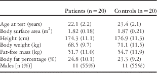
Overall, the basic characteristics of the two groups (Table 1) were similar. No race-/ethnicity-based differences were present. The participants were asymptomatic, and post-surgery check-ups had been concluded for the entire group of patients. Only two patients had traces of a residual shunt.
Demographics and clinical characteristics.

Data presented as mean with standard deviation or a total numbers with percentages
Self-reported exercise habits
Data on self-reported exercise habits are shown in Table 2. The weekly amount of moderate- and high-intensity exercise did not differ between the groups, but patients were more physically active than controls in terms of low-intensity exercise.
Self-reported levels of physical activity.

Data from the International Physical Activity Questionnaire in young adults operated for ventricular septal defect and healthy controls
Cardiac MRI flow measurements
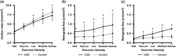
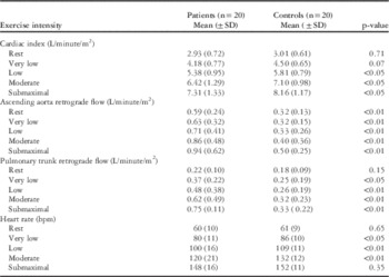
Cardiac index and retrograde flow during rest and exercise are shown in Figure 1 and detailed in Table 3. At rest, cardiac index and heart rate did not differ between patients and controls; however, the retrograde aortic flow was significantly higher in patients (0.59 (±0.24) L/minute/m2) than in controls (0.32 (±0.13) L/minute/m2) (p<0.01) at rest and remained higher throughout exercise.
Hemodynamic flow of the ascending aorta and the pulmonary trunk in VSD-patients and controls at increasing exercise intensity. (a) Cardiac Index related to exercise intensity. (b) Ascending aorta retrograde flow related to exercise intensity. (c) Pulmonary trunk retrograde flow related to exercise intensity. VSD, ventricular septal defect. *, p<0.05 significant difference between patients and controls. Submax., submaximal intensity. Very low intensity (> 12.5%≤37.5%); Low intensity (> 37.5%≤62.5%); Moderate intensity (> 62.5%≤87.5%); Submaximal intensity (> 87.5%).

Haemodynamic measurements by MRI.

Blood flow in the ascending aorta and the pulmonary trunk with corresponding heart rates at different exercise intensities. very low intensity (>12.5% ⩽37.5%); low intensity (>37.5% ⩽62.5%); moderate intensity (>62.5% ⩽87.5%); and submaximal intensity (>87.5%)
As seen in Figure 1a, the cardiac index increased less in the patient group during exercise, with more prominent differences between the groups when they approached the submaximal-intensity exercise level. The patient group’s cardiac index was significantly lower at low, moderate, and submaximal exercise intensities, p<0.05. The cardiac index was 10% lower in patients than in controls at submaximal exercise, p<0.05.
This growing difference in cardiac index corresponded, in part, to a similar pattern of increasing retrograde pulmonary flow with increasing exercise intensity in the patient group. The average difference in cardiac index at maximal exercise was 0.85 L/minute/m2, and the average difference in retrograde pulmonary flow was 0.42 L/minute/m2, thereby accounting for ~50% of the net reduction in cardiac index.
Vessel cross-sectional area and diameter
The patient group had significantly larger aorta dimensions with a mean cross-sectional area of 803±234 mm2 compared with 601±117 mm2 in controls, p<0.01, and a diameter of 32±5 mm compared with 28±3 mm in controls, p<0.01. There was no difference in pulmonary trunk dimensions: the patient group had a mean cross-sectional area of 591±181 mm2 compared with 610±121 mm2 in controls, p=0.70, and a diameter of 27±4 mm compared with 28±3 mm in controls, p=0.59.
Inter- and intra-observer reliability
We found an intra-observer, intra-class correlation coefficient of 0.77 and an inter-observer, intra-class correlation coefficient of 0.79 in post-hoc MRI analysis. The two calculations consisted of 37 and 41 pairs of observations, respectively.
Correlations and subgroup analysis
Using regression analysis, we assessed the relationship between the measured differences in cardiac index at submaximal exercise and the following parameters: age at surgery, coexisting presence of right bundle branch block, closure with patch or direct suture, and preoperative banding. No correlations were found.
Discussion
We demonstrated a significantly lower cardiac index during exercise among patients operated for ventricular septal defects compared with healthy, matched controls in the present study with more than 20 years of follow-up. Similar to other studies, we found a significant retrograde flow in the ascending aorta at rest, which remained unchanged during exercise; however, unexpectedly, we found an increase in retrograde blood flow in the pulmonary trunk during exercise that accounted for ~50% of the reduction in cardiac index at peak exercise.
There are several novel aspects to our study. We are the first to directly measure reduced cardiac output responses to progressive exercise in these patients. Our findings are compatible with our previous demonstration of a 20% impairment in peak exercise capacity,Reference Heiberg, Laustsen, Petersen and Hjortdal 12 which is directly correlated with decreased right ventricular peak systolic velocity during exercise and abnormal right ventricular force–frequency relationships.Reference Heiberg, Schmidt, Redington and Hjortdal 16 The mechanisms behind these abnormalities and their consequences have yet to be fully explored. The present study, however, provides a potential missing link to our understanding of the abnormal right ventricular responses observed in these patients and the underlying mechanisms.
We have previously demonstrated chronotropic incompetence at maximal exercise levels in this patient group; however, this cannot by itself explain the abnormal cardiac index responses at submaximal exercise, as there was no difference in heart rate between patients and controls. Similarly, it seems unlikely that the differences in cardiac index can be explained by simple deconditioning in the patient group. Indeed, the self-reported levels of physical activity were similar or perhaps even higher in the patient group.Reference Heiberg, Laustsen, Petersen and Hjortdal 12
The reason for the abnormal cardiac index response at submaximal exercise appears to lie in the physiological responses to exercise and a hitherto unrecognised pathological response in our patients after ventricular septal defect closure. We demonstrated a substantially higher retrograde aortic flow in the patient group with a relatively stable magnitude in response to exercise. Under normal conditions, retrograde aortic flow occurs in diastole in the ascending aorta, partly reflecting the coronary artery blood flow.Reference Bogren and Buonocore 22 , Reference Bogren, Klipstein and Firmin 23 The normal value for coronary blood flow is 122 ml/minute.Reference Ganz, Tamura and Marcus 24 Indexed to an average-sized human being with a body surface area of 1.81 m2 Reference Verbraecken, Van de Heyning, De Backer and Van Gaal 25 , this corresponds to 0.07 L/minute/m2, which is ~12% of the retrograde flow measured in our patients at rest. The increased flow in our patients is presumably attributed to co-existing aortic regurgitation,Reference Ambrosi, Faugere and Desfossez 26 even though none of the patients had “significant” aortic or pulmonary valve insufficiencies at their last clinical follow-up. Our results correspond to those reported in a newly published follow-up study by Menting et al,Reference Menting, Cuypers and Opi 15 showing that the incidence of mild-to-moderate aortic regurgitation nearly doubled from 11 to 21% over a 20-year follow-up period in a group of 178 patients operated for ventricular septal defects. Furthermore, a recently published follow-up study by Gabriels et al demonstrated the presence of dilated ascending aorta in 15% of 47 patients operated for ventricular septal defects.Reference Gabriels, Van De Bruaene and Helsen 27 This predisposition to aortic regurgitation and dilated ascending aorta after early ventricular septal defect repair is not easily explained; nevertheless, we believe that it may be related to the increased aortic diameters that we observed in the patient group. Given that the amount of aortic retrograde flow remains essentially constant throughout exercise, it is unlikely to be directly related to our observation of an impaired cardiac output response and would not explain the findings of impaired right ventricular responses in other studies.Reference Möller, Brun and Fredriksen 11 , Reference Menting, Cuypers and Opi 15 , Reference Heiberg, Schmidt, Redington and Hjortdal 16
The key to understanding these responses may lie in our unexpected observation of progressive retrograde pulmonary flow with increasing exercise workload in the patient group. Indeed, at least 50% of the difference in cardiac index between controls and patients was directly attributable to the amount of retrograde flow in the pulmonary trunk. As this is an unexpected and novel finding, we have no mechanistic data to explain this phenomenon. There are, however, different pathophysiological mechanisms that could explain the altered pulmonary trunk blood flow, either individually or in conjunction. In patients with pulmonary incompetence, a backward blood flow into the right ventricle occurs during diastole. The backward flow is likely to cause an increase in retrograde flow in the pulmonary trunk. Our findings could be caused by a progressive pulmonary incompetence with increasing exercise intensity, which affects the pulmonary trunk blood flow. In patients with repaired tetralogy of Fallot, however, the amount of pulmonary regurgitation decreases during exercise.Reference Roest, Helbing and Kunz 28 Despite the fact that the pathophysiology of patients with tetralogy of Fallot differs from that of patients with ventricular septal defect, we find it contradictory that a simultaneous increase in retrograde pulmonary flow would be present during exercise. A second potential explanation could lie in the fact that pulmonary trunk flow is altered as demonstrated in patients with pulmonary hypertension. These patients have an increased relative retrograde flowReference Bogren, Klipstein and Mohiaddin 29 , Reference Helderman, Mauritz, Andringa, Vonk-Noordegraaf and Marcus 30 that coincides with the appearance of a rotational blood flow vortex in the pulmonary trunk.Reference Reiter, Reiter and Kovacs 31 The vortex occurs in the primary flow direction, typically below the right pulmonary artery at the posterior wall of the pulmonary trunk.Reference Reiter, Reiter and Kovacs 31 Clearly, ventricular septal defect patients are not to be compared with patients with pulmonary hypertension, but a similar backward-bounce phenomenon is a conceivable explanation for the increased retrograde pulmonary flow in ventricular septal defect patients. In the present study, however, we used a real-time phase-contrast MRI sequence that does not allow for further data quantification or flow visualisation needed to confirm the existence of a vortex.
Even though we are not able to fully describe the mechanistic background of diverging haemodynamic parameters demonstrated in the patient group, this does not diminish the importance of the underlying fundamentals of our findings. Several studies have previously pointed to a slightly increased pulmonary vascular resistance as the main culprit in explaining right ventricular dysfunction in these patients.Reference Möller, Brun and Fredriksen 11 , Reference Heiberg, Ringgaard, Schmidt, Redington and Hjortdal 14 – Reference Heiberg, Schmidt, Redington and Hjortdal 16 , Reference Heiberg, Redington and Hjortdal 32 As an example, Möller et alReference Möller, Brun and Fredriksen 11 demonstrated an abnormal right ventricular systolic pressure response to exercise despite a normal right ventricular pressure at rest, suggesting exercise-induced dysfunction in the pulmonary artery endothelium. Therefore, it seems logical to consider the possibility that the increased retrograde pulmonary flow reflects an exercise-induced increase in pulmonary vascular resistance. This, in turn, would not only directly affect total forward flow in the pulmonary artery, and hence cardiac output, but also reduce right ventricular contractile efficiency, adding to the net loss in cardiac index that we observed. Interestingly, the pulmonary artery dilation seen in childhood in the presence of an intra-cardiac shunt is remodelled in adult life.
Limitations
The abnormal retrograde flow patterns and the increased aortic diameter were secondary and unexpected outcomes of this study. Future follow-up studies should include echocardiographic evaluation of aortic and pulmonary valve functions during exercise to confirm these indirect observations. Furthermore, an assessment of pulmonary vascular resistance during exercise and its relationship with our observed flow patterns would help clarify individual responses and the relationship between resistance and flow in these patients. It should also be noted that our observations were made at submaximal exercise levels. In a previous study,Reference Heiberg, Laustsen, Petersen and Hjortdal 12 we showed a markedly lower peak oxygen uptake of ~20% among patients operated for ventricular septal defects at maximal exercise. Later, this was revealed to be directly correlated with an abnormal right ventricular force–frequency relationship.Reference Heiberg, Schmidt, Redington and Hjortdal 16 In the present study, the exercise tests were terminated at a submaximal exercise level because of practical circumstances related to exercising in an MRI scanner. It is therefore feasible that an even larger difference in cardiac output could have been demonstrated at higher exercise levels.
A real-time, phase-contrast MRI sequence was chosen for the exercise test instead of an electrocardiography-triggered MRI sequence because of an unreliable electrocardiography signal during exercise. This entailed that exercise could be completed without pauses, breath holds, or consequent decreases in heart rate; however, the imaging quality was lower than during regular electrocardiography-triggered MRI sequences. Moreover, because of the shorter length of the pulmonary trunk compared with the aorta, measurement of the pulmonary trunk flow was more susceptible to cardiac movements due to heavy breathing during exercise.
Thorough considerations were made to reduce participant discomfort and at the same time obtain measurements during exercise, causing the real-time, phase-contrast MRI sequence to be shortened to lower the overall MRI scan time. Consequently, this limited the temporal resolution that could influence the assessment of dynamic changes during exercise; however, the implemented MRI technique was sensitive enough to identify significant haemodynamic differences between groups.
Finally, these patients were operated in the 1990s. Today’s management with earlier surgery and optimised treatment may have eliminated the problems described in this article; however, thousands of patients were operated in the last century and they live with the consequences. Thus, not only do we have an obligation to pay attention to any potential late complications in this cohort of patients, but also have implications for the late follow-up of patients operated upon in a more contemporary era.
Conclusion
We demonstrated an exercise-related impairment in cardiac index in young adults operated for ventricular septal defects in childhood. The reduced cardiac index appears to be related to increasing retrograde flow in the pulmonary artery with progressive exertion. Our findings link a previously demonstrated reduction in peak exercise capacity to cardiac performance and provide intriguing new evidence to support a direct and indirect effect of exercise-induced pulmonary vascular abnormalities on global cardiovascular performance.
Acknowledgements
The authors are profoundly thankful to Christine Bauer-Jensen for her patience, dedication, and solid contributions to the data analysis.
Financial Support
The study was supported by Aarhus University and by grants from the following Danish Foundations: Eva & Henry Fraenkels Memorial Foundation; The Kids Heart Fund; Helga & Peter Korning’s Fund; and the Kirsten Anthonius Memorial Trust.
Conflicts of Interest
None.
Ethical Standards
The authors assert that all procedures contributing to this work comply with the ethical standards of The Regional Committee on Biomedical Research Ethics of the Central Denmark Region and with the Helsinki Declaration of 1975, as revised in 2008, and the study has been approved by the institutional committees.