Introduction
The International Space Station (ISS) provides an ideal research platform for astrobiology experiments devoted to the studies of central problems, such as:
-
• origin and evolution of life (e.g. Fridlund et al. Reference Fridlund2010);
-
• possibility of interplanetary life transport in the harsh space environment (Weber & Greenberg Reference Weber and Greenberg1985; Nicholson Reference Nicholson2009).
The scientific consortium Response of Organisms to the Space Environment was established by the scientists interested in studying astrobiology questions by use of the ISS, more exactly the EXPOSE facility (Horneck et al. Reference Horneck1999; Rabbow et al. Reference Rabbow, Horneck, Rettberg, Schott, Panitz, Hatton, Dettmann, Demets and Reitz2009) to be mounted on external pallets of the ISS. The experiment Phage and Uracil Response (PUR) joined the consortium. The environmental factors of outer space (temperature, pressure/vacuum, various types of radiations, etc.) strongly differ from the environment known on the Earth. Their effects on living systems, applied either separately or combined, have been investigated in space and at ground-based space simulation facilities (Horneck et al. Reference Horneck, Bücker, Reitz, Reinhardt, Dose, Martens, Menningmann and Weber1984; Rabbow et al. Reference Rabbow, Rettberg, Panitz, Drescher, Horneck and Reitz2005; Nicholson et al. Reference Nicholson2011).
The main goal of the PUR experiment was to study the solar ultraviolet (UV) climate in Earth's orbit in terms of the biological risk i.e. to measure the biologically effective UV dose in long-term exposure. The basic principle of the biological UV dosimetry is to detect and quantify the lesions induced by UV photons either in the nucleic acid (NA) or in the NA bases e.g. uracil (Rontó et al. Reference Rontó, Gáspár, Gróf, Bérces and Gugolya1994; Gróf et al. Reference Gróf, Gáspár and Rontó1996; Kerékgyártó et al. Reference Kerékgyártó, Gróf and Rontó1997). Namely, in living systems NAs/NA bases are the main target molecules for UV photons. Accordingly, we used in our biological UV-dosimeter bacteriophage T7 and polycrystalline uracil as detectors (sensors) of UV damage.
Applying biological UV dosimetry in space allows us to answer the following questions:
-
1. Are the biological dosimeters that have been successfully used on the Earth's surface (Rontó et al. Reference Rontó, Gáspár, Gróf, Bérces and Gugolya1994; Gróf et al. Reference Gróf, Gáspár and Rontó1996; Fekete et al. Reference Fekete, Vink, Gáspár, Bérces, Módos, Rontó and Roza1998; Bérces et al. Reference Bérces, Fekete, Gáspár, Gróf, Rettberg, Horneck and Rontó1999; Munakata et al. Reference Munakata, Kazadzis, Bais, Hied, Rontó, Rettberg and Horneck2000) suitable for UV dosimetry in low Earth orbit (LEO), too, and do they function correctly in the harsh extraterrestrial environment?
-
2. To what extend can the UV dosimetry results, coming from the solar electromagnetic waves, be affected by cosmic particle radiation present in space, too?
-
3. Can the damage efficiency of the extraterrestrial UV radiation in space be different from that experienced on the Earth's surface (Setlow & Setlow Reference Setlow and Setlow 1965 ; Lindberg & Horneck Reference Lindberg and Horneck1991; Munakata et al. Reference Munakata, Saito and Hieda1991; Douki et al. Reference Douki, Zalizniak and Cadet1997; Kovács et al. Reference Kovács, Fekete, Bérces and Rontó2007)?
Experimental section
Composition of the detectors
The UV dosimeter developed in the Research Group for Biophysics consists of a detector/sensor system containing either a nucleoprotein (bacteriophage T7; ATTCC11303-B7) or polycrystalline uracil (Sigma-Aldrich Co.U 0750). For the ISS experiment PUR both detector materials/samples were used in homogeneous thin films sedimented (phage T7) or evaporated (uracil; Kerékgyártó et al. Reference Kerékgyártó, Gróf and Rontó1997) on the inner side of a small calcium–fluoride plate of 2 mm thickness. This plate was the upper window of a vacuum-tightly closed stainless steel case of a diameter of 16 mm. Before accommodation, the quality of all 32 thin films was controlled by their absorption spectra obtained by use of a Unicam UV–Vis spectrophotometer. The transmission of the windows had a constant value (90%) for the wavelengths between 200 nm and 7 μm, which was taken into account in the evaluation of the physical UV exposure. To prevent spurious oxydization of the samples, the cases were filled with the inert gas (argon).
In the experiment, PUR 2×16 samples were accommodated in the ESA's astrobiological exposure facility attached to the external platform of Zveszda (EXPOSE-R)Footnote 1 facility on the external pallet of the ISS in compartment 4 of tray 2 (Rabbow et al. Reference Rabbow2014). On the surface of the compartment, 4×4 samples were accommodated: these were the ‘irradiated’ samples; and 4×4 samples were located in the second layer beneath the irradiated samples: these were the ‘dark’ samples.
Function of the detectors
The biological UV dosimetry is based on damage of the NA bases induced by UV photons: as a consequence of the absorption of a UV photon a variety of photolesions can be formed, such as cis–syn cyclobutadipyrimidines (CPDs) and pyrimidine (6–4) pyrimidone photoproducts (6–4 PPs), the latter can photoisomerize into related Dewar valence isomers upon exposure to wavelengths about 320 nm. Other photolesions induced by UV radiation are photohydration of cytosine, oxidation of guanine into 8-oxo-7,8-dihydroguanine and the formation of adducts between either two adjacent adenine bases or between adenine and a vicinal thymine (Fisher & Johns Reference Fisher, Johns and Wang1976; Douki et al. Reference Douki, Court, Sauvaigo, Odin and Cadet2000; Cadet et al. Reference Cadet, Sage and Douki2005). In dry DNA and bacterial endospores 5-(5-thymyl)-5,6-dihydrothymine, the so-called ‘spore photoproduct’ (SP) is the main photoproduct induced by UV, and upon UV exposure of DNA or cells in vacuum trans–syn CPDs are induced as additional pyrimidine photoproduct (Lindberg & Horneck 1991; Hieda et al. Reference Hieda, Suzuki, Hirono, Suzuki and Furuzawa1994). UV at wavelengths λ<130 nm induces in addition single- and double-strand breaks, as demonstrated after synchrotron irradiation of pBR322 DNA (Hieda et al. Reference Hieda, Suzuki, Hirono, Suzuki and Furuzawa1994).
In our biological dosimeters, such pyrimidine dimers can be formed either inside the NA of the phage particle or in the polycrystalline uracil film (thin layer). The measurement of the biologically weighted UV radiation (biological UV dosimetry) is based on the damage of the NA bases induced by UV photons.
The formation of cyclobutane pyrimidine dimer is the leading UV-photoproduct in the uracil or phage T7 detector (Rontó et al. Reference Rontó, Gáspár, Fekete, Kerékgyártó, Bérces and Gróf2002) and it is indicated by a decrease of the absorbance (optical density (OD)), the extent of the decrease is proportional to the biologically effective dose (Gróf et al. Reference Gróf, Gáspár and Rontó1996; Bérces et al. Reference Bérces, Fekete, Gáspár, Gróf, Rettberg, Horneck and Rontó1999). In their arrangement on the ISS the dosimeters were passive, i.e. the OD-measurements could only be performed before and after the irradiation in space. After finishing the irradiation and retrieval of the samples the directly weighted biological UV dose/health risk was measured by spectroscopy in the UV range of 200–400 nm. The extent of UV damage efficiency to uracil and phage T7 were expressed by the change of the OD at 288 nm wavelength after exposure (ΔOD) related to the OD value before irradiation (ODo).
Arrangement for measuring the dose–effect relation
Our dosimeters function passively; thus the dosimetry results represent the biological dose which is cumulated from the start till finishing of the exposure. To have some information on the time course of the dose accumulation, the following experimental arrangement was used. In EXPOSE-R, the compartment of experiment PUR was divided into four quarters, each of them contained four samples: two uracil and two phage T7 thin films. The quarters were covered by neutral-density filters of different UV transmission properties: the first filter 100%, the second, third and fourth 1, 0.01 and 0.0001% transmission for the UV radiation, respectively. This arrangement allowed us to study the kinetics of the development of UV effects both on uracil and phage T7 in dependence of the exposure in a dose range of six orders of magnitude.
Results
The EXPOSE-R facility flew around the Earth's orbit approximately 2 years long. Taking into account the model calculation of RedShift BVBAFootnote 2 this exposure time corresponded to an equivalent exposure of 2687 h or 16 700 MJ m−2 for the full solar radiation and 11 000 MJ m−2 for the UV components at a range of 100–400 nm.
Dark samples
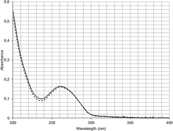
The dark samples were not exposed to UV radiation, however, one cannot exclude the exposure to cosmic/particle radiation present in LEO. The ionizing radiation can destroy the samples; the effect can be detected by UV spectroscopy. Figure 1 represents the typical results, where the spectrum of phage T7 after flight (dashed line) coincides practically with that before flight (full curve). The uracil spectra (not shown) before and after flight did not change significantly; however, a slight change occurred which can be interpreted by different water content of the polycrystalline uracil samples. The difference can be eliminated by a slight heating (40 °C) of the samples. The second derivatives of the two spectra correspond each other indicating that the uracil bases remained intact.
Absorption spectrum of bacteriophage T7 dark sample before (full line) and after (dashed line) 2 years long space flight.

Exposed samples
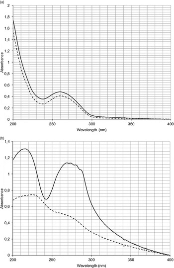
A typical change of the phage T7 spectrum (dashed line) is demonstrated in Fig. 2(a) induced by an exposure on the EXPOSE-R facility below the neutral filter of 0.01% transparency. Taking into account the model calculation of RedShift (RedShift 2011) and the attenuation power of the filter in this case the physical UV dose was 1.10 MJ m−2.
(a) A typical absorption spectrum of a solar UV irradiated bacteriophage T7 sample after the flight, that was located beneath a neutral density filter of 0.01% transparency (dashed line); the calculated physical dose was 1.1 MJ m−2 for total UV. For comparison the starting spectrum, obtained before flight, is also demonstrated (full line). (b) Absorption spectra of a solar UV irradiated uracil sample before (full line) and after flight (dashed line) at a filter transparency of 0.01%; the calculated physical dose was 1.1 MJ m−2 for total UV.

For comparison the original spectrum before the flight of the same sample (full line) is indicated too. The irradiation of the uracil samples is presented in Fig. 2(b). The decrease of the OD values is more pronounced than for phage T7 at the same attenuation (at 0.01% transparency). The significant decrease in the absorption band about 260 nm may be suggestive of a notable photo-induced degradation of uracil. This is confirmed by the uracil samples that were exposed without any attenuation (100% transparency), where the absorption maxima completely disappeared indicating the total destruction of the uracil structure (data not shown).
Dose–effect relations
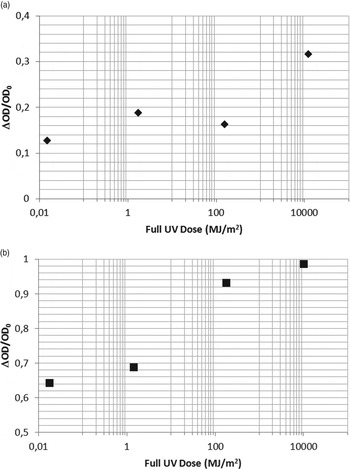
Summarizing the 32 initial (before flight) and 32 final (after flight) spectra of the dark and irradiated samples, two UV dose–NA-damaging effect functions were calculated. The graphics for uracil damage in dependence on UV dose is presented in Fig. 3(a), whereas in Fig. 3(b), the dose–effect function for phage T7 is demonstrated. The damage to uracil as well as to phage T7 are expressed as change of the OD at 288 nm wavelength after exposure (ΔOD) related to the OD value before irradiation (ODo). Comparing the slopes of the two graphics the uracil proved to be more sensitive to the extraterrestrial UV radiation.
(a) Dose – dependence of damage to bacteriophage T7 by extraterrestrial solar UV (110–400 nm) radiation, measured in the EXPOSE-R facility. (b) Dose – dependence of damage to uracil by extraterrestrial solar UV (110–400 nm) radiation measured in EXPOSE-R facility.

Discussion
Based on the obtained results we can try to answer the questions enumerated in the Introduction. Dale Warren Griffin (Reference Griffin2013) started his recent article with the citation: ‘The study of viruses has not been applied in astrobiology to the extent of other disciplines’ (Stedman & Blumberg Reference Stedman and Blumberg2008). However, the use of viruses, specifically bacterial viruses (bacteriophages), seems to be profitable in astrobiology for searching the limits of life. Viruses, among them bacterial viruses too, can survive extreme environmental conditions (temperature, pressure, chemical composition and various types of radiations) and specific bacteriophages can be applied for quantification of the biological effects (Rontó et al. Reference Rontó, Bérces, Fekete, Kovács, Gróf and Lammer2004). Phage T7 on the Earth's surface is used in solution/suspension, whereas for the ISS experiment PUR sedimented phage T7 samples (Hegedüs et al. Reference Hegedüs, Módos, Rontó and Fekete2003) were used in vacuum-tightly closed sample cases. They survived the space environment without measurable damage, if shielded from solar UV radiation.
Concerning Uracil thin film as detector, it has been shown that the evaporated polycrystalline substance can function correctly both on the Earth's surface (Bérces et al. Reference Bérces, Fekete, Gáspár, Gróf, Rettberg, Horneck and Rontó1999; Munakata et al. Reference Munakata, Kazadzis, Bais, Hied, Rontó, Rettberg and Horneck2000) and in simulated space conditions (Fekete et al. Reference Fekete, Módos, Hegedüs, Kovács, Rontó, Péter, Lammer and Panitz2005); however, because of the high vacuum in space, the sample holders containing the evaporated uracil samples were vacuum-tightly closed.
The dark samples, more exactly, their spectra or second derivatives either for uracil or for phage T7, did not show significant changes indicating that no or very few destructions in the uracil ring or of the phage DNA were induced by cosmic ray particles during the 2 years long flight. Berger et al. (Reference Berger, Hajek, Bilski, Vanhavere, Horwacik, Körner and Reitz2012) measured in the ESA's Astrobiological Exposure facility attached to the Columbus module facility a total dose range for ionizing radiation of 121±6–214±16 mGy during the 1.5-year flight time, depending on shielding by external structures. For the slightly longer lasting EXPOSE-R mission a total dose range of 225–320 mGy was measured (Berger et al. Reference Berger, Hajek, Bilski and Reitz2014; Rabbow et al. Reference Rabbow2014). In agreement with our results, it was shown in the SESLO experiment (Nicholson et al. Reference Nicholson2011) that the ionizing radiation environment did not influence significantly the growth characteristics neither of the wild-type nor of radiation sensitive Bacillus subtilis strains. On the ISS, the ionizing radiation dose rates are expected to be 12–15 times lower than those in the Organism/Organic Exposure to Orbital Stresses free-flying nanosatellite of NASA mission. It should be noted that DNA components in the solid state are highly resistant to chemical effects of ionizing radiation. Therefore, measurable effects caused by cosmic radiation are not expected to be found in our biological dosimeters.
Considering the dose–effect relations obtained for uracil and phage T7 samples, the slope of the uracil dose–effect curve was higher than that of the phage T7 curve, indicating that uracil is more sensitive for extraterrestrial UV at wavelengths λ>110 nm than bacteriophage-DNA inside the phage particle. The higher resistance of the phage-NA can be one of the factors in the protection of living systems from the harsh space environmental conditions. The higher resistance of the phage-NA than uracil can be interpreted by several factors, e.g. by presence of the protein envelop and the additional presence of purine bases in the phage-DNA (50%) which are less sensitive to UV photons than pyrimidine bases (Blackburn et al. Reference Blackburn, Gait, Loakes and Williams2006). In addition, one cannot exclude the spectral composition of the extraterrestrial solar radiation, too. In the extraterrestrial solar UV radiation, the role of the short wavelengths (<280 nm) can partly contribute to the inactivation/destruction and partly to protection/ preservation. The microorganisms, containing NA can be damaged by solar UV radiation, but the damage can be reverted with a certain probability by the same or similar UV photons (Setlow & Setlow Reference Setlow and Setlow1965). The photoreversion can be added to the protective effect of water ice and various minerals surrounding the microorganisms (Horneck et al. Reference Horneck, Rettberg, Reitz, Wehner, Eschweiler, Strauch, Panitz, Starke and Baumstark-Kahn2001), if they participate in interplanetary transport. For the protective effect of short wavelength UV a further argument is given by Horneck et al. (Reference Horneck, Eschweiler, Reitz, Wehner, Willimek and Strauch1995), who found in the inactivation action spectrum of B. subtilis HA 101 spores (in the wavelength range 210–290 nm) a decrease of the killing efficiency of the UV radiation at about 230 nm. This effect coincides with our results obtained concerning the photoreversion in simulation experiments (Kovács et al. Reference Kovács, Fekete, Bérces and Rontó2007). Namely the highest reversion efficiency of the photoproducts was found at the wavelengths 220–240 nm. Significant secondary photoreactions may have occurred upon exposure of uracil and bacteriophage T7 to the huge fluence of UV radiation, as was the case during the EXPOSE-R mission. This may include the photo-induced reversal of CPDs as well as the conversion of 6–4 PPs into the Dewar valence isomers. The latter reaction may also lead to changes in the distribution of dimeric pyrimidine photoproducts and makes it difficult to interpret the effective biological dose data. Similarly, a reversion of the SP at extremely high UV doses has been observed in the DNA of spores of B. subtilis 168, that were exposed to solar extraterrestrial UV radiation (λ>110 nm) within the parallel SPORES experiment of the EXPOSE-R mission (Panitz et al. Reference Panitz, Horneck, Rabbow, Rettberg, Moeller, Cadet, Douki and Reitz2014).
The kinetics of the UV dose accumulation was detected and measured indirectly by using neutral density filters of different attenuation power. For the passive dosimeters like ours, the only possible approach was the application of the filters; however, recently the precursor of a continuous measuring system was constructed and manufactured in cooperation with the student group of Budapest Technical University (Goldschmidt et al. Reference Goldschmidt, Kovaliczky, Szabó, Rontó and Bérces2012; Grósz et al. Reference Grósz, Gorócz, Futó, Vatali, Szabó and Bérces2013), which functioned correctly in a student experiment sponsored by European Space Agency.
Acknowledgement
This work was supported by ESA-PRODEX and ESA-PECS No. 98006, 98008 and 98070.