INTRODUCTION
An extensive tissue migration before arriving at the intestine is one of the life-cycle characteristics shared by a number of intestinal nematodes. Despite the fact that animal parasitism has arisen several times in the phylum Nematoda independently (Blaxter, 2003), intestinal nematodes in different clades exhibit a similar migration pattern which involves connective tissues or liver and lungs, e.g. Ascaris and Toxocara in Clade III (Spiruria), Strongyloides in Clade IV (Tylenchia), and Necator and Nippostrongylus in Clade V (Rhabditia). Although the meaning of tissue migration remains a puzzle to date, tissue migration should be associated with larval development. Larvae do not proceed to development when they are trapped in connective tissues (Daly, Mayrhofer and Dent, 1999; Dent et al. 1999).
Strongyloides venezuelensis is a rodent intestinal nematode native to rats. In S. venezuelensis infection, third-stage infective larvae penetrate skin, migrate through connective tissues, and enter the circulation. Then larvae break into the airway and reach the small intestine (Takamure, 1995), where they lodge themselves in mucosal epithelial layer (Maruyama et al. 2000, 2003). It should be noted that from skin to intestinal mucosa, S. venezuelensis larvae continue invading host tissues. If larval development and tissue migration is associated, larvae in one tissue component might be clearly distinguishable in invasion specificity and protein expression related to tissue invasion from ones in other tissues. In addition, such changes would be irreversible if they were developmentally regulated. Identification of stage-specific developmental markers will lead us to discover specific factors that stimulate larval development.
In the present study, we examined tissue invasion abilities and expression of stage-specific proteins of larvae along the migration stages. Our study indicated that S. venezuelensis experiences major changes in connective tissues and in lungs, with marked alteration of tissue migration ability associated with protein expressions.
MATERIALS AND METHODS
Parasites and animals
Strongyloides venezuelensis has been maintained in our laboratory in male Wistar rats as previously described (Maruyama, Nawa and Ohta, 1998). A NZW rabbit, ICR mice, and C57BL/6 mice were purchased from SLC Japan (Hamamatsu, Japan). All animals were kept and handled under the guidelines of the Center for Experimental Animal Science, Nagoya City University Graduate School of Medical Sciences.
Preparation of larvae in different developmental stages
Infective larvae (L3i) were prepared by a fecal culture method. Lung-stage larvae were collected as follows; male ICR mice were subcutaneously inoculated with 30000 L3i, and lungs were removed 75 h post-infection (p.i.), homogenized with a Polytron PT-MR3000 (Kinematica AG, Littau, Switzerland) at 20000 rmp for a few sec. Lung homogenates were wrapped with Kimwipe papers and incubated in phosphate-buffered saline (PBS) at 37 °C for 1·5 h. Emerging worms were collected and extensively washed with sterile PBS. Mucosal worms were collected from the small intestine of infected ICR mice 85 h p.i. They consisted mainly of fourth-stage larvae with a few young adult worms (Wertheim, 1970). In this article, larvae recovered from small intestines 85 h p.i. are referred to as mucosal larvae. For the preparation of larvae migrating through connective tissues, L3i were injected into the peritoneum of ICR mice, and recovered 20h p.i. Larvae recovered from the peritoneum were checked for tissue migration ability to make sure that they could be used as connective tissue-migrating larvae (see Results section).
Infections
For percutaneous infection, 1500 L3i suspended in PBS were applied onto the shaved abdominal skin of mice under anaesthesia.The L3i suspension was then covered with a 1·0 cm GF/D filter paper (Whatman, Maidstone, UK), and left for 10 min to allow larvae to penetrate. The number of larvae that penetrated skin was estimated by counting the larvae remaining in the filter paper. On average, 70–80% of applied larvae completed skin penetration in 10 min. Intraduodenal implantation was performed as previously described (Maruyama et al. 2000). Briefly, the abdomen of recipient mice was opened under anaesthesia, and 1000 larvae suspended in 0·3 ml of PBS were injected into the duodenum. Subcutaneous injection, intraperitoneal injection, and oral infection were performed according to the standard protocol.
Zymography
Protease activities of parasite antigens were analysed in substrate gel electrophoresis (zymography) with Novex gel and buffer system (Invitrogen, Carlsbad, CA, USA). Worm antigens were separated on 12% Tris-glycine polyacrylamide gels containing 0·05% casein, and incubated overnight with zymogram developing buffer (Invitrogen, Carlsbad, CA, USA). Gels were then stained with Coomassie Brilliant Blue, in which protease activities were visualized as clear bands in a blue background. In order to determine the type of proteases, the inhibitors, 1 mM 4-(2-aminoethyl) benzenesulfonyl fluoride (AEBSF), 10 μM E-64, 10 mM ethylenediaminetetraacetic acid (EDTA), or 1 mM 1,10-phenanthroline were added to the developing buffer. All protease inhibitors were purchased from Sigma-Aldrich (St Louis, MO, USA).
Antibody against S. venezuelensis adhesion molecules
Antisera against adhesion molecules secreted from adult worms were prepared as has been described (Maruyama et al. 2003). Adult worms recovered from infected rats were washed extensively with PBS, and then incubated (approximately 15000/dish) in 100 mm tissue-culture plastic dishes (Nunc, Denmark) in PBS at 37 °C overnight. After the incubation, dishes were vigorously washed with PBS by repeated pipetting until the worms and eggs were completely removed. Then adhesion molecules, which formed visible insoluble complexes on the bottom of the dishes, were scraped off with a cell scraper (Cell Lifter; Costar, USA). A female NZW rabbit was immunized by injecting adhesion molecules emulsified in Freund's complete adjuvant (CFA) for the first immunization, and Freund's incomplete adjuvant (IFA) for 2 subsequent immunizations. Antigen was administered at 3-week intervals. Blood samples were collected from the rabbit before and after immunization, and the specificity of the antibodies was confirmed.
Fluorescent immunohistochemistry
Mice were infected with 5000 of S. venezuelensis L3i, and tissue samples (skin; 25 h p.i., lung; 75 h p.i., small intestine; 85 h p.i.) were collected, and fixed with 10% formalin. Paraffin-embedded tissue sections were dewaxed, blocked with 5% BSA in PBS, and incubated with rabbit antisera diluted to 1 to 1000, followed by FITC-conjugated goat anti-rabbit IgG (Santa Cruz Biotechnology, Santa Cruz, CA, USA). Binding of antisera was observed and photographed with an Olympus BX51 fluorescent microscope and DP70 digital camera (Olympus, Tokyo, Japan). To stain adhesion molecules secreted in vitro, lung larvae, mucosal larvae, and adult worms were incubated in culture slides (LAB-TEK II; Nunc, Denmark) overnight at 37 °C. Culture slides are slide glasses equipped with plastic chambers for cell culture. Slides were washed vigorously with PBS and fixed with methanol. Antibody staining with rabbit antisera was performed as described above.
Quantification of secreted adhesion molecules
The amount of secreted adhesion molecules was semi-quantified by using an enzyme-linked immunosorbent assay (ELISA) with rabbit antisera. Larvae and adult worms were incubated in wells of microtitre plates (100 worms/well) at 37 °C overnight, to allow worms to secrete adhesion molecules on the bottom of the wells. Wells were then washed vigorously and blocked with PBS containing 3% BSA, and incubated with rabbit antisera. Antibodies bound to the wells were detected with horse-radish peroxidase (HRP)-conjugated goat anti-rabbit IgG (Kirkegaard & Perry Laboratories, USA), followed by substrate ABTS (2,2-azino-di (3-ethyl-benzthiazoline sulfonate); Kirkegaard & Perry Laboratories). Optical densities were read using a Bio-Rad microtitre plate reader.
RESULTS
Skin penetration and connective tissue migration
S. venezuelensis infection starts with skin penetration by third-stage infective larvae (L3i). We first examined larvae of different developmental stages for percutaneous and subcutaneous infections. We found that only L3i but not lung larvae nor mucosal larvae penetrated skin. The reason that lung larvae could not reach the intestine was not due to the failure of connective tissue migration, because they could infect successfully by subcutaneous inoculation (Fig. 1). Mucosal larvae were no longer able to migrate through connective tissues. These findings indicated that third-stage larvae lost skin penetrating ability in connective tissues, then they further lost connective tissue migrating ability until arriving at the small intestine after passing through lungs.

Fig. 1. Fecal egg count of mice infected either percutaneously or subcutaneously with infective larvae (circle), lung larvae (triangle), and mucosal larvae (rectangular). Each mouse was inoculated with 1000 worms, and the number of eggs per gram feces was measured daily. All values are mean±S.D. (n=5).
We then determined when larvae became unable to penetrate skin. Because it was difficult to obtain enough fresh larvae from connective tissues by incubating minced muscles and skin after subcutaneous inoculation, we injected L3i into the peritoneal cavity, recovered them and used them as connective tissue migrating larvae. Intraperitoneally inoculated L3i completed the development normally (Fig. 2A), and larvae recovered from the peritoneum 20 h after inoculation were fully capable of migrating through the connective tissue (Fig. 2B). Based on these data, we considered the use of intraperitoneally injected L3 as a substitute for connective tissue migrating larvae should be justified. We then tested L3i and L3 recovered from the peritoneum 3 h and 20 h after intraperitoneal injection for skin penetration. Skin penetration was significantly reduced 3 h after intraperitoneal injection, and completely lost after 20 h (Fig. 3).

Fig. 2. (A) Fecal egg count of mice after subcutaneous (closed circle) or peritoneal (open circle) infections. Each mouse was inoculated with 1000 infective larvae and the number of eggs per gram feces was measured daily. (B) Fecal egg count of mice after subcutaneous infections with infective larvae (closed circle) or larvae recovered from the peritoneum 20 h after injection (open circle). All values are mean±S.D. (n=5).

Fig. 3. Percutaneous invasion by Strongyloides venezuelensis larvae. Approximately 1500 infective larvae, and larvae recovered from the peritoneum 3 h and 20 h after injection were applied on the abdominal skin of mice for 10 min. Larvae that remained in the filter papers, and larvae recovered from lungs 3 days after infection were counted. All values are mean±S.D. (n=5). *P<0·00001.
Metalloprotease from infective larvae
In S. stercoralis, infective larvae are known to produce a matrix metalloprotease that facilitates skin penetration (McKerrow et al. 1990; Brindley et al. 1995). Although no such enzymes have been reported in S. venezuelensis, we anticipated a similar enzyme in S. venezuelensis because species of the genus Strongyloides are closely related each other (Dorris, Viney and Blaxter, 2002). We performed casein zymography with S. venezuelensis antigens (Fig. 4). It turned out that L3i of S. venezuelensis had a protease activity at 40 kDa, which was similar in molecular weight as S. stercoralis protease, Ss40 (Brindley et al. 1995). To determine the class of this protease, inhibitors including AEBSF (serine protease inhibitor), E-64 (cysteine protease inhibitor), EDTA (divalent cation chelator), and 1,10-phenanthroline (metalloprotease inhibitor) were added to the reaction buffer (Fig. 4B). Protease activity at 40 kDa was completely abolished by EDTA and 1,10-phenanthroline, but not by AEBSF and E-64, indicating that this enzyme was a metalloprotease. Then we checked the metalloprotease activity in antigens of L3i and L3 recovered from the peritoneum 3 h and 20 h after intraperitoneal inoculation. Zymograms clearly showed that the metalloprotease activity at 40 kDa significantly reduced 3 h after inoculation, and completely disappeared after 20 h, which was apparently associated with skin penetration (Fig. 4C).

Fig. 4. Metalloprotease activity in Strongyloides venezuelensis infective larvae. (A) Metalloprotease at 40 kDa was expressed in infective larvae, but not in other developmental stages (Lane 1, infective larvae; Lane 2, lung larvae; Lane 3, mucosal larvae; Lane 4; adult worms). (B) Sensitivity to protease inhibitors. (C) Disappearance of metalloprotease activity after intraperitoneal injection.
Mucosal invasion and adhesion molecules
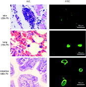
Oral and intraduodenal inoculation showed similar infection patterns, in which lung larvae and mucosal larvae, but not L3i, were capable of settling down in intestinal mucosa (Fig. 5). Adult S. venezuelensis secrete adhesion molecules from the mouth that play an important role in mucosal invasion (Maruyama et al. 2000, 2003). Therefore we stained tissue sections of connective tissues, lungs, and intestinal mucosa of infected mice with antisera against S. venezuelensis adhesion molecules (Fig. 6). Fluorescent immunohistochemistry revealed that lung larvae and intestinal worms were positive for adhesion molecules, which was in accordance with invasion capacity. As expected, no connective tissue larvae stained positive. We further examined if lung larvae were capable of secreting adhesion molecules. Adult worms, mucosal larvae, and lung larvae were incubated overnight in culture wells, and secreted adhesion molecules were stained with the antiserum. We found that lung worms secreted adhesion molecules, although the amount was less than that secreted by mucosal larvae and adult worms (Fig. 7).

Fig. 5. Fecal egg count of mice infected either orally or intraduodenally with infective larvae (circle), lung larvae (triangle), and mucosal larvae (rectangle). Each mouse was inoculated with 1000 worms, and the number of eggs per gram feces was measured daily. All values are mean±S.D. (n=5).

Fig. 6. Expression of adhesion molecules in vivo. Tissue sections of infected mice were stained with antisera against Strongyloides venezuelensis adhesion molecules. In lung sections, the oesophageal portion of the larvae was positive.

Fig. 7. Secretion of adhesion molecules. (A) Lung larvae, mucosal larvae, and adult worms were incubated in wells of culture slides, and secreted adhesion molecules were stained with antisera. Mucosal larvae and adult worms secreted large amounts of adhesion molecules. (B) Infective larvae, lung larvae, mucosal larvae, and adult worms were incubated in wells of microtitre plates overnight, and secreted adhesion molecules were detected with serially diluted rabbit antisera followed by HRP-conjugated anti-rabbit IgG.
DISCUSSION
From skin to intestinal mucosa, S. venezuelensis larvae invade several different tissues that have distinct physiological and biochemical characteristics. Our present study clearly demonstrates that S. venezuelensis larvae experience major changes in invasion specificity during tissue migration. Infective larvae (L3i) were able to penetrate skin, but once inside the host tissues, they lost this ability within a few hours. Skin-penetration was associated with an infective larva-specific metalloprotease activity, suggesting that this enzyme was essentially important in skin penetration.
Recent analysis of S. stercoralis infective larva transcripts demonstrates that Ss40 is an astacin-like metalloproteinase (Gomez Gallego et al. 2005). From the similarities in molecular weight and expression pattern observed in the present study, S. venezuelensis L3i metalloprotease seems to be a homologue of Ss40. Infective larvae of Ancylostoma caninum also secrete a developmentally regulated astacin family metalloprotease upon host stimulation (Zhan et al. 2002). It is interesting that both Strongyloides (Clade IV) and Ancylostoma (Clade V) use astacin family metalloproteases for host entry. Free-living Caenorhabditis elegans has exceptionally large numbers of astacin genes, at least one of which is thought to be a digestive enzyme (Mohrlen, Hutter and Zwilling, 2003). The use of astacine-like metalloproteases might have been the first step for animal parasitism in free-living soil nematodes.
Studies on Strongyloides infections have shown major changes taking place after larvae enter the host. The protein composition of S. venezuelensis larvae, analysed by 2D gel electrophoresis, markedly changes after skin penetration (Tsuji et al. 1993), in which infective L3 were different from lung and adult-stage worms. Expression of daf-7, a key ligand in one of the three pathways that control dauer larva development in Caenorhabditis elegans, peaks in the L3i stage with little or no expression in the parasitic stages. RNA level of daf-7 in L3i is reduced by larval penetration of host skin or development in the host (Crook et al. 2005).
Connective tissue larvae further develop to become lung-stage larvae that secrete adhesion molecules. Adhesion molecules are important in attachment to the intestinal epithelium and are used to construct the wall of worm tunnels in the intestinal epithelium (Maruyama et al. 2003). It is not known what kind of stimuli elicit the expression of adhesion molecules in S. venezuelensis larvae. Obviously there is no need for transport through the stomach and exposure to bile, which is required for excystation of metacercariae and protozoan cysts (Gold, Stein and Tzipori, 2001).
The exact mechanism by which larvae break into the air spaces from the circulation is not known. However, considering that the average width of larvae (15–20 μm; Wertheim, 1970) is larger than the size of lung capillaries (7–10 μm), larvae in the circulation would embolize and break lung capillaries. Intra-alveolar bleeding in S. venezuelensis infection is obvious, which could be fatal in humans (Kinjo et al. 1998). Once inside the air spaces, larvae might be passively carried by cilliar movement of the bronchial epithelium, or they may actively crawl up the airway.
Our present study revealed successive changes in migration ability in developing S. venezuelensis larvae. The changes probably involve not only invasion-related mechanisms but many other biological processes as well. A major question now would be what promotes transformation of larvae: from free-living L3i to connective tissue larvae, lung larvae, and to mucosal larvae. It has been reported that a temperature shift from 25 °C to 37 °C stimulates development of free-living L3i to the parasitic stage, judged by morphology and protein composition (Tsuji and Fujisaki, 1994). However, RNA level of daf-7 is not influenced by a temperature shift (Crook et al. 2005). One single factor would not be enough to make sufficient changes in developing larvae. Because larvae migrate through blood and air spaces, physiological conditions such as oxygen pressure (Kita and Takamiya, 2002) should be taken into account in addition to host-derived biomolecules.