Introduction
Head and neck squamous cell carcinoma (SCC) is the sixth leading cause of cancer and cancer-related deaths worldwide.Reference Jemal, Bray, Center, Ferlay, Ward and Forman1 Tobacco and alcohol use are major risk factors for head and neck cancer development. Additionally, human papilloma virus (HPV) is a causative factor in a growing number of oropharyngeal head and neck cancers, and thus another important risk factor.Reference Gillison, Koch, Capone, Spafford, Westra and Wu2–Reference Fakhry and Gillison4 Human papillomavirus is present in up to 50 per cent of all oropharyngeal cancers; there is currently an epidemic of oropharyngeal cancer related to high-risk HPV infection, despite an overall decrease in head and neck cancer incidence worldwide. Given the improved response of HPV-associated tumours to surgical and non-surgical treatment options, appropriate and accurate initial clinical staging is becoming increasingly important for managing head and neck cancer patients.
Despite advances in surgical and non-surgical treatments, the prognosis for head and neck SCC patients continues to be poor, usually because of early regional lymphatic spread of disease. The overall five-year survival rate for head and neck SCC patients is less than 50 per cent.Reference Kim, King and Agulnik5 Neck status is the most important prognostic factor and an important driver of overall tumour–node–metastasis (TNM) staging.Reference Spiro, Alfonso, Farr and Strong6, Reference Layland, Sessions and Lenox7
Overall staging and TNM classification are based on the primary tumour size, the size, number and sidedness of the involved cervical nodes and evidence of distant metastasis. Involvement of the cervical lymph nodes, even in the absence of distant metastasis, has grave prognostic significance and greatly decreases the overall survival rate. Accurate and reliable evaluation of the neck for regional metastasis is therefore a key prognostic and therapeutic factor in head and neck cancer. Tools used for tumour staging include physical examination, fine needle aspiration (FNA) biopsy with pathological evaluation and various imaging modalities. Regional metastasis to the neck can be difficult to ascertain on physical examination. Ultrasound, magnetic resonance imaging (MRI), computed tomography (CT) and positron emission tomography (PET) with contrast-enhanced CT have been used extensively. Reported sensitivities and specificities for MRI and CT range from 36 per cent to 94 per cent and from 50 per cent to 98 per cent, respectively.Reference Roh, Yeo, Kim, Lee, Cho and Choi8 The ability to combine PET with CT made PET-CT a popular modality for initial cancer staging and post-treatment evaluation of head and neck cancer patients.
Although PET-CT is commonly used to evaluate head and neck cancer before and after treatment, the indications for using this imaging technique are still under debate. Positron emission tomography computed tomography imaging can be a powerful, highly sensitive and highly specific diagnostic tool, with the highest reported sensitivity (96 per cent) and specificity (98.5 per cent) for evaluating cervical lymph node involvement.Reference Schwartz, Ford, Rajendran, Yueh, Coltrera and Virgin9 However, PET-CT is easily affected by the metabolic, inflammatory and infectious changes associated with many head and neck tumours. These may introduce false positive results and decrease PET-CT specificity, thus incorrectly increasing tumour stage and leading to potential over-treatment with multiple therapeutic modes such as surgery, radiation and chemotherapy. Most reports on the use of PET-CT for head and neck SCC examine its ability to evaluate the treatment response; however, few studies have evaluated the ability of PET-CT to correctly determine the initial tumour stage.Reference Roh, Yeo, Kim, Lee, Cho and Choi8, Reference Schwartz, Ford, Rajendran, Yueh, Coltrera and Virgin9 Furthermore, the extent to which the primary tumour site and clinical factors such as HPV and/or p16 tumour positivity, tobacco and alcohol use, age, sex, and race affect the ability of PET-CT to determine the initial head and neck SCC stage is poorly understood. This study aimed to evaluate the ability of PET-CT to determine initial cancer staging compared with the ‘gold standard’ of pathological evaluation of gross surgical specimens, and to evaluate the effect of tumour and clinical characteristics on PET-CT staging.
Materials and methods
Patient selection
The study protocol was approved by the institutional review board of Johns Hopkins Hospital, Baltimore, Maryland, USA. Head and neck SCC patients who underwent pre-operative PET with contrast-enhanced CT and primary surgery between July 2010 and January 2013 at the Johns Hopkins Medical Institutions were included in the retrospective analysis. Exclusion criteria were a lack of pre-operative PET-CT, PET-CT performed at an external facility, or a previous history of surgery, radiation or chemotherapy to the head and neck region. Pre-operative PET-CT imaging reports were retrospectively compared with operative pathology reports for primary tumour resection and neck dissection specimens. Tumour–node–metastasis classification and overall stage as determined by PET-CT were compared with those based on the pathological evaluation of surgical specimens.
Clinical data
Medical records of all eligible study patients were retrospectively reviewed. Clinicopathological and demographic information were collected, including age, sex, race, smoking and alcohol exposure, tumour site, HPV tumour status, TNM classification and overall stage based on pathological and PET-CT findings (as defined by the American Joint Committee on Cancer). Human papillomavirus status was determined by both p16 immunohistochemical analysis and in situ hybridisation for high-risk HPV DNA detection.
Staging by diagnostic imaging
General practice is for patients evaluated at Johns Hopkins Hospital, including those who do not form part of a prospective clinical trial, to undergo initial PET-CT and other anatomical scans (CT and/or MRI) for staging, especially those with oral cavity and oropharynx tumours or those with an advanced stage tumour. All pre-operative PET-CT scans were performed at the Johns Hopkins Medical Institutions using a commercially available PET-CT scanner (Discovery LS; GE Healthcare, Little Chalfont, UK). Contrast enhancement was used in all cases and a standard PET-CT protocol was followed. Patients fasted for at least four hours before PET acquisition and received an intravenous injection of fluorine-18 fluoro-deoxyglucose (8.14 MBq/kg). A tracer uptake phase of approximately 60 minutes was allowed and 68Ge transmissions scans were used to generate a transmission map. The CT portion used a multidetector helical CT scanner (LightSpeed Plus; GE Healthcare). The parameters for CT acquisition were: 140 kV, 80 mA, 0.8 seconds per CT rotation and a pitch of 2; CT images were reconstructed at 2.5-mm intervals. All CT examinations were performed after intravenous administration of iodinated contrast material unless contraindicated by a prior allergic reaction or renal insufficiency. Acquired PET and CT data were combined into a single superimposed image and analysed by a dedicated team of fellowship-trained head and neck neurological and nuclear medicine radiologists.
Surgical pathology staging
All patients underwent surgery comprising primary tumour resection and neck dissection. Neck dissection involved the surgical resection of all clinically appropriate cervical lymph node levels, including those with PET-CT determined or suspicious positive nodes. Neck dissection specimens were analysed by head and neck pathologists. Pathology reports were reviewed and data was collected retrospectively. Pathological TNM classification and overall stage were determined according to American Joint Committee on Cancer definitions.
Statistical analysis
The PET-CT determined TNM classification and overall stage were compared with the pathological evaluation of the surgical specimen. The sensitivity, specificity, positive predictive value, negative predictive value and accuracy of this staging method were calculated for each primary tumour site. Agreement between PET-CT and pathological TNM classification and overall staging was examined by univariate and multivariate analyses both overall and for each primary tumour site. Descriptive statistics were used to identify important variables in the study population. The association of age, sex, race, smoking, alcohol, tumour classification and primary tumour site with agreement between PET-CT and pathological stage investigated by univariate and multivariate logistical regression. For multivariate analysis, the final model was selected using a backward selection technique that eliminated variables whose p values were greater than 0.1. All statistical tests were two sided and statistical significance was set at a p value greater than 0.05. All analyses were performed using R (version 2.15.1; Vienna, Austria; http://cran.r-project.org) and SAS (version 9.3, SAS Institute, Cary, North Carolina, USA).
Results
Tumour staging in the study population
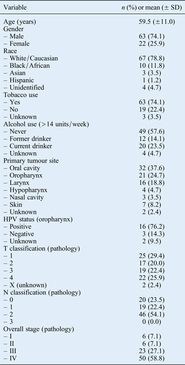
Of the 328 patients diagnosed with head and neck SCC who received surgical treatment, 99 were excluded because of previous chemotherapy or radiotherapy and 144 because pre-operative PET with contrast-enhanced CT was either not obtained at our institution or not obtained at all, with investigation involving other imaging modalities. The remaining 85 patients who underwent pre-operative PET-CT and primary surgery without pre-operative radiotherapy or chemotherapy were included in the analysis. Summary clinicopathological and demographics data are shown in Table I. The patient cohort had an average age of 59.5 years (range 30–87 years) and 74.1 per cent were male. Most patients were Caucasian (78.8 per cent) and used tobacco (74.1 per cent) but not alcohol (37.6 per cent). Primary anatomical sites included the oral cavity (32 patients; 37.6 per cent), oropharynx (21 patients; 24.7 per cent), larynx (16 patients; 18.8 per cent), hypopharynx (4 patients; 4.7 per cent), nasal cavity (3 patients; 3.5 per cent) and skin (7 patients; 8.2 per cent); the primary site was unknown in 2 patients (2.4 per cent). Cutaneous and sinonasal SCCs were specifically included because the aim was to correlate PET-CT and pathology of the head and neck. Of the 21 patients with primary oropharyngeal tumours, 16 (76.2 per cent) were positive for both human papillomavirus (HPV) DNA and p16. Tumour classification by pathology was T1 for 25 (29.4 per cent), T2 for 17 (20.0 per cent), T3 for 19 (22.4 per cent), T4 for 22 (25.9 per cent) and TX (i.e. unknown) for 2 (2.4 per cent). Node classification by pathology was 46 patients with N2 (54.1 per cent), 20 with N0 (23.5 per cent) and 19 with N1 (22.4 per cent) regional disease. No patients had N3 disease verified by pathology. Overall staging by pathology was 6 stage I tumours (7.1 per cent), 6 stage II tumours (7.1 per cent), 23 stage III tumours (27.1 per cent) and 50 stage IV tumours (58.8 per cent).
Study population characteristics

SD = standard deviation; HPV = human papillomavirus; T = tumour; N = node
Accuracy of imaging for detecting regional metastases
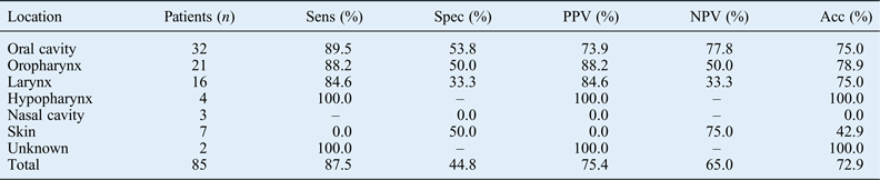
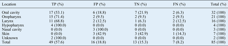
For all primary anatomical sites, PET-CT identified regional cervical metastasis with a sensitivity of 87.5 per cent and a specificity of 44.8 per cent, compared with the gold standard of pathological review of surgical resections. The positive predictive value was 75.4 per cent, the negative predictive value was 65.0 per cent and the overall accuracy (defined as the percentage of cases correctly determined to be positive or negative) was 72.9 per cent (Table II). There were 13 true negatives (15.3 per cent), 7 false negatives (8.2 per cent), 49 true positives (57.6 per cent) and 16 false positives (18.8 per cent) for PET-CT (Table III).
Utility of PET-CT for detecting regional metastases
PET-CT = positron emission tomography computed tomography; Sens = sensitivity; Spec = specificity; PPV = positive predictive value; NPV = negative predictive value; Acc = accuracy
Accuracy of PET-CT for detecting regional metastases
PET-CT = positron emission tomography computed tomography; TP = true positive; FP = false positive; TN = true negative; FN = false negative
Tumour and node staging: comparison
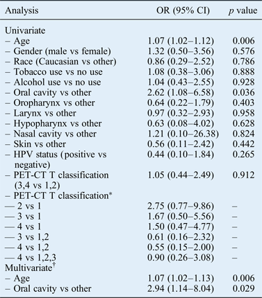
Univariate analysis of the agreement between PET-CT and pathological node classification revealed significant odds ratios (ORs) for age (1.07, 95 per cent confidence interval (CI) 1.02 to 1.12) and oral cavity site (2.62, 95 per cent CI 1.08 to 6.58). Gender, race, tobacco or alcohol use, other primary anatomical tumour site, HPV status, and high PET-CT tumour classification (T3 or T4) were not significant. Analysis of PET-CT and pathological node classification agreement by pathological tumour classification revealed no significant difference for smaller and larger primary tumours. Backward variable selection of age and oral cavity tumours by multivariate modelling revealed significant ORs for age (1.07, 95 per cent CI 1.02 to 1.13) and oral cavity site (2.94, 95 per cent CI 1.11 to 7.74; Table IV).
Pathology vs PET-CT node classification
*Wald confidence intervals. †Backward variable selection, only age and oral cavity are selected into the multivariate model. PET-CT = positron emission tomography computed tomography; OR = odds ratio; CI = confidence interval; HPV = human papillomavirus; T = tumour
Overall staging: comparison
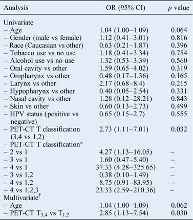
Univariate analysis of the agreement between overall PET-CT stage and overall pathological stage revealed significant ORs for PET-CT tumour classification of greater than or equal to T3 (2.73, 95 per cent CI 1.11 to 7.01), T2 vs T1 (4.27, 95 per cent CI 1.13 to 16.05), T4 vs T1 (37.33, 95 per cent CI 4.28 to 325.65) and T4 vs T1–3 (23.33, 95 per cent CI 2.59 to 210.36), and a nearly significant OR for age (1.04, 95 per cent CI 1.00 to 1.09). Multivariable modelling with backward variable selection of age and high PET-CT tumour classification (T3 or T4) revealed a significant OR for high PET-CT tumour stage (2.85, 95 per cent CI 1.11 to 7.31) and a nearly significant OR for age (1.04, 95 per cent CI 1.00 to 1.09). All other anatomical tumour sites and clinical factors such as HPV DNA and/or p16 tumour positivity, sex, race, smoking, and alcohol intake were not significantly associated with PET-CT and pathological staging agreement (Table V).
Pathology vs PET-CT for overall staging
*Wald confidence intervals. †Backward variable selection, only age and oral cavity are selected into the multivariate model. PET-CT = positron emission tomography computed tomography; OR = odds ratio; CI = confidence interval; HPV = human papillomavirus; T = tumour
Discussion
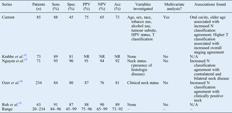
This study aimed to evaluate the ability of PET with contrast-enhanced CT to determine the initial cancer stage compared with the gold standard of pathological evaluation of gross surgical specimens and to evaluate the extent to which tumour and clinical characteristics affect its ability to do so. Positron emission tomography computed tomography is commonly used for managing head and neck cancer, especially for evaluating the tumour response to treatment. The utility and ideal timing of PET-CT to evaluate the post-treatment response has been an area of active research; many studies found that 12 weeks after completion of therapy was the most appropriate time point.Reference Gupta, Master, Kannan, Agarwal, Ghsoh-Laskar and Rangarajan10, Reference Lango, Myers and Garden11 However, the evaluation of PET-CT for initial head and neck SCC staging has been less thoroughly investigated, and the indication for this imaging modality and its appropriateness are still under debate. This series addresses important gaps in the literature by reporting the staging performance of PET-CT and investigating by multivariate analysis which variables might affect this (Table VI). This study found PET-CT to have a sensitivity of 87.5 per cent, specificity of 44.8 per cent, positive predictive value of 75.4 per cent, negative predictive value of 65.0 per cent and accuracy of 72.9 per cent for detecting regional metastases. It also demonstrated a greater level of agreement between PET-CT and pathological evaluation in determining node classification in older patients and in oral cavity primary tumours in both univariate and multivariate analyses. Finally, there was a greater level of agreement between PET-CT and pathological evaluation in determining the overall stage of primary tumours with a higher pathological tumour classification in both univariate and multivariate analyses. Patient age may also influence the agreement for overall staging, but a larger cohort size is necessary to investigate this in more depth.
Literature comparing PET-CT with histology
PET-CT = positron emission tomography computed tomography; Sens = sensitivity; Spec = specificity; Acc = accuracy; PPV = positive predictive value; NPV = negative predictive value; T = tumour; N = node; NR = not reported; N/A = not applicable
Our analysis of the ability of PET-CT to detect regional metastases revealed an overall false positive rate of 18.8 per cent. Given the extent to which positive regional metastasis affects the prognosis and treatment of head and neck SCC, PET-CT should be used judiciously for initial staging in head and neck cancer patients. Because the false positive rate is so high, clinicians should not rely on this modality alone for clinical staging, but should instead integrate their clinical acumen, physical examination findings, other imaging modalities, pathological evaluation of FNA biopsy and other clinical prognostication factors to inform their clinical judgement. The most valuable use of PET-CT during initial staging remains the detection of distant metastasis or second primary cancer, for which the negative predictive value is high (99.7 per cent).Reference Kim, Roh, Yeo, Kim, Lee and Choi16, Reference Yi, Kim, Lee, Choi, Nam and Kim17
Data from our cohort of 85 patients suggest that PET-CT staging is more closely correlated with the gold standard of pathological evaluation in older patients, a higher tumour classification and oral cavity cancers. Advanced age might cause fewer false positives because older patients have less lymphoid tissue in the head and neck region as lymphoid tissue atrophies with age: reduced lymphoid tissue mass would lead to less inflammatory drainage and, thus, fewer false positives. Primary tumours with a higher tumour classification are larger with a greater potential for regional lymphatic spread and seeding into the drainage system; they therefore have a higher pre-test probability of nodal involvement, which reduces the proportion of false positives. The oral cavity is well supplied with lymphatic vessels, and occult lymphatic metastasis has been shown to occur in up to 34 per cent of oral cavity tumours.Reference Shah18, Reference Duvvuri, Simental, D'Angelo, Johnson, Ferris and Gooding19 Furthermore, the incidence of contralateral or bilateral lymph node metastasis of oral cavity cancer was reported to be as high as 30 per cent in some studies,Reference Olzowy, Tsalemchuk, Schotten, Reichel and Harreus20, Reference Kowalski, Bagietto, Lara, Santos, Tagawa and Santos21 leading to a high pre-test probability and fewer false positives. Myers et al. demonstrated similar results, with PET accurately detecting the presence or absence of cervical metastasis in all oral cavity SCC patients in a limited cohort of eight patients.Reference Myers, Wax, Nabi, Simpson and Lamonica22
It is possible that the imaging and pathology reports used in this retrospective analysis may be less standardised than those obtained using a prospective methodology. For this analysis, all pathology reports were reviewed by senior head and neck pathologists and all PET-CT studies were analysed by board-certified, fellowship-trained, head and neck neurological and nuclear medicine radiologists. Hopkins criteria for interpreting head and neck PET-CT have been established but not yet validated in the pre-operative setting.Reference Marcus, Ciarallo, Tahari, Mena, Koch and Wahl23 Additionally, because these PET-CT and pathology reports were in fact used to determine disease staging and influence oncology treatment, we feel that their use is appropriate for this analysis, which aimed to evaluate the performance of PET-CT in determining the initial pre-operative staging. Our institutional PET-CT protocol used high injection doses of contrast agent, which should improve sensitivity. Despite this, the analysis demonstrated a false negative rate of almost 10 per cent. Given the high false positive and negative rates for evaluating locoregional involvement, PET-CT scans may be better used for staging patients by primarily evaluating the presence of distant metastases. However, these data also justify studying this subject further with prospective analyses.
• Positron emission tomography computed tomography has not been adequately investigated for initial head and neck squamous cell carcinoma staging
• Regional metastasis classification was most reliable in older patients and oral cavity primary tumours
• Overall staging was most reliable for primary tumours with a higher pathological tumour classification
• Multimodality staging should be used at the initial patient evaluation
Conclusion
Although PET with contrast-enhanced CT can be valuable for initial patient staging, pre-operative PET-CT has substantial false positive (18.8 per cent) and false negative (8.2 per cent) rates for evaluating regional metastasis in head and neck SCC. Compared with the gold standard of pathological evaluation of neck dissection specimens, PET-CT imaging evaluation of regional metastasis in head and neck SCC is most reliable in older patients and for oral cavity primary tumours. Furthermore, overall PET-CT staging is most reliable in patients with higher grade T3 and T4 tumours. Despite its utility, PET-CT should remain only one of the many tools used to evaluate patients, and we should continue to use multimodality staging, including clinical examination, ultrasound, MRI and FNA biopsy of the neck to verify the initial staging and more completely evaluate the potential for regional spread at patient presentation.
Larger prospective studies are required to evaluate the role of PET-CT in head and neck SCC in greater detail. Currently, several Radiation Therapy Oncology Group and Eastern Cooperative Oncology Group trials are being performed for head and neck SCC. These could prospectively evaluate the utility of these modalities for initial cancer staging. Thus, subgroup analysis of these larger trials is warranted to provide more useful prospective data.
Acknowledgements
We gratefully thank two clinical biostatisticians, X Zhou and H Wang, for performing all statistical analyses, including univariate and multivariate analyses.