




No CrossRef data available.
Article contents
RAPD method useful for distinguishing Leishmania species: design of specific primers for L. braziliensis
Published online by Cambridge University Press: 05 December 2003
Abstract
The technique of Random Amplification Polymorphic DNA allows fragments of the genome to be amplified by means of polymerase chain reaction (PCR) without previous knowledge of their sequences. The protozoa of the genus Leishmania present great genetic variability, making it difficult to characterize the different species. A method is developed with a single 10-mers long primer, which allows the species L. braziliensis, L. mexicana, L. infantum, L. tropica, L. chagasi, L. amazonensis and L. major to be differentiated. These products amplified by RAPD have also facilitated the design of some primers that amplify L. braziliensis DNA exclusively.
Keywords
Information
- Type
- Research Article
- Information
- Copyright
- 2003 Cambridge University Press
INTRODUCTION
Human pathologies due to different Leishmania species include localized cutaneous lesions (L. major, L. tropica and L. aethiopica, or the L. mexicana complex), cutaneous lesions that may later become mucosal lesions (L. braziliensis complex), or disseminated infections that affect various organs (L. infantum, L. donovani, L. chagasi).
Characterization of the different species based on isoenzymatic or immunological methods is complicated due to the additional genetic variability that exists among strains (Banuls, Hide & Tibayrenc, 1999). Several different molecular methods have so far been used to identify the different Leishmania species. These include characterization of DNA by RAPD (Hanofi et al. 2001), polymerase chain reaction (PCR) (Rodrigues et al. 2002), PCR-ELISA (Piñero et al. 1999), simple sequence repeat anchored polymerase chain reaction (SSR-PCR) (Volpini et al. 2001), or permissively primed intergenes polymorphic-polymerase chain reaction (PPIP-PCR) that allows Old World Leishmanias to be differentiated (Eisenberger & Jaffe, 1999).
In this study, a useful method was developed for both primer design and for characterizing L. braziliensis, L. mexicana, L. infantum, L. chagasi, L. tropica, L. amazonensis and L. major by RAPD.
MATERIALS AND METHODS
Parasites
The parasite species used were L. braziliensis (MHOM/PE/95/LQ8), L. mexicana (MNYC/BZ/62/M397), L. infantum (MON183/LEM2572), L. chagasi (MCAN/BR/98/ARAςATUBA), L. tropica (MON58/LEM2578), L. amazonensis (MHOM/BR/77/LTB0016) and L. major (LM 252). Four L. braziliensis strains isolated from the ulcers of patients from Cuzco (Peru) diagnosed as suffering from L. braziliensis were submitted by Dr Pacheco (University of San Antonio de Cuzco, Peru). Four strains isolated from patients in Barcelona (Spain) infected with L. infantum were submitted by Dr Gallego (University of Barcelona, Spain).
DNA preparation
The Leishmania culture and DNA preparation was performed following the protocols described by Gonzalez et al. (2002). The resulting DNA concentration was measured with a Gene Quant spectrophotometer (Pharmacia Biotech, Cambridge, UK).
In order to isolate single colonies of each Leishmania spp., the Goldberg & Chiari (1980) protocol was followed (except that enriched RPMI medium was used instead of the LIT medium used with trypanosomes). Human DNA was obtained from skin biopsies of healthy individuals.
DNA amplification
Twenty 10-mer primers were tested with the Random Amplified Polymorphic DNA KIT C (Operon Technologics, Alameda, CA, USA) by PCR assay. Then, 10 ng DNA of each Leishmania strain, 1U Taq polymerase (Roche, New Jersey, USA), 20 mM of each nucleotide and 20 pmol of each primer (only 1 by PCR) were dissolved in PCR buffer (Roche, New Jersey, USA) to a final volume of 30 μl. The amplification protocol for RAPD was a denaturation step at 95 °C for 3 min, followed by 30 cycles at 95 °C for 1 min, at 40 °C for 1 min at 72 °C for 1 min 30 s and a final elongation step at 72 °C for 7 min (Williams et al. 1990). Finally, the PCR products were separated on 1% agarose gels.
When the OPC 20 primer-Cy5 was used, the amplifying conditions were the same; however, PCR products were processed in the automatic system ALFexpress (Amersham Pharmacia Biotech, Cambridge, UK) and the results were processed with the Fragment Manager program (Amersham Pharmacia Biotech, Cambridge, UK), which provides a graph version of the results.
The PCR conditions for primers L-28a TTTATGCGGTGGTCTGT and L-28b GCTACCTGCTTGCATT were 30 ng of leishmania DNA, 1U Taq polymerase (Roche, New Jersey, USA), 20 mM of each nucleotide and 30 pmoles of each Lb-28 primer in PCR buffer (Roche, New Jersey, USA), to a final volume of 50 μl. The visualization was in 1·5% agarose gels. The primers were designed by the Gene-Runner programme.
DNA fragment labelling
DNA fragments from RAPD and PCR amplification of L. braziliensis were isolated from an agarose gel (Qiagen gel extraction kit, Qiagen, Hilden, Germany) and labelled with digoxygenin by PCR. The reaction conditions were the same as used for OPC20 primer and Lb28 primers but with the nucleotide mix also containing dUTP-digoxygenin (Roche Diagnostics, Mannheim, Germany). After the labelling reaction, the product was electrophoresed again in 1% agarose gel and the probe isolated with the Qiagen gel extraction kit (Qiagen, Hilden, Germany).
Hybridization
Amplified products from L. braziliensis, L. mexicana, L. infantum, L. chagasi, L. tropica, L. amazonensis and L. major were blotted on a nylon positive-charged membrane (Roche Diagnostics, Mannheim, Germany) by alkaline transfer. After air drying, the membrane was hybridized by the established protocol (Sambrook, Fritsch & Maniatis, 1989). The hybridization was run for 16 h at 65 °C, then washed twice in 2X SSC and 0·1% (w/v) SDS for 15 min at room temperature, once in 1X SSC/0·1% (w/v) SDS at 60 °C and once in 0·1X SSC/0·1% (w/v) SDS at 60 °C. It was then developed with anti-digoxygenin antibody following the manufacturer's protocol (Roche Diagnostics, Mannheim, Germany).
Cloning of PCR products
A fragment of 840 bp amplified by PCR after purifying with the Qiagen gel extraction kit (Qiagen Hilden, Germany) was cloned in the vector pGEMT Easy, following the manufacturer's instructions, and then transformed into E. coli TOPP 3 competent cells (Invitrogen, Groningen, The Netherlands). The clones were selected by picking 14 colonies and amplifying with the primers OPC 20 or Lb28 according to the case. The plasmids were then purified by Quantum Prep Plasmid Miniprep (Bio-Rad, Richmond, CA, USA) following the manufacturer's protocol.
Sequencing
Plasmids from suitable colonies were sequenced by PCR using T7-cyanine and M13-cyanine as primers and the AutoCycle Sequencing Kit (Amersham Pharmacia Biotech) in a Gene Amp 9600 Perkin Elmer following the manufacturer's instructions. For DNA electrophoresis and sequence analysis ALFexpress DNA sequencer was used (Amersham Pharmacia Biotech, Cambridge, UK).
RESULTS
The optimal result was obtained with OPC-20 (ACTTCGCCAC) (Fig. 1) because of the differences between the band patterns for each species.

Fig. 1. Diagram of different Leishmania PCR amplified products after processing with the Fragment Manager programme. (A) L. braziliensis; (B) L. mexicana; (C) L. infantum; (D) L. tropica; (E) L. amazonensis; (F) L. chagasi; (G) L. major. The peaks belonging to the pattern of each species are indicated with arrows. The X-axis shows the fragment sizes in base pairs.
Because of the difficulties in analysing the different band patterns in agarose gels, processing was done with the primer OPC-20 marked with cyanine and the ALFexpress Sequencer with the fragment manager program, obtaining a electropherogram that allowed the results to be analysed more objectively.
The results show a differential peak pattern for each Leishmania species studied. The L. braziliensis peak group pattern included peaks at 300–330, 550–630 and 840 bp (Fig. 1A). L. mexicana showed 1 peak at 355 and another at 590 bp (Fig. 1B), whereas L. infantum and L. chagasi had 2 peaks at 730–740 bp and 1 at 790 and at 860 (Fig. 1C and F). L. tropica had 1 important single peak at 345 (Fig. 1D), L. amazonensis had 1 peak at 415, another at 760 and at 790 (Fig. 1E) and L. major had 3 peaks between 800 and 850 bp (Fig. 1G).
These patterns were repeated using DNA from different colonies of the same strains cultured on Petri dishes (figure not shown) and also when using Leishmania isolated from different leishmaniasis patients (Figs 2 and 3).

Fig. 2. Diagram of products of PCR made with Leishmania braziliensis isolated from ulcerates of different patients. (A) MHOM/PE/95/LQ8; (B) MHOM/PE/LQ1; (C) MHOM/PE/95/LQ2; (D) MHOM/PE/95/LQ5; (E) MHOM/PE/95/LQ7. The L. braziliensis pattern is a group of peaks between 300–330 bp, at 550–630 bp and 1 peak at 840 bp. The X-axis shows the fragment sizes in base pairs.

Fig. 3. Leishmania PCR amplified product diagram of Leishmania infantum isolated from different patients. (A) MON183/LEM2572; (B) MHOM/ES/1999/BCN224; (C) MHOM/ES/1999/BCN113; (D) MHOM/ES/1999/BCN225; (E) MHOM/ES/1999/BCN221. The X-axis shows the fragment sizes in base pairs. The L. infantum pattern is a group of peaks among 730–740 bp, 1 peak at 790 bp and another at 860 bp.
With the purpose of making some diagnostic primers for L. braziliensis we selected the fragment corresponding to 840 bp. This fragment of L. braziliensis was purified from the agarose gel and cloned obtaining 14 colonies that were checked by PCR, confirming that each had the same size. Sequencing of 3 of them with M13 and T7 primers revealed the same sequence (Accession No. AJ575133). Blasta analysis (Altschul et al. 1997) of the 840 bp fragment sequence did not find any homology with sequences described in the databases.
In order to discover if the different products amplified with L. braziliensis DNA were included in the fragments amplified by the other species, the 840 bp fragments from the L. braziliensis amplified product hybridized with the products from L. mexicana, L. infantum, L. tropica, L. amazonensis and L. major were fixed in a nylon positive-charged membrane (Roche Diagnostics, Mannheim, Germany). In none of the cases was there hybridization with products from other species, nor with other fragments from the same species (photo not shown).
Two primers were designed starting from the L. braziliensis 840 bp fragment sequence, called Lb28-A and Lb28-B. The amplified DNA fragment obtained with these primers is 500 bp long. To adjust the annealing temperature these primers were tested at 55 °C, 60 °C and 65 °C, the best result being at 60 °C. As shown in Fig. 4, amplification only took place with the expected size when DNA from L. braziliensis was used. Negative results were obtained for the other Leishmanias as well as for human DNA.

Fig. 4. Leishmania PCR amplification with Lb28 primers. The reactions were done with 30 ng of leishmania DNA of each species, 30 pmoles of each Lb28 primer in a final volume of 50 μl. (A) The amplified products were separated by electrophoresis in 1·5% agarose gel. Lane (a) molecular weight marker; lane (b) Leishmania braziliensis; lane (c) L. infantum; lane (d) L. tropica; lane (e) L. mexicana; lane (f) L. amazonensis. (B) Hybridization using the amplified fragment of L. braziliensis with the Lb28 primers labelled with digoxygenin, as probe. Amplified products were blotted on a nylon membrane from the agarose gel. Only DNA of L. braziliensis (lane b) reacted with the probe.
These results were confirmed by hybridization, using the amplified fragment of L. braziliensis with the Lb28 primers labelled with digoxygenin, as probe. The Lb28 primers were also used with DNA from the Peruvian Leishmania isolates and in every case results were positive.
DISCUSSION
The utility of the RAPD for differentiation of species has been very controversial (Dos Santos, Nienhuis & Slocum, 1994), but the present results show clearly visible peak patterns for each of the studied species.
The conditions used for the RAPD-PCR were standard, with an annealing temperature corresponding to the theoretical temperature, although some authors propose the use of higher temperatures. Results show that these conditions are highly specific and highly repeatable for identifying the different species used as controls.
The results for L. infantum and L. chagasi coincide with those of other authors who were also unable to distinguish between these species included in the L. donovani complex (Mauricio, Stothard & Miles, 2000; Hide, Bañuls & Tibayrenc, 2001). The diagrams obtained from single colonies of strains cultured on Petri dishes demonstrate that the peaks were not due to combination of DNA from different strains or species (diagrams not shown). When using DNA from L. braziliensis isolated from the ulcers of patients in Cuzco the L. braziliensis pattern was maintained. The same result was obtained with the L. infantum pattern when this was repeated with strains isolated from patients infected with L. infantum, which demonstrates the repeatability of the method.
There are no signs of hybridization with the selected fragment of L. braziliensis in those amplified from the other species. This leads us to think that amplified regions in each species have no homology with the amplified fragments of other species, so we did not apply this method to phylogeny studies as other researchers have done (photo not shown).
The specificity of the primers Lb28 to identify L. braziliensis is demonstrated, since they do not amplify with other Leishmania species, nor when human DNA was used (photo not shown). It is necessary to check with biological samples like blood and skin, because this would be a rapid specific diagnostic method for people with cutaneous lesions in South America.
In conclusion, this method should be highly useful in Leishmania species characterization, due to the high level of reproducibility and specificity that the method shows with this OPC-20 primer, and the simplicity of recognition of each species' characteristic pattern.
References
REFERENCES
ALTSCHUL, S. F.,
MADDEN, T. L.,
SCHAFFER, A. A.,
ZHANG, J.,
ZHANG, Z.,
MILLER, W. &
LIPMAN, D. J.
(
1997).
Gapped BLAST and PSI-BLAST: a new generation of protein database search programs.
Nucleic Acids Research 25, 3389–3402.CrossRefGoogle Scholar
BANULS, A. L.,
HIDE, M. &
TIBAYRENC, M.
(
1999).
Molecular epidemiology and evolutionary genetics of Leishmania parasites.
International Journal for Parasitology 29, 1137–1147.CrossRefGoogle Scholar
DOS SANTOS, J. B.,
NIENHUIS, J. &
SLOCUM, M. K.
(
1994).
Comparison of RAPD and RFLP genetic markers in determining genetic similarity among Brassica oleracea L. genotypes.
Theoretical and Applied Genetics 87, 909–915.CrossRefGoogle Scholar
EISENBERGER, C. L. &
JAFFE, C. L.
(
1999).
Leishmania: identification of Old World species using a Permissively Primed Intergenic Polymerase Chain Reaction.
Experimental Parasitology 91, 70–77.CrossRefGoogle Scholar
GOLDBERG, S. S. &
CHIARI, E.
(
1980).
Growth and isolate of single colonies of Trypanosoma cruzi on solid medium.
Journal of Parasitology 66, 677–679.CrossRefGoogle Scholar
GONZALEZ, A. C.,
MARTINEZ, E.,
CARMELO, E.,
PIÑERO, J. E.,
ALONSO, V.,
DEL CASTILLO, A. &
VALLADARES, B.
(
2002).
Analysis of NLS and rRNA binding motifs in L25 ribosomal protein from Leishmania (Viannia) braziliensis: investigation of its diagnostic capabilities.
Parasitology 125, 51–57.CrossRefGoogle Scholar
HANOFI, R.,
BARRHOUMI, M.,
BELHDJ ALI, S. &
GUIZANI, I.
(
2001).
Molecular analyses of old world Leishmania RAPD markers and development of a PCR assay selective for parasites of the L. donovani species.
Experimental Parasitology 98, 90–99.CrossRefGoogle Scholar
HIDE, M.,
BAÑULS, A. L. &
TIBAYRENC, M.
(
2001).
Genetic heterogeneity and phylogenetic status of Leishmania (Leishmania) infantum zymodeme MON-1: epidemiological implications.
Parasitology 123, 425–432.CrossRefGoogle Scholar
MAURICIO, I. L.,
STOTHARD, J. R. &
MILES, M. A.
(
2000).
The strange case of Leishmania chagasi.
Parasitology Today 16, 188–189.CrossRefGoogle Scholar
PIÑERO, J. E.,
MARTINEZ, E.,
PACHECO, R.,
ARAGÓN, Z.,
DE ARMAS, F.,
DEL CASTILLO, A. &
VALLADARES, B.
(
1999).
PCR-ELISA for diagnosis of mucocutaneous leishmaniasis.
Acta Tropica 25, 21–29.CrossRefGoogle Scholar
RODRIGUES, E. H.,
FELINTO DE BRITO, M. E.,
MENDOÇA, M. G.,
WERKHAUSER, R. P.,
COUTINHO, E. M.,
SOUZA, W. V.,
MILITAO DE ALBUQUERQUE, M. DE F.,
JARDIM, M. L. &
ABATH, F. G.
(
2002).
Evaluation of PCR for diagnosis of American cutaneous leishmaniasis in an area of endemicity in northeastern Brazil.
Journal of Clinical Microbiology 40, 3572–3576.CrossRefGoogle Scholar
SAMBROOK, J.,
FRITSCH, E. F. &
MANIATIS, T.
(
1989).
Molecular Cloning: A Laboratory Manual.
Cold Spring Harbor Laboratory Press,
NY.
VOLPINI, A. C.,
DE AZEREDO PASSOS, V. M. &
ROMANHA, A. J.
(
2001).
Attempt to differentiate Leishmania (Leishmania) amazonensis, L. (L.) chagasi, Leishmania (Viannia) braziliensis and L. (V.) guyanensis using the SSR-PCR technique.
Parasitology Research 87,
1056–1059.Google Scholar
WILLIAMS, J. G.,
KUBELIK, A. R.,
LIVAK, K. J.,
RAFALSKI, J. A. &
TINGEEY, S. V.
(
1990).
DNA polymorphins amplified by arbitrary primers are useful as genetic markers.
Nucleic Acids Research 18, 6531–6535.CrossRefGoogle Scholar
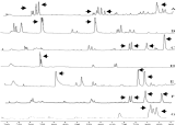
Fig. 1. Diagram of different Leishmania PCR amplified products after processing with the Fragment Manager programme. (A) L. braziliensis; (B) L. mexicana; (C) L. infantum; (D) L. tropica; (E) L. amazonensis; (F) L. chagasi; (G) L. major. The peaks belonging to the pattern of each species are indicated with arrows. The X-axis shows the fragment sizes in base pairs.

Fig. 2. Diagram of products of PCR made with Leishmania braziliensis isolated from ulcerates of different patients. (A) MHOM/PE/95/LQ8; (B) MHOM/PE/LQ1; (C) MHOM/PE/95/LQ2; (D) MHOM/PE/95/LQ5; (E) MHOM/PE/95/LQ7. The L. braziliensis pattern is a group of peaks between 300–330 bp, at 550–630 bp and 1 peak at 840 bp. The X-axis shows the fragment sizes in base pairs.

Fig. 3. Leishmania PCR amplified product diagram of Leishmania infantum isolated from different patients. (A) MON183/LEM2572; (B) MHOM/ES/1999/BCN224; (C) MHOM/ES/1999/BCN113; (D) MHOM/ES/1999/BCN225; (E) MHOM/ES/1999/BCN221. The X-axis shows the fragment sizes in base pairs. The L. infantum pattern is a group of peaks among 730–740 bp, 1 peak at 790 bp and another at 860 bp.

Fig. 4. Leishmania PCR amplification with Lb28 primers. The reactions were done with 30 ng of leishmania DNA of each species, 30 pmoles of each Lb28 primer in a final volume of 50 μl. (A) The amplified products were separated by electrophoresis in 1·5% agarose gel. Lane (a) molecular weight marker; lane (b) Leishmania braziliensis; lane (c) L. infantum; lane (d) L. tropica; lane (e) L. mexicana; lane (f) L. amazonensis. (B) Hybridization using the amplified fragment of L. braziliensis with the Lb28 primers labelled with digoxygenin, as probe. Amplified products were blotted on a nylon membrane from the agarose gel. Only DNA of L. braziliensis (lane b) reacted with the probe.
 You have
Access
- 13
- Cited by