Introduction
The family Catostylidae comprises six genera and 23 species (Collins et al., Reference Collins, Jarms and Morandini2020). The genus Crambionella was first described by Stiasny (Reference Stiasny1921) and is characterized by the presence of three-winged, stout and pyramidal oral arms lacking whip-like filaments but with terminal clubs (Stiasny, Reference Stiasny1921; Rao, Reference Rao1931). Medusae also possess an intra-circular network of anastomosing canals that communicates with the ring canal but not the stomach, and occasionally with adjacent radial canals (Stiasny, Reference Stiasny1922; Kramp, Reference Kramp1961). Lappets are divided by a deep furrow and are free of any anastomosing canals (Stiasny, Reference Stiasny1922, Reference Stiasny1937; Rao, Reference Rao1931).
The genus comprises four species, viz. Crambionella orsini (Vanhoffen, Reference Vanhoffen1888), Crambionella stuhlmanni (Chun, Reference Chun1896), Crambionella helmbiru Nishikawa, Mulyadi & Ohtsuka, 2014 and Crambionella annandalei Rao, Reference Rao1931 (Collins et al., Reference Collins, Jarms and Morandini2020). Although these four species have distinct, if overlapping distributions in coastal waters, our knowledge about them differs greatly. Crambionella orsini is the type species for the genus and has been widely reported from the northern Indian Ocean: from Assab, Red Sea (Vanhoffen, Reference Vanhoffen1888); off Madras (Menon, Reference Menon1930); around Krusadai Island, Gulf of Mannar (Menon, Reference Menon1936); in the Gulf of Oman and in the Straits of Hormuz (Stiasny, Reference Stiasny1937; Billett et al., Reference Billett, Bett, Jacobs, Rouse and Wigham2006; Daryanabard & Dawson, Reference Daryanabard and Dawson2008) as well as the Iranian Gulf (Kramp, Reference Kramp1956); off Pondicherry and Travancore along the east (Ranson, Reference Ranson1945) and west (Nair, Reference Nair1946) coasts of India, respectively. Crambionella stuhlmanni was originally described from material collected around Zanzibar (Chun, Reference Chun1896), but has also been recorded from Mozambique (Stiasny, Reference Stiasny1922) and Madagascar (Ranson, Reference Ranson1945), and from the east coast of South Africa (Neethling et al., Reference Neethling, Channing, Gershwin and Gibbons2011; Perissinotto et al., Reference Perissinotto, Taylor, Carrasco and Fox2013). Crambionella helmbiru is a recently recognized species from Cilacap and Karang Duwur, central Java, Indonesia (Nishikawa et al., Reference Nishikawa, Thu, Yusoff, Lindsay, Mulyadi, Mujiono, Ohtsuka and Nishida2009; Kitamura & Omori, Reference Kitamura and Omori2010; Nishikawa et al., Reference Nishikawa, Ohtsuka, Mulyadi, Mujiono, Lindsay, Miyamoto and Nishida2014).
Crambionella annandalei was originally collected from the Andaman Sea near Port Blair by Annandale and Prashad in 1923, and kept in the Museum of the Zoological Survey of India, only to be formally described in 1931 by Rao (Reference Rao1931). In that paper, Rao (Reference Rao1931) mentioned that the species could also be found along the coasts of Puri (Odisha), Vizagapatam (Andhra Pradesh) and Madras (Tamil Nadu). It has subsequently been recovered from the mouth of the Kalatan River, Myanmar (Kitamura & Omori, Reference Kitamura and Omori2010) and the Digha coast, west Bengal (Sarkar et al., Reference Sarkar, Talukdar, Dutta and Ramakrishna2002). Whilst Menon (Reference Menon1930) identified what he thought at the time was C. stuhlmanni from the Madras coast, this has subsequently been regarded as C. annandalei (Kramp, Reference Kramp1961).
Our knowledge of the diversity of Scyphozoa is changing, as we come to understand that cryptic speciation in the group is relatively common and that the descriptions on which species definitions are based are incomplete (e.g. Ras et al., Reference Ras, Neethling, Engelbrecht, Morandini, Bayha, Skrypzeck and Gibbons2020). Here, we provide a detailed taxonomic re-description of the Crambionella annandalei from the Western Bay of Bengal.
Materials and methods
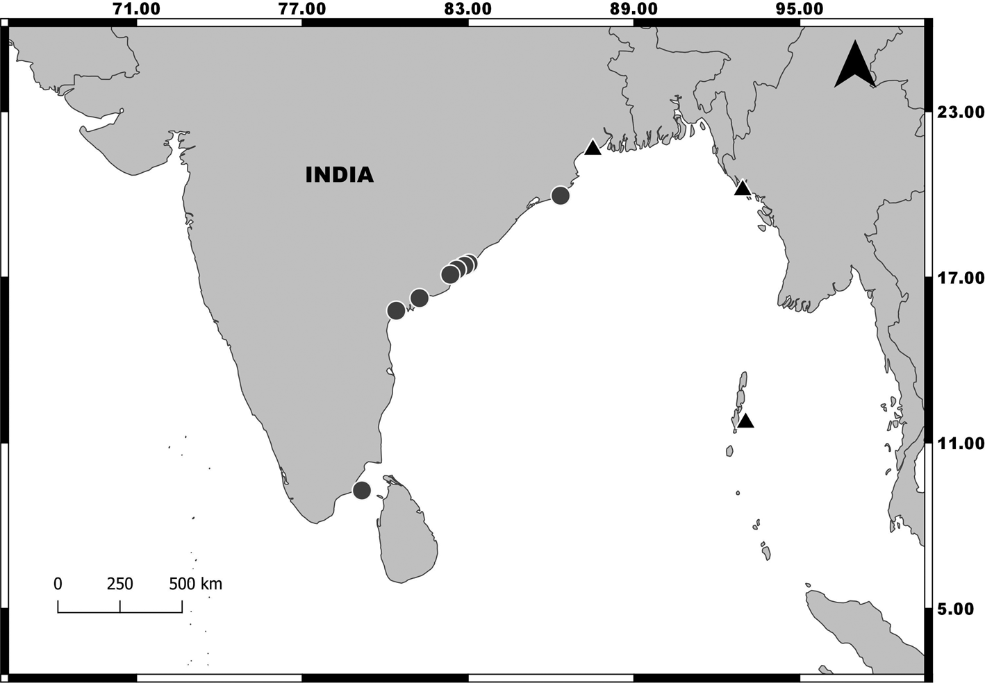
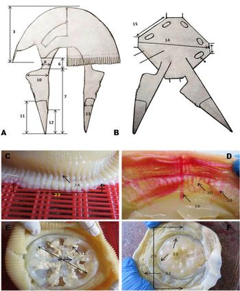
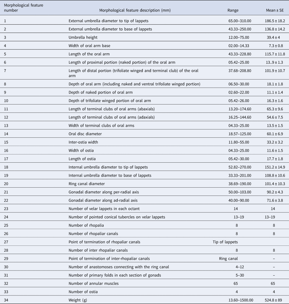
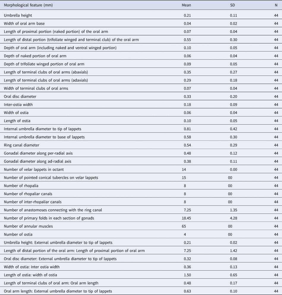
Specimens (Figure 1) were collected from various locations around India: 40 specimens from gillnet landings along the coast of Andhra Pradesh between March and July 2018; two specimens from trawl by-catch at Astaranga Fishing Harbour, Puri, Odisha coast on 23 March 2019 and two specimens from gillnet landings at Gulf of Mannar, Mandapam, Tamil Nadu coast on 15 July 2018 (Figure 2). The specimens were brought to the laboratory and photographed, and the umbrella diameter (mm) and wet weight (g) individually determined. A total of 33 morphological features were measured from the fresh specimens using digital Vernier callipers (Table 1 & Figure 3A–F), following Neethling et al. (Reference Neethling, Channing, Gershwin and Gibbons2011) and Nishikawa et al. (Reference Nishikawa, Ohtsuka, Mulyadi, Mujiono, Lindsay, Miyamoto and Nishida2014). The canal systems were injected with Rose Bengal to highlight their arrangement, after which specimens were preserved in 10% formalin.
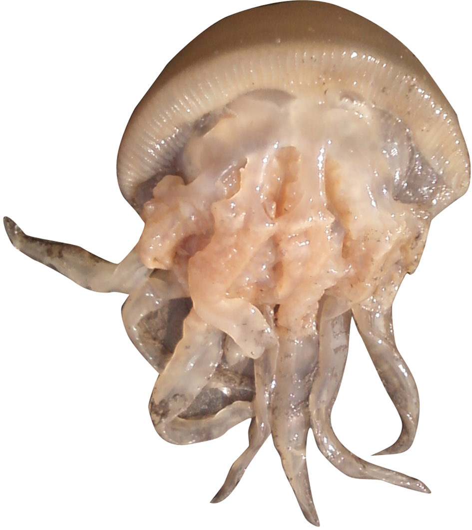
Crambionella annandalei Rao (Reference Rao1931) from Molapeta landing centre, Kakinada.

Distribution pattern of Crambionella annandalei in the Indian Ocean (triangle: reported from Andaman Island, Myanmar and West Bengal; circle: occurrence in the present study along the east coast of India).

Morphological features measured on Crambionella annandalei collected from the east coast of India. (A) Schematic diagram of a longitudinal section along per-radial axis of a specimen (adapted from Neethling et al., Reference Neethling, Channing, Gershwin and Gibbons2011); (B) schematic diagram of the oral disc, from a subumbrella view (adapted from Neethling et al., Reference Neethling, Channing, Gershwin and Gibbons2011); photograph showing the exumbrella view (C) and subumbrella view (E & F) of C. annandalei demonstrating various morphological measurements; subumbrella view showing the intra-circular anastomosing canal networks, after injecting Rose Bengal solution (D). Only two of the eight oral arms are shown in A and B; for details see Table 1.

Morphological measurements of C. annandalei from Andhra Pradesh

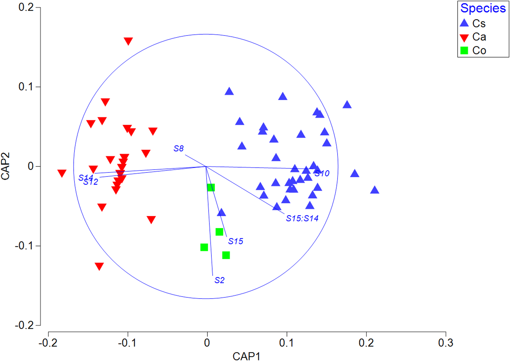
As the size of individuals change, the measures on morphological features may also change, which makes it difficult to compare the morphological features between individuals. Therefore, all the morphometric variables were converted into proportions relative to external bell diameter (BD) for size independent comparison (Table 2). In order to compare the multivariate morphology of the specimens collected here with that of Crambionella stuhlmanni and Crambionella orsini, a Canonical Analysis of Principal Coordinates (CAP) routine was executed in PRIMER-7, using comparable data provided by MJ Gibbons (see Neethling et al., Reference Neethling, Channing, Gershwin and Gibbons2011). Standardized data were first correlated against BD for each species and those features that remained size-dependent were removed. Data were then normalized and a resemblance matrix based on Euclidean distance was constructed between all specimens. Following Anderson et al. (Reference Anderson, Gorley and Clarke2008), we used the ‘leave one out’ routine to determine the subset of PCO axes to be used in the CAP (e.g. Ras et al., Reference Ras, Neethling, Engelbrecht, Morandini, Bayha, Skrypzeck and Gibbons2020). It was not possible to include Crambionella helmbiru in this analysis owing to a lack of data.
Standardized morphological features of C. annandalei from Andhra Pradesh

Results
SYSTEMATICS
Order DISCOMEDUSAE Haeckel, 1880
Suborder DAKTYLIOPHORAE Stiasny, Reference Stiasny1920
Family CATOSTYLIDAE Gegenbaur, 1857
Genus CRAMBIONELLA Stiasny, Reference Stiasny1921
Crambionella annandalei Rao, Reference Rao1931
(Figures 1 & 3–7; Tables 1–3)
Crambionella annandalei: ZMIP.492/1, Zoological Survey of India (Indian Museum), bell diameter: 170 mm, locality: Andaman Island, Port Blair, December, 1923, Annandale and Prashad, p. 50–55, figs 4–8, pls III, IV, fig. 1; Kitamura & Omori, Reference Kitamura and Omori2010, fig. 4, pp. 109–110; Sarkar et al., Reference Sarkar, Talukdar, Dutta and Ramakrishna2002, pp. 31–33.
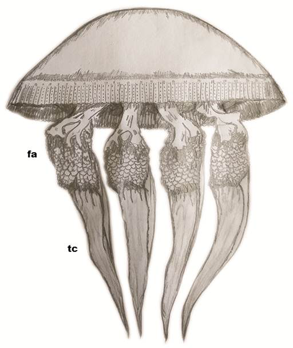
Schematic diagram of the whole body in lateral view. Only four of the eight oral arms are shown. fa, foliaceous appendage; tc, terminal club.

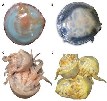
Colour variation of exumbrella and its margin in C. annandalei. (A) Exumbrella translucent white with single reddish brown margin (BD: 98 mm); (B) exumbrella and its margin translucent white (BD: 102 mm); (C) exumbrella and its margin faintly brown (BD: 285 mm); (D) exumbrella and its margin faintly yellow (BD: 275 mm).

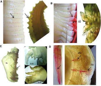
(A) Presence of pointed conical tubercle on the umbrella margin (left figure) and microscopic view (right figure); (B) vellar and rhopaliar lappets, exumbrella view (left figure) and microscopic view of rhopalium with rhopaliar canal (right figure); (C) microscopic view of foliaceous appendages on trifoliate winged portion of oral arms showing their morphological variation; (D) canal system of terminal club of oral arm and subumbrella view after injecting Rose Bengal solution: axial canal, distal part of oral arm showing the distribution of canals in the terminal appendage (left figure) and point of termination of rhopaliar and inter-rhopaliar canals (subumbrella view) (right figure). vl, velar lappet; rhl, rhophaliar lappet; rh, rhophalia; rhc, rhophaliar canal; fa, foliaceous appendage; irhc, inter-rhophaliar canal.

CAP plot showing the distribution of Crambionella specimens with respect to the two CAP axes  $\lpar \delta _1^2 = 0.92\comma \;\,\delta _2^2 = 0.17\rpar $. Vector overlay showing changes in standardized base variables with respect to CAP axes. S2, umbrella height; S8, depth of winged portion of oral arm; S10, length of terminal clubs of oral arm (adaxials); S12, oral disc diameter; S14, width of ostia; and S15, length of ostia and the ratio between length of ostia: width of ostia (S15:S14); Cs, Crambionella stuhlmanni; Co, Crambionella orsini; Ca, Crambionella annandalei.

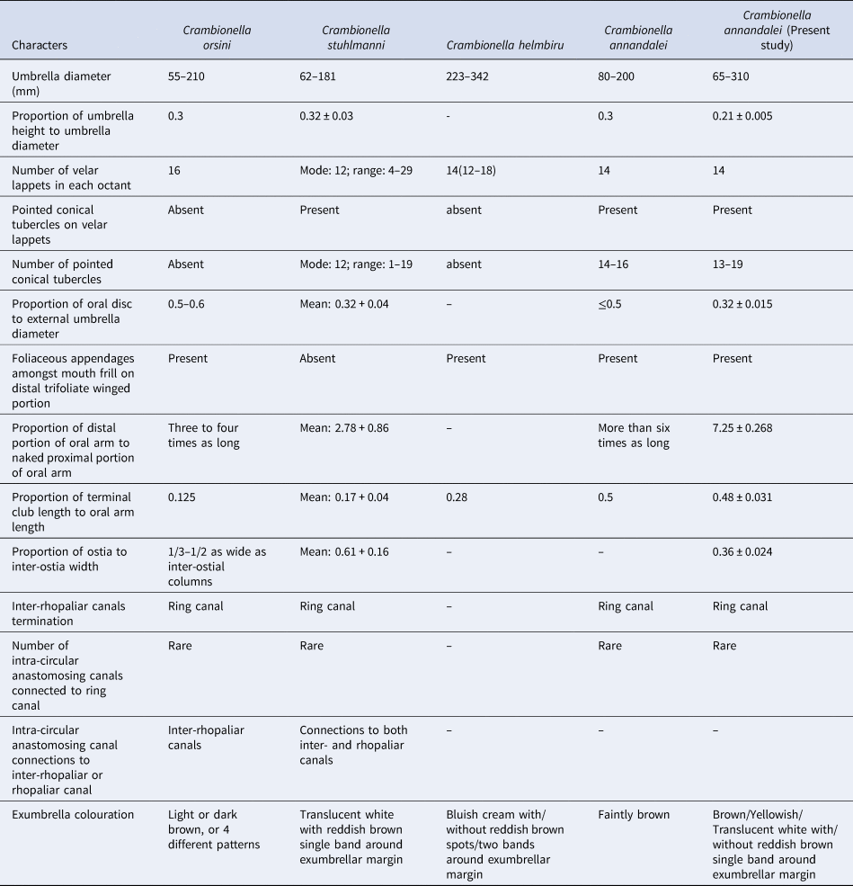
A character matrix highlighting morphological features that differ among the four Crambionella species (Neethling et al., Reference Neethling, Channing, Gershwin and Gibbons2011; Nishikawa et al., Reference Nishikawa, Ohtsuka, Mulyadi, Mujiono, Lindsay, Miyamoto and Nishida2014) and the Crambionella material under the present study

Crambionella stuhlmanni: Menon M.G.K. (Reference Menon1930), Bulletin of Madras Government Museum (N.S), III (I), p. 18, pl. III, figs 14 a, c & e.
MATERIALS EXAMINED
Forty specimens (bell diameter: 65–310 mm and weight: 13.6–1500 g), March to July, 2018, Pudimadaka, Rajanagaram, Molapeta, Bangarammapalem, Manginapudi (08°20 03.54N 77°58 24.48E), coll. P. R. Behera, depth 7–50 m, gear: gillnet; two specimens (bell diameter: 120–125 mm and weight: 450–460 g), 23 March 2019, Astaranga Fishing Harbour, Puri, Odisha coast (19°58′27.102′′N 86°20′20.9544′′E), coll. S.K. Roul, depth: 30–40 m; gear: multiday bottom trawl; two specimens (bell diameter: 150–160 mm and weight: 500–550 g), 15 July 2018, Gulf of Mannar, Mandapam, Tamil Nadu (9°16′21.85′′N 79°08′13.88′′E), coll. R. Saravanan, depth 5–10 m, gear: gillnet.
Eight specimens collected by P.R. Behera from the Pudimadaka landing centre were deposited in the Designated National Repository (DNR) of ICAR-CMFRI with accession numbers CA 3.1.1.1–CA 3.1.1.8. Two specimens collected from the Gulf of Mannar, Tamil Nadu were deposited in the Marine Biodiversity Museum of Regional Centre of ICAR-CMFRI, Mandapam with accession numbers MMM-CMFRI 18001 and MMM-CMFRI 18002.
Description
Umbrella hemispherical in shape, diameter 65–310 mm, thickened centrally, thinning to margin (Figure 4). Exumbrella surface smooth, finely granulated apically. Colour in life, variable: exumbrella of six specimens cream, with single reddish brown spot on each velar lappet (Figure 5A); 10 uniformly cream (Figure 5B); balance either light brown (Figure 5C) or light yellow (Figure 5D); light brown in colour on preservation.
Umbrella margin lacking tentacles, divided into 14 narrow velar lappets per octant, separated by deep furrows and bearing a single row of (13–19) pointed tubercles along the length of their mid-line on a prominent ridge: tubercles vary in size, largest proximally and smallest at bell margin (Figure 6A). With eight rhopalia situated equidistantly around bell margin, set in deep notches; each with a short, stout stalk with a spherical mass at its free end enclosed in a membrane; flanked by a pair of small, narrow and elongated rhopaliar lappets that diverge distally and do not extend beyond the velar lappets (Figure 6B).
Eight oral arms arise in pairs from the oral disc of the sub-umbrella. Each oral arm is divided into a naked proximal portion, cylindrical in cross section and without frills (ratio to bell diameter: 0.07) and a distal, three-winged portion supporting foliaceous appendages amongst mouthlets (more numerous on adaxial sides) (Figure 6C) and with terminal club (ratio to bell diameter: 0.55). Terminal clubs long, stout, tapering distally, terminated with pointed tips without mouth frills or appendages, longer abaxially than adaxially (Figures 4 & 3A).
Oral disc octagonal (centre cross-shaped), diameter less than half of the external bell diameter (Table 1). Four genital ostia present, crescent-shaped, elevated and thickened along the edge. Ostia and inter-ostia width not equal. Gonads rounded in shape, cream in colour; observed in specimens >110 mm bell diameter (Figure 3E). Subumbrella with 65 well-developed annular muscles, interrupted into two distinct regions by a groove along the ring canal. The outer is wide and has well defined ‘curtain-like’ muscles and the inner is narrow, and its muscles are comparatively less conspicuous.
With 16 radial canals, eight rhopaliar (four per-radial and four ad-radial) and eight inter-rhopaliar (inter-radial); rhopaliar canals extend to umbrella margin, inter-rhopaliar canals end at the ring canal (Figure 6D). Intra-circular anastomosing network extends over less than half the area between radial canals and connects to the ring canal only (Figure 3D); rhopaliar lappets each with a short, blind extension of the rhopaliar canal (Figure 6C). Eight canals extend from the central gastric cavity down the middle of the oral arms; axial canal in terminal club giving rise to narrow transverse canals penetrating into each wing (Figure 6D).
Discussion
The CAP plot is shown in Figure 7, and is based on the following standardized features: umbrella height, depth of winged portion of oral arm, length of terminal clubs of oral arm (adaxials), oral disc diameter, width of ostia and length of ostia and the ratio between length of ostia: width of ostia. All other features were correlated with specimen size and were excluded. The plot shows a clear separation of the three species using two CAP axes; the canonical correlation associated with the first is high  $\lpar \delta _1^2 = 0.92\comma \;\,\delta _2^2 = 0.17\rpar $. Using the ‘leave-one-out’ procedure, four orthonormal PCO axes were used in the final CAP analysis, and 94.5% of all specimens were correctly assigned to the correct a priori taxon (two specimens each of C. annandalei and C. stuhlmanni were misidentified as C. orsini).
A character matrix highlighting the morphological features that differ among the four Crambionella species and the Crambionella material described here is given in Table 3. Whilst it is clear that there is overlap in some measures and meristics, C. annandalei can be separated from the other species by a combination of the following features: (1) the presence of pointed conical tubercles on the velar lappets, (2) the presence of foliaceous appendages amongst the mouth frills, (3) the high ratio of the distal portion of the oral arm to the naked proximal portion (7.25 ± 0.268) and (4) the high ratio of the terminal club length to the oral arm length (0.48 ± 0.031). It should be noted that although the latter two features can be useful to separate species, they do vary with individual size.
Based on the present study and earlier descriptions, it is evident that the distribution pattern of Crambionella annandalei is confined to the Eastern Indian Ocean, in the Bay of Bengal and the Andaman Sea (Figure 2). Locally it is called ‘Munthakaya’ (in Telugu), and it is caught by gillnets operated by motorized craft at depths less than 50 m along the coast of Andhra Pradesh. This species supports an active jellyfish fishery during its peak swarming season (March to July) and processed jellyfishes are generally exported to South-east Asian countries (Behera et al., Reference Behera, Raju, Jishnudev, Shubhadeep and Saravanan2020).
The colour of living C. annandalei has previously been reported as faintly brown (Sarkar et al., Reference Sarkar, Talukdar, Dutta and Ramakrishna2002; Kitamura & Omori, Reference Kitamura and Omori2010), whereas our observations show the existence of four distinct colour morphs. Similar observations have been made for C. helmbiru from central Java, where Nishikawa et al. (Reference Nishikawa, Ohtsuka, Mulyadi, Mujiono, Lindsay, Miyamoto and Nishida2014) reported that the umbrella varied from a uniform (blue) colour to individuals possessing two reddish-brown bands around the margin. A single reddish-brown band on the umbrella margin has been observed for some C. stuhlmanni from the St Lucia Estuary, South Africa (Neethling et al., Reference Neethling, Channing, Gershwin and Gibbons2011 cf. Perissinotto et al., Reference Perissinotto, Taylor, Carrasco and Fox2013). Although four colour morphs of C. orsini were observed by Stiasny (Reference Stiasny1937), all with a marginal band of pigment, Daryanabard & Dawson (Reference Daryanabard and Dawson2008) have reported uniformly pigmented individuals from the Gulf of Oman. Scyphozoans are known to display a relatively high level of colour and pattern variation (e.g. Morandini and Marques, Reference Morandini and Marques2010), and it could be argued that the relatively high level of localized colour variation observed within the genus Crambionella could be an adaptation for concealing this large gelatinous zooplankton from visual predators. Such adaptive phenotypic plasticity would be beneficial for globally distributed scyphozoan jellyfish species which are subjected to significant temperature variation (Nawroth et al., Reference Nawroth, Feit, Colin, Costello and Dabiri2010).
Acknowledgements
The authors are grateful to the Director, ICAR-Central Marine Fisheries Research Institute, Kochi for his encouragement and support. We are grateful to the reviewers of previous drafts of the manuscript for their useful comments on the text; especially Mark J Gibbons (University of the Western Cape, South Africa). This study was carried out under the ICAR-CMFRI in-house project (MBD/JLY/32).