I. INTRODUCTION
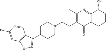
Paliperidone (trade name Invega) is a benzisoxazole derivative and the principal active metabolite of risperidone. As a dopamine antagonist and serotonin type 2A antagonist of the atypical antipsychotic class of medications, it is approved for the treatment of schizophrenia (Dolder et al., Reference Dolder, Nelson and DeyoAm2008). The systematic name (CAS Registry Number 144598-75-4) is (RS)-3-[2-[4-(6-fluorobenzo[d]isoxazol-3-yl)-1-piperidyl]ethyl]-7-hydroxy-4-methyl-1,5-diazabicyclo[4.4.0]deca-3,5-diene-2-one. A two-dimensional molecular diagram is shown in Figure 1.
The molecular structure of paliperidone.

The presence of high-quality reference powder patterns in the Powder Diffraction File (PDF; ICDD, 2014) is important for phase identification, particularly by pharmaceutical, forensic, and law enforcement scientists. The crystal structures of a significant fraction of the largest dollar volume pharmaceuticals have not been published and thus calculated powder patterns are not present in the PDF-4 databases. Sometimes experimental patterns are reported, but they are generally of low quality. This structure is a result of the collaboration among ICDD, Illinois Institute of Technology (IIT), Poly Crystallography Inc., and Argonne National Laboratory to measure high-quality synchrotron powder patterns of commercial pharmaceutical ingredients, include these reference patterns in the PDF, and determine the crystal structures of these active pharmaceutical ingredients (APIs).
Even when the crystal structure of an API is reported, the single-crystal structure was often determined at low temperature. Most powder measurements are performed at ambient conditions. Thermal expansion (often anisotropic) means that the peak positions calculated from a low-temperature single-crystal structure often differ significantly from those measured at ambient conditions. These peak shifts can result in failure of default search/match algorithms to identify a phase, even when it is present in the sample. High-quality reference patterns measured at ambient conditions are thus critical for easy identification of APIs using standard powder diffraction practices.
II. EXPERIMENTAL
Paliperidone was a commercial reagent, purchased from Santa Cruz Biotechnology, and was used as-received. The white powder was packed into a 1.5 mm diameter Kapton capillary and rotated during the measurement at ~50 cycles s−1. The powder pattern was measured at 295 K at beam line 11-BM (Lee et al., Reference Lee, Shu, Ramanathan, Preissner, Wang, Beno, Von Dreele, Ribaud, Kurtz, Antao, Jiao and Toby2008; Wang et al., Reference Wang, Toby, Lee, Ribaud, Antao, Kurtz, Ramanathan, Von Dreele and Beno2008) of the Advanced Photon Source at Argonne National Laboratory using a wavelength of 0.413 891 Å from 0.5° to 50°2θ with a step size of 0.001° and a counting time of 0.1 s step−1. The pattern was indexed using Jade 9.5 (MDI, 2014) on a primitive monoclinic unit cell having a = 14.153, b = 21.534, c = 6.913 Å, β = 92.3°, and V = 2105.1 Å3. The space group was suggested to be P21/n, which is consistent with the racemic nature of commercial material. The unit-cell volume is consistent with Z = 4. After solution and refinement of the structure, a reduced cell search in the Cambridge Structural Database (Allen, Reference Allen2002) yielded Refcode YAGRIJ for paliperidone (Betz et al., Reference Betz, Gerber, Hosten, Dayananda, Yathirajan and Thomas2011).
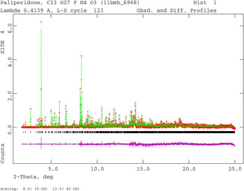
The structure in this study was solved by direct methods (including the Resolution Bias Modification) using EXPO2009 (Altomare et al., Reference Altomare, Camalli, Cuocci, Giacovazzo, Moliterni and Rizzi2009). The Rietveld refinement was carried out using General Structure Analysis System (GSAS) (Larson and Von Dreele, Reference Larson and Von Dreele2004). Only the 1.8°–25.0° portion of the pattern was included in the refinement (d min = 0.96 Å). All non-H-bond distances and angles were subjected to restraints, based on a Mercury/Mogul Geometry Check (Bruno et al., Reference Bruno, Cole, Kessler, Luo, Motherwell, Purkis, Smith, Taylor, Cooper, Harris and Orpen2004; Sykes et al., Reference Sykes, McCabe, Allen, Battle, Bruno and Wood2011) of the molecule. The Mogul average and standard deviation for each quantity were used as the restraint parameters. The restraints contributed 3.31% to the final χ 2. Isotropic displacement coefficients were refined, grouped by chemical similarity. The hydrogen atoms were included in calculated positions (Materials Studio; Accelrys, 2013), which were recalculated during the refinement. The U iso of each hydrogen atom was constrained to be 1.3× that of the heavy atom to which it is attached. The peak profiles were described using profile function #4 (Thompson et al., Reference Thompson, Cox and Hastings1987; Finger et al., Reference Finger, Cox and Jephcoat1994), which includes the Stephens (Reference Stephens1999) anisotropic strain broadening model. The background was modeled using a nine-term shifted Chebyshev polynomial. The final refinement of 111 variables using 23 282 observations yielded the residuals R wp = 0.086, R p = 0.070, and χ 2 = 2.496. The largest peak (1.24 Å from C26) and hole (0.29 Å from F7) in the difference Fourier map were 0.48 and −0.68 e (Å)−3, respectively. The Rietveld plot is included as Figure 2. The largest errors are in the positions of the low-angle peaks, and may reflect subtle changes in the specimen during the measurement.
(Color online) The Rietveld plot for the refinement of paliperidone. The red crosses represent the observed data points, and the green line is the calculated pattern. The magenta curve is the difference pattern, plotted at the same vertical scales as the other patterns. The vertical scale has been multiplied by a factor of 10 for 2θ > 8.0°, and by a factor of 40 for 2θ > 13.5°.

The single crystal structure YAGRIJ has the hydroxyl group (in our numbering) C27–O31–H58 disordered over two positions, with occupancies 0.86 and 0.14. In addition, the methyl hydrogens H48, H49, and H50 are disordered over two positions. Refinement of this disordered model (126 variables) using the same strategy yielded the residuals R wp = 0.086, R p = 0.070, and χ 2 = 2.526. While it is impossible to know if the single crystal and powder samples were exactly the same, this result points out that it may be difficult to detect small levels of disorder using powder data, even synchrotron powder data.
A density functional geometry optimization (fixed experimental unit cell) was carried out using CRYSTAL09 (Dovesi et al., Reference Dovesi, Orlando, Civalleri, Roetti, Saunders and Zicovich-Wilson2005). The basis sets for the H, C, N, and O atoms were those of Gatti et al. (Reference Gatti, Saunders and Roetti1994), and the basis set for F was that of Nada et al. (Reference Nada, Catlow, Pisani and Orlando1993). The calculation used eight k-points and the B3LYP functional, and took ~15 days on a 3.0 GHz PC.
III. RESULTS AND DISCUSSION
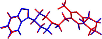
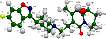
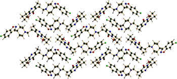
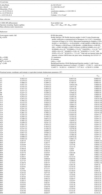
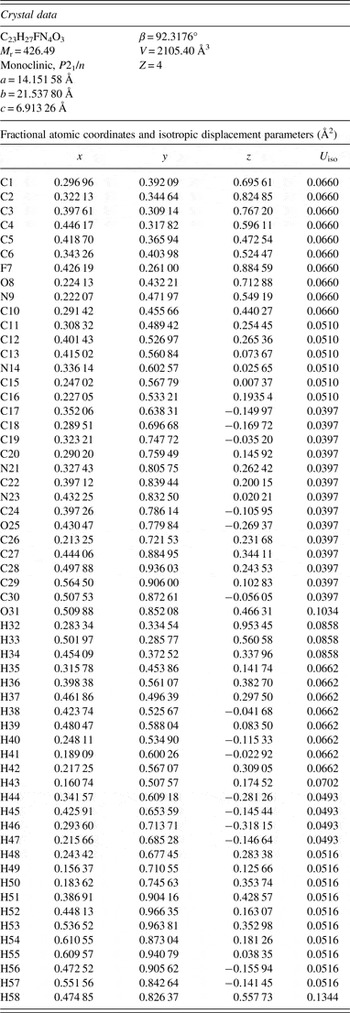
The refined atom coordinates of paliperidone are reported in Table I, and the coordinates from the density functional theory (DFT) optimization in Table II. The root-mean-square deviation of the non-hydrogen atoms is 0.068 Å, and the maximum deviation is 0.154 Å, at C43 (Figure 3). The excellent agreement between the refined and optimized structures is strong evidence that the structure is correct (van de Streek and Neumann, Reference van de Streek and Neumann2014). The discussion of the geometry uses the DFT-optimized structure. The asymmetric unit (with atom numbering) is illustrated in Figure 4, and the crystal structure is presented in Figure 5.
(Color online) Comparison of the refined and optimized structures of paliperidone. The Rietveld refined structure is colored red, and the DFT-optimized structure is in blue.

(Color online) The molecular structure of paliperidone, with the atom numbering. The atoms are represented by 50% probability spheroids.

(Color online) The crystal structure of paliperidone, viewed down the c-axis.

Rietveld refined crystal structure of paliperidone.

DFT-optimized (CRYSTAL09) crystal structure of paliperidone.

All the bond distances, angles, and torsion angles fall within the normal ranges indicated by a Mercury Mogul Geometry Check. A Mercury Molecule Overlay of the current structure and the single-crystal structure YAGRIJ failed, because the hydroxyl group O31–H58 (in our numbering) is disordered 86/14% over two positions in the single-crystal structure. The displacement coefficient of O31 is much higher than that of the other atoms, suggesting that disorder is also present in the room-temperature powder structure. A Mercury Structure Overlay indicated that the root-mean-square difference between the two molecules was only 0.033 Å, and thus the expansion between 200 and 295 K involves intermolecular separations.
A quantum mechanical (DFT/B3LYP functional/6-31G* basis set/vacuum) energy analysis using Spartan ‘14 (Wavefunction, 2013) suggests that the observed conformation is with 3.4 kcal mole−1 of a local minimum. A molecular mechanics (MMFF) conformational analysis indicates that the observed conformation is 43.08 kcal mole−1 higher in energy than the minimum-energy conformation, which curls up on itself to generate parallel stacking of the fused ring systems. This difference in energy indicates that intermolecular interactions are important in determining the solid-state conformation.
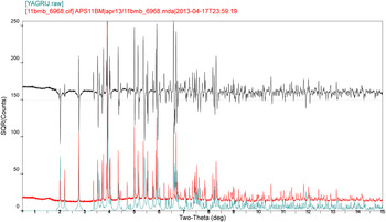
Determination of the single-crystal structure at 200 K and the powder structure at 295 K provides an opportunity to assess the thermal expansion of paliperidone. The lattice parameters (transformed into the current P2 1/n setting) are reported in Table III. The unit-cell volume at 295 K is 1.5% larger than at 200 K, but the expansion is anisotropic. The b-axis is nearly constant at the two temperatures, while the a- and c-axes expand by 0.71 and 0.87%, respectively, from 200 to 295 K. Comparison of the observed powder pattern with that calculated from YAGRIJ (Figure 6) shows that the positions of some peaks differ significantly. Some of the differences are large enough that a room-temperature powder diffraction pattern would not be identified as paliperidone in a default search/match, even if a pattern calculated from YAGRIJ were present in the PDF. The differences provide strong evidence for the desirability of including high-quality ambient-condition patterns in the PDF™.
(Color online) Comparison of the observed 295 K powder pattern of paliperidone (red) with the pattern calculated from the 200 K structure in YAGRIJ (cyan). The black curve is the difference, and indicates the significant peak shifts between the two temperatures.

Lattice parameters of paliperidone; space group P21/n.

There is only one significant hydrogen bond in the crystal structure of paliperidone (Table IV). This H-bond is between the hydroxyl group O31–H58 and the ketone oxygen O25. The result is a chain along the c-axis with graph set C1,1(7) (Etter, Reference Etter1990; Bernstein et al., Reference Bernstein, Davis, Shimoni and Chang1995; Shields et al., Reference Shields, Raithby, Allen and Motherwell2000). Using the correlation between the H···Acceptor distance for O–H···O hydrogen bonds developed by Rammohan, A. and Kaduk, J. A. (2016, Unpublished data), this hydrogen bond contributes 12.6 kcal mole−1 to the crystal energy. In the single-crystal structure YAGRIJ, the major occupancy hydrogen bond corresponds to 12.6 kcal mole−1, while the minor occupancy orientation is reasonable for two hydrogen bonds with energies of 6.9 and 4.9 kcal mole−1. The multiple potential hydrogen bonds and their relative energies may explain the disorder in the single-crystal structure. A very weak intermolecular C25–H50···F7 hydrogen bond may contribute to the crystal packing, and an intramolecular C30–H57···O25 hydrogen bond may influence the conformation of the molecule.
Hydrogen bonds in paliperidone.

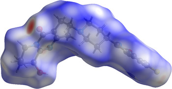
The volume of the Hirshfeld surface (Figure 7; Hirshfeld, Reference Hirshfeld1977; McKinnon et al., Reference McKinnon, Spackman and Mitchell2004; Spackman and Jayatilaka, Reference Spackman and Jayatilaka2009; Wolff et al., Reference Wolff, Grimwood, McKinnon, Turner, Jayatilaka and Spackman2012) is 518.54 Å3, 98.52% of one-fourth the unit-cell volume (526.349 Å3). The molecules are thus not tightly packed. The only significant close contacts (red in Figure 7) involve the O–H···O hydrogen bonds. An analysis of the contributions to the total crystal energy using the Forcite module of Materials Studio (Accelrys, 2013) suggests that angle distortion terms are a major intramolecular contribution to the crystal energy, and that van der Waals interactions dominate the intermolecular forces.
(Color online) The Hirshfeld surface of paliperidone. Intermolecular contacts longer than the sums of van der Waals radii are colored blue, and contacts shorter than the sums of the radii are colored red. Contacts equal to the sums of the radii are white.

The Bravais–Friedel–Donnay–Harker (Bravais, Reference Bravais1866; Friedel, Reference Friedel1907; Donnay and Harker, Reference Donnay and Harker1937) morphology suggests that we might expect an elongated morphology for paliperidone, with {001} as the long axis. A second-order spherical harmonic preferred orientation model was included in the refinement; the texture index was 1.043, indicating a small but significant preferred orientation in this rotated capillary specimen.
The powder pattern of paliperidone is included in the PDF as entry 00-064-1497.
SUPPLEMENTARY MATERIAL
To view supplementary material for this article, please visit http://dx.doi.org/10.1017/S0885715616000087
ACKNOWLEDGEMENTS
The use of the Advanced Photon Source at Argonne National Laboratory was supported by the U. S. Department of Energy, Office of Science, Office of Basic Energy Sciences, under Contract No. DE-AC02-06CH11357. This work was partially supported by the International Centre for Diffraction Data. We thank Lynn Ribaud for his assistance in data collection.