Introduction
Pendred syndrome is an autosomal recessive syndrome which accounts for 10 per cent of syndromic hearing loss cases. It is the second most common cause of syndromic hearing loss after Usher's syndrome. It is caused by bi-allelic mutations in the SLC26A4 gene, which is responsible for encoding pendrin (an anion exchanger) and is expressed in the inner ear, thyroid gland and kidneys.
The majority of patients present with profound, prelingual deafness; others develop deafness later in life. A goitre develops, usually during childhood, but there is substantial interfamilial and regional variation. Dietary iodide intake appears to be a significant modifier of the thyroid phenotype.Reference Kopp, Pesce and Solis1
Pendrin is found in the apical membrane of a subpopulation of intercalated cells in the cortical part of the renal collecting duct. Pendrin appears to play a critical role in the process of renal bicarbonate secretion. Thus, in the absence of pendrin there is the potential for bicarbonate retention and metabolic alkalosis. However, there have been no previous reports of metabolic alkalosis in a Pendred syndrome case.
Case report
A 46-year-old woman was referred to our cochlear implant centre with progressive, profound, bilateral, sensorineural hearing loss since childhood. She had been recently diagnosed with mild hypothyroidism.
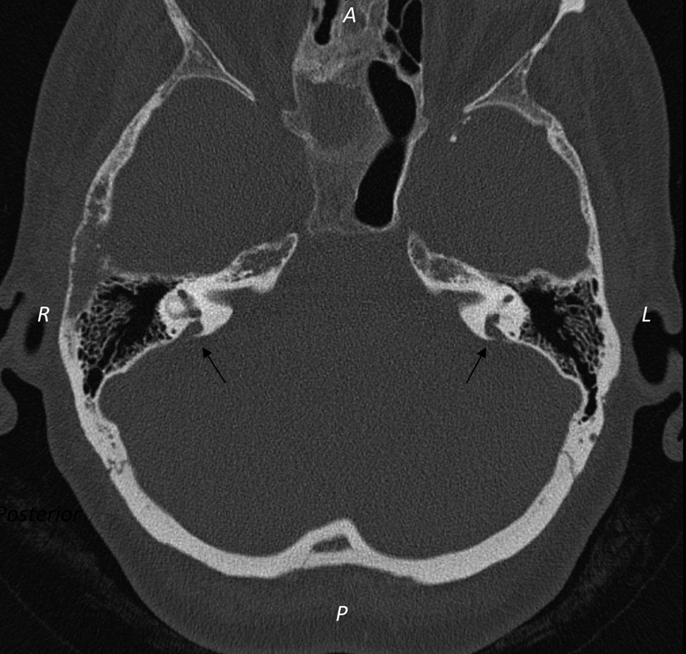
Subsequent investigation, as part of the patient's assessment for cochlear implant candidacy, included temporal bone computerised tomography and genetic studies. These revealed bilaterally enlarged vestibular aqueducts (Figure 1) and cochlear Mondini defects, consistent with a diagnosis of Pendred syndrome. This was confirmed by genetic studies showing a SLC26A4 gene mutation.
Axial computed tomography scan (high definition) of temporal bones, showing bilateral enlarged vestibular aqueducts (arrows), A(Anterior) , P(Posterior), L(Left), R(Right)
The patient met audiological candidacy requirements; however, selecting the most appropriate ear to implant was hampered by the patient's inability to comply with caloric vestibular nystagmography testing to assess labyrinthine function. The patient recalled that she had always felt that her left ear was the better hearing ear, possibly since a head injury during childhood. The patient's lifestyle was also of concern, as she had an introverted personality and a history of alcohol abuse.
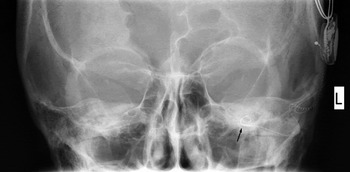
Following multidisciplinary assessment and funding approval, the patient underwent left cochlear implantation (Figure 2). A standard Nucleus device was used, with full insertion into the scala tympani via a cochleostomy approach. Good impedance and telemetry values were recorded at the time.
Postero-anterior X-ray of the facial bones taken post-implant, showing the position of the electrodes (black arrow). L = left

Post-operatively, the patient appeared to suffer from deterioration in her balance, but was able to be discharged home on day one.
A week later, she had to be readmitted for conservative management of a wound infection. Although discharged home successfully, she was noted to be increasingly reliant on aids for walking.
By the time she was seen in the post-operative review and implant programming clinic, she was relying on a wheelchair for mobility. She was very reluctant to get out of her wheelchair due to concerns over her balance and a sensation of nausea, as well as bouts of vomiting. She also complained bitterly of an increased perception of tinnitus in both ears, as a result of surgery.
At the time the implant was switched on, the patient developed an adverse reaction to sound. Her tinnitus was so problematic that she had resorted to creating loud noises to try to drown it out. The local authority's environmental officers were called to intervene on several occasions. She had also relapsed into alcohol abuse.
Despite the best efforts and support of the implant team and the patient's general practitioner, she became an infrequent attender to subsequent programming and clinic appointments, and was labelled a poor implant user.
Some two years later, the patient was brought in as an emergency case to the accident and emergency department, having been found by her sons at home unwell, lethargic and confused. It was reported that she had been drinking alcohol and vomiting over the preceding few days. It was noted that she had had two similar episodes since her cochlear implantation, and had been prescribed potassium to treat hypokalaemia.
Examination revealed that the patient was acutely confused. Her temperature was normal and she had an oxygen saturation of 91 per cent on air, with tachycardia of 120/minute. Further investigation indicated metabolic alkalosis: arterial blood gas sampling revealed a pH of 7.59, bicarbonate concentration of 45 mmol/L and base excess of +20.4 mmol/L. The patient also had severely deranged electrolytes, in particular a potassium concentration of 1.4 mmol/l, despite a normal urinary potassium concentration.
Because of the patient's profoundly low potassium levels, she was admitted to the high dependency unit for fluid resuscitation and electrolyte replacement. As her potassium levels began to rise to normal levels, she suffered multiple cardiac ventricular fibrillation arrests. She was successfully resuscitated and transferred to the cardiac care unit.
While undergoing rehabilitation in the cardiac care unit, an ENT consultation was sought as the patient complained of nausea and occasional vomiting on any movement. She still complained of tinnitus, and was proving a challenge to the physiotherapists in their attempts to encourage mobilisation. It was feared that any further potassium loss through vomiting was likely to put her at risk of further life-threatening hypokalaemia.
Extensive psychological, vestibular and cochlear implant rehabilitation followed, and she became a good user of her implant. She also stopped drinking.
At the time of writing, the patient was using her cochlear implant as an aid to lip-reading. She had also become mobile, to the level of her pre-morbid functioning.
Discussion
Pendred syndrome was first described by Vaughan Pendred in 1896.Reference Kopp, Pesce and Solis1, Reference Cremers, Bolder, Admiraal, Everett, Joosten and van Hauwe2 It is an autosomal recessive condition defined by familial sensorineural hearing loss and goitre.Reference Kopp, Pesce and Solis1 Pendred syndrome comprises a constellation of clinical features, including defective organification of iodide (which may present as goitre and/or hypothyroidism) and temporal bone defects (i.e. enlarged vestibular aqueduct and Mondini cochlear saccular dysplasia).Reference Kopp, Pesce and Solis1 It is thought that Pendred syndrome accounts for 10 per cent of all syndromic hearing loss cases, making it the second commonest syndromic cause of hearing loss after Usher's syndrome.
The hallmark of Pendred syndrome is sensorineural hearing loss, which may present as either profound prelingual deafness or progressive hearing loss.Reference Kopp, Pesce and Solis1, Reference Cremers, Bolder, Admiraal, Everett, Joosten and van Hauwe2
Pendred syndrome is cause by bi-allelic mutations in the SLC26A4 gene, which is responsible for encoding pendrin. Pendrin is an anion exchanger and is expressed in the inner ear, thyroid gland and kidneys.Reference Kopp, Pesce and Solis1, Reference Wangemann, Nakaya, Wu, Maganti, Itza and Sanneman3
In the inner ear, studies in mice models have found SLC26A4 messenger RNA predominantly in the endolymphatic duct and sac, areas of the utricule and saccule, and in the external cochlear sulcus region.Reference Kopp, Pesce and Solis1, Reference Wangemann, Nakaya, Wu, Maganti, Itza and Sanneman3 This expression pattern involves regions that are important for endolymphatic fluid resorption, and it is postulated that defects in the pendrin protein lead directly to endolymphatic hydrops.
In a mouse model of Pendred syndrome, it was found that the inner ear appeared to develop normally until embryonic day 15, after which there was progressive dilatation of the endolymphatic system, resulting in complete degeneration of sensory cells and malformation of otoconia and otoconial membranes.Reference Kopp, Pesce and Solis1 This process is thought to lead to enlarged vestibular aqueduct and Mondini defects in Pendred syndrome patients. It is worth noting that both enlarged vestibular aqueduct and Mondini defects are not specific to Pendred syndrome, and can be found in other disorders.
In thyroid tissue, pendrin is present in a limited subset of cells within the thyroid follicles, exclusively in the apical membrane of the follicular epithelium. Several research findings have suggested that pendrin could be involved in mediating iodide transport into the follicular lumen. There is substantial interfamilial and regional variation. Nutritional iodide intake appears to be a significant modifier of the thyroid phenotype. Under conditions of high iodide intake, thyroid enlargement may be very mild or absent.
More recently, investigators have examined the expression of pendrin in the kidney.Reference Wangemann, Nakaya, Wu, Maganti, Itza and Sanneman3 Pendrin has been found in the apical membrane of a subpopulation of intercalated cells in the cortical part of the renal collecting duct.Reference Kopp, Pesce and Solis1, Reference Wangemann, Nakaya, Wu, Maganti, Itza and Sanneman3, Reference Royaux, Wall, Karniski, Everett, Suzuki and Knepper4 It seems that pendrin plays a critical role in the process of renal bicarbonate secretion. In the absence of pendrin, there is the potential for bicarbonate retention and hence metabolic alkalosis. However, pendrin-deficient mice and human Pendred syndrome patients do not generally develop metabolic alkalosis.Reference Royaux, Wall, Karniski, Everett, Suzuki and Knepper4 This is presumably because other kidney buffer mechanisms are in place and compensate for this mild impairment. However, if for any reason these compensatory mechanisms are removed (e.g. renal failure), then the risk of metabolic alkalosis may be increased.
• A patient with Pendred syndrome is presented, who developed life-threatening hypokalaemia due to implant-induced imbalance and alcohol dependency, leading to multiple cardiac arrests
• Pendred syndrome is caused by bi-allelic mutations in the SLC26A4 gene, which encodes for the pendrin protein
• Patients have enlarged vestibular aqueducts and cochlear Mondini defects (although these features are not specific to Pendred syndrome)
• Pendrin is an anion exchanger mainly found in the inner ear, thyroid gland and kidneys
• Pendrin occurs in the cortical part of the renal collecting duct, and plays a critical role in renal bicarbonate secretion
• Without pendrin, there is the potential for bicarbonate retention and metabolic alkalosis
Metabolic alkalosis is often associated with hypokalaemia. This occurs as a result of K+ redistribution and excessive renal K+ loss. Loss of gastric contents results in volume depletion and metabolic alkalosis, both of which promote kaliuresis. Hypovolaemia stimulates aldosterone release, which augments K+ secretion by the principal cells. In addition, the filtered load of HCO3− exceeds the reabsorptive capacity of the proximal convoluted tubule, thereby increasing distal delivery of NaHCO3, which enhances the electrochemical gradient favouring K+ loss in the urine.
In our patient, we believed that frequent vomiting, due to balance impairment and/or increased alcohol intake, led to an emesis-associated potassium loss. This, combined with a renal inability to reabsorb urinary potassium losses, led to metabolic alkalosis and life-threatening hypokalaemia.Reference Fauci, Braunwald, Kasper, Hauser, Longo, Jameson, Anthony and Fauci5
Conclusion
Cochlear implantation is a burgeoning therapy for patients with bilateral, profound, sensorineural hearing loss, and results in proven (and often strongly publicised) substantial improvements in patient quality of life. It is tempting to be over-confident about implant candidates' successful outcomes. The presented case highlights the need to ensure that patients, and their care networks, have a realistic expectation of implant outcomes. Our patient's case also reinforces the need for dedicated rehabilitation teams. One must also bear in mind the potential psychological, physiological and metabolic disturbances that implantation, and any underlying metabolic conditions, may trigger.