INTRODUCTION
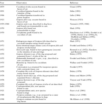
Although coccidia are one of the first parasitic protozoa discovered, little was known of their importance in cats and dogs until 1970 (Levine and Ivens, Reference Levine and Ivens1981). An explosion of information has occurred on this topic in the last 40 years and I had the fortune of being associated with many of the studies in this field (Table 1). In the present special issue of the journal some of these personal experiences are recalled. My objective is not to discuss controversies regarding nomenclature or to seek or give priorities to works by different groups. The present discussion relates to faecally transmitted coccidians of domestic cats (Felis domesticus) and domestic dogs (Canis familiaris).
Knowledge of the evolution of coccidia of cats and dogs

EARLY HISTORY OF CAT AND DOG COCCIDIA
Coccidia split into three groups
Until the report of Wenyon (Reference Wenyon1923), only 1 species of coccidium was thought to be present in the faeces of cats and dogs. Wenyon (Reference Wenyon1923, Reference Wenyon1926) reviewed earlier literature and concluded that Grassi (Reference Grassi1879) probably named a coccidium in the cat now regarded as Coccidium rivolta, and Stiles (Reference Stiles1891) named a parasite Coccidium bigemina first from the dog (Table 1). Lühe (Reference Lühe and Mense1906) transferred these parasites to the genus Isospora.
Wenyon (Reference Wenyon1923) made the first distinction that there were at least 3 species of coccidia in cats and dogs, based on the oocyst size: (1) large oocyst measuring approximately 40 long μm; (2) medium oocyst size measuring 25 μm long; (3) small oocyst size measuring 10 μm long.
Wenyon (Reference Wenyon1923) named the coccidium with large oocysts as Isospora felis and partly described its endogenous stages in the intestine of the cat.
The large oocyst size coccidium split into two species
Nemeséri (Reference Nemeséri1959, Reference Nemeséri1960) probably was the first to conduct cross-transmission of dog and cat coccidia. He could not transmit the large coccidium from the dog to the cat and named the dog parasite as a new species, Isospora canis.
Shah (Reference Shah1970, Reference Shah1971), who was my teacher in the veterinary school, made the first definitive study in this field. He micro-isolated a single I. felis oocyst, produced a cloned isolate by feeding the oocyst to a coccidia-free kitten, re-described its oocysts, and described the asexual and sexual stages of the parasite in the intestine of the kittens. He also showed that the parasite was not transmissible to dogs. Lepp and Todd (Reference Lepp and Todd1974) described asexual and sexual stages of I. canis using cloned parasites.
The medium oocyst size coccidium split into several species
Coccidia are ubiquitous and it is difficult to raise cats and dogs coccidia-free. In the 1960s, it became possible to raise parasite-free cats and dogs by using Caesarian section and germ-free isolators. Fortunately for me, I got a professorship in veterinary parasitology in 1973 at the Ohio State University, Columbus, Ohio, where cats and dogs were raised in isolators. I used these pets for cross-transmission studies with cat and dog coccidia. I was unable to infect dogs with Isospora rivolta-like parasite from cats, and named the dog parasite, Isospora ohioensis (Dubey, Reference Dubey1975), and retained the designation I. rivolta for the cat parasite. I also described the first complete life cycles of a canine (I. ohioensis in the dog, Dubey, Reference Dubey1978) and a feline coccidium (I. rivolta in the cat, Dubey, Reference Dubey1979).
Before the recognition of I. rivolta and I. ohioensis as separate species, Mahrt (Reference Mahrt1967, Reference Mahrt1968) had very carefully studied the life cycle of an I. rivolta-like parasite of the dog. During the studies on the development of I. ohioensis, I realized that there were differences in the endogenous stages of I. ohioensis and I. rivolta in the dog, the former located in the surface epithelium and the latter in the lamina propria. I re-examined all histological sections from dogs that Professor Mahrt made available to me and we named the parasite studied by Mahrt as Isospora neorivolta (Dubey and Mahrt, Reference Dubey and Mahrt1978). Trayser and Todd (Reference Trayser and Todd1978) described another species, Isospora burrowsi, from the dog. There is an overlap in size of the oocysts and in the location in the intestine of dogs among I. ohioensis, I. neorivolta, and I. burrowsi, therefore, this group is sometimes called I. ohioensis-like parasites.
THE SMALL-SIZED OOCYSTS, COCCIDIUM BIGEMINA, AND EXPLOSION OF INFORMATION
As said earlier, this parasite was first named by Stiles (Reference Stiles1891) who found it as a fully sporulated oocyst in the lamina propria of the dog. Wenyon (Reference Wenyon1923, Reference Wenyon1926) reviewed earlier literature and stated that both forms (in the epithelium and in the lamina propria) were found in dogs and cats; both unsporulated and sporulated forms were regarded as the same parasite, I. bigemina.
Discovery of Toxoplasma gondii life cycle
The most thrilling discovery in my life was made in 1970, when cats fed Toxoplasma gondii tissue cysts shed I. bigemina-like oocysts, and animals fed these oocysts died of toxoplasmosis. Several groups of workers (Table 1) independently and around the same time (see Dubey, Reference Dubey2008) found T. gondii oocysts in cat faeces (Hutchison et al. Reference Hutchison, Dunachie, Siim and Work1970; Frenkel et al. Reference Frenkel, Dubey and Miller1970; Dubey et al. Reference Dubey, Miller and Frenkel1970a, Reference Dubey, Miller and Frenkelb; Sheffield and Melton, Reference Sheffield and Melton1970; Overdulve, Reference Overdulve1970).
Although the discovery of the oocyst stage in the life cycle of a coccidian parasite would be expected to prove T. gondii to be a coccidian parasite, this was a big challenge at that time for the following reasons. First, faecal infectivity (oocyst) was linked with Toxocara infectivity (Hutchison, Reference Hutchison1965, Reference Hutchison1967). Second, the oocyst had been called a new cyst (Work and Hutchison, Reference Work and Hutchison1969). Toxoplasma gondii oocysts were morphologically identical to oocysts of the previously described coccidian parasite (I. bigemina) of cats and dogs. In light of these facts, the evidence provided by us to link T. gondii oocysts to faecal infectivity is noteworthy (Frenkel et al. Reference Frenkel, Dubey and Miller1970; Dubey et al. Reference Dubey, Miller and Frenkel1970a). We subjected the newly discovered faecal stage (oocyst) to the following mutually independent tests to accumulate critical evidence for or against its being identical to T. gondii (Dubey et al. Reference Dubey, Miller and Frenkel1970a). (i) Newborn kittens and littermate controls were used to avoid as far as possible pre-existing coccidial infections. (ii) Comparison of the development of oocysts and of infectivity in relation to heat, cold, oxygenation and chemicals. (iii) Comparison by filtration of the size of the infectious entity with oocyst size. (iv) Comparison of the density characteristics of oocysts and of infectivity. (v) Comparison of the electrophoretic characteristics of oocysts and infectivity. (vi) Antigenic comparison of oocysts with the standard RH strain of Toxoplasma by means of the fluorescent antibody test. (vii) Identification of the endogenous cycle preceding the development of oocysts, and linking it antigenically to Toxoplasma infectivity of oocysts before and after excystation. (viii) Comparison of the appearance of oocysts and Toxoplasma infectivity in the faeces of cats after feeding of cysts, trophozoites, and oocysts (Dubey et al. Reference Dubey, Miller and Frenkel1970a).
In retrospect, the discovery and characterization of the T. gondii oocyst in cat faeces was also delayed because until 1970 coccidian oocysts were sporulated in 2·5% potassium dichromate. Chromation of the oocyst wall interfered with excystation of the sporozoites when oocysts were fed to mice and thus the mouse infectivity titre of the oocysts was lower than expected from the number of oocysts administered (Dubey et al. Reference Dubey, Miller and Frenkel1970a). These findings led to the use of 2% sulfuric acid as the best medium for sporulation and storage of T. gondii oocysts. Unlike dichromate, which was difficult to remove from oocysts, sulfuric acid could be easily neutralized and the oocysts could be inoculated directly into mice (Dubey and Frenkel, Reference Dubey and Frenkel1973). Unlike other coccidians, T. gondii oocysts were found to excyst efficiently when inoculated parenterally into mice. This observation alleviated the need for oral inoculation for the bioassay of oocysts (Dubey and Frenkel, Reference Dubey and Frenkel1973).
The medical and biological significance of the discovery of the life cycle of T. gondii described above is that unlike the previous view that coccidia were host specific (confined to 1 host) and site specific (confined to intestine and faeces), T. gondii infected virtually all warm-blooded animals including humans and virtually any cell of the body. However, it retained an evolutionary characteristic of completing the oocyst phase only in 1 host (cats, not only domestic but other felids as well).
Although T. gondii has a worldwide distribution and perhaps the widest host range of any parasite, there is only 1 species, gondii in the genus, Toxoplasma. Why some people develop clinical toxoplasmosis whereas most remain asymptomatic is largely unknown. During the 1980s and 1990s methods were developed to recognize genetic differences among T. gondii isolates from humans and animals (Pfefferkorn and Pfefferkorn, Reference Pfefferkorn and Pfefferkorn1980; Dardé et al. Reference Dardé, Bouteille and Pestre-Alexandre1987; Tibayrenc et al. Reference Tibayrenc, Kjellberg, Arnaud, Oury, Brenière, Dardé and Ayala1991; Sibley et al. Reference Sibley, Leblanc, Pfefferkorn and Boothroyd1992; Howe and Sibley, Reference Howe and Sibley1995). Mapping of T. gondii genes was achieved recently (Khan et al. Reference Khan, Taylor, Su, Mackey, Boyle, Glover, Tang, Paulsen, Berriman, Boothrooyd, Pfefferkorn, Dubey, Ajioka, Roos, Wootton and Sibley2005), and undoubtedly will help in the search for better antigens for diagnosis and protection, and mechanism of disease. Until recently, T. gondii was considered clonal with very little genetic variability (Howe and Sibley, Reference Howe and Sibley1995). Lehmann et al. (Reference Lehmann, Marcet, Graham, Dahl and Dubey2006) made the first indepth study of genetic variability among more than 275 T. gondii isolates obtained worldwide from one host (free-range chicken) and in one laboratory (Dubey et al. Reference Dubey, Graham, Blackston, Lehmann, Gennari, Ragozo, Nishi, Shen, Kwok, Hill and Thulliez2002c) and found geographical differences, with some isolates being confined to Brazil whereas others being worldwide in distribution. Recent studies using a large numbers of isolates from Brazil and the USA indicated higher genetic diversity than previously recognized (Dubey and Su, Reference Dubey and Su2009). It is hoped that further genetic studies on T. gondii isolates from various sources may lead to identification of sources of infections for humans.
Discovery of Sarcocystis life cycle
Soon after the discovery of the life cycle of T. gondii, Heydorn and Rommel (Reference Heydorn and Rommel1972) and Rommel et al. (Reference Rommel, Heydorn and Gruber1972) found that dogs and cats fed Sarcocystis cysts (sarcocysts) developed gamonts and oocysts in the lamina propria, and these oocysts, unlike previously known coccidia, sporulated in situ. Thus, the mystery of the sporulated oocysts in the lamina propria previously found by many scientists earlier became clear. To my knowledge there are at least 10 species of Sarcocystis that are found in cat faeces, and at least 21 in dog faeces (Dubey et al. Reference Dubey, Speer and Fayer1989, and many more would be found in the future (Dubey and Odening, Reference Dubey, Odening, Samuel, Pybus and Kocan2001). It is almost impossible to distinguish Sarcocystis species based on the morphology of oocysts (Dubey et al. Reference Dubey, Speer and Fayer1989). Therefore, it is futile to speculate which species was present in the sample examined by Stiles (Reference Stiles1891). On a personal note, Dr J. K. Frenkel and I also attempted to search for Sarcocystis oocysts in the faeces of cats in 1970 but were unsuccessful because we fed sarcocysts from the heart of cattle to cats. In retrospect, now we know that cattle have 3 species of Sarcocystis, S. cruzi (transmitted only through canids), S. hirsuta (transmissible via cats), and S. hominis (transmissible via humans and primates) and S. hirsuta transmitted by cats does not form sarcocysts in the heart of cattle (Dubey et al. Reference Dubey, Speer and Fayer1989).
Discovery of new genera, Hammondia, Neospora, and finding of oocysts in cats and dogs
Frenkel and Dubey (Reference Dubey1975) described a new parasite, Hammondia hammondi. This parasite is morphologically similar to T. gondii but with a different life cycle. Unlike, T. gondii, it is non-pathogenic, and has an obligatory 2-host life cycle. Its oocysts resemble T. gondii in size and structure (Dubey and Sreekumar, Reference Dubey and Sreekumar2003; Schares et al. Reference Schares, Vrhovec, Pantchev, Herrmann and Conraths2008). A similar parasite was found to cycle through dogs (Heydorn, Reference Heydorn1973; Dubey and Fayer, Reference Dubey and Fayer1976) and was named Hammondia heydorni (Dubey, Reference Dubey and Kreier1977; Dubey et al. Reference Dubey, Barr, Barta, Bjerkås, Björkman, Blagburn, Bowman, Buxton, Ellis, Gottstein, Hemphill, Hill, Howe, Jenkins, Kobayashi, Koudela, Marsh, Mattsson, McAllister, Modrý, Omata, Sibley, Speer, Trees, Uggla, Upton, Williams and Lindsay2002b). Although T. gondii and H. hammondi oocysts are produced by cats, dogs do ingest cat faeces, and thus all three species, T. gondii, H. hammondi, and H. heydorni can be present in faeces of naturally-infected dogs (Schares et al. Reference Schares, Pantchev, Barutzki, Heydorn, Bauer and Conraths2005).
A new parasite, Neospora caninum, was described in 1988 (Dubey et al. Reference Dubey, Carpenter, Speer, Topper and Uggla1988). Until 1988, this parasite was misdiagnosed as T. gondii (Dubey et al. Reference Dubey, Barr, Barta, Bjerkås, Björkman, Blagburn, Bowman, Buxton, Ellis, Gottstein, Hemphill, Hill, Howe, Jenkins, Kobayashi, Koudela, Marsh, Mattsson, McAllister, Modrý, Omata, Sibley, Speer, Trees, Uggla, Upton, Williams and Lindsay2002b). Its veterinary importance became known a few years later when it was found to cause abortion in cattle and clinical disease in many other species of animals (Dubey et al. Reference Dubey, Schares and Ortega-Mora2007). Its oocyst was discovered in 1998 when dogs, but not cats, fed tissue cysts shed oocysts (McAllister et al. Reference McAllister, Dubey, Lindsay, Jolley, Wills and McGuire1998). Cats are not definitive hosts for N. caninum (McAllister et al. Reference McAllister, Dubey, Lindsay, Jolley, Wills and McGuire1998). Unlike, T. gondii, N. caninum is regarded as a major pathogen of cattle and dogs (Dubey et al. Reference Dubey, Schares and Ortega-Mora2007).
Discovery of the life cycle of Besnoitia
Besnoitia spp. are parasites of livestock and wild animals. Some species of Besnoitia cause economic losses to farmers in some parts of the world. The transmission of Besnoitia spp. remained a mystery until Wallace and Frenkel (Reference Frenkel and Dubey1975) reported that cats fed tissues of rodents infected with a rodent Besnoitia (B. wallacei) shed Toxoplasma-like oocysts (Frenkel, Reference Frenkel1977). Earlier, Peteshev et al. (Reference Peteshev, Galuzo and Polomoshnov1974) from Kazakhstan had reported that cats fed tissues of cows infected with B. besnoiti had shed Besnoitia oocysts. However, their findings could not be confirmed (Rommel, Reference Rommel1975). Subsequently, cats were shown to be the definitive hosts for B. darlingi of the American opossum (Smith and Frenkel, Reference Frenkel1977; Dubey et al. Reference Dubey, Lindsay, Rosenthal, Sreekumar, Hill, Shen, Kwok, Rickard, Black and Rashmir-Raven2002a) and for a new species, B. oryctofelisi that we isolated from a rabbit in Argentina (Dubey et al. Reference Dubey, Sreekumar, Lindsay, Hill, Rosenthal, Venturini, Venturini and Greiner2003). Thus, 3 species of Besnoitia, B. wallacei, B. darlingi, and B. oryctofelisi have T. gondii-like oocysts in the faeces of cats.
CRYPTOSPORIDIAL OOCYSTS IN FAECES OF CATS AND DOGS
Cryptosporidia spp. are coccidian parasites. Tyzzer originally described 2 species, C. parvum, and C. muris, as parasites of rodents (Tyzzer, Reference Tyzzer1910, Reference Tyzzer1912). After the discovery of the Human Immunodeficiencry Virus and the disease Acquired Immunodeficiency Syndrome (AIDS), cryptosporidia were recognized as an important opportunistic pathogen for AIDS. Currently there are numerous species and genotypes of Cryptosporidium, including C. felis (Iseki, Reference Iseki1979) and C. canis (Fayer et al. Reference Fayer, Trout, Xiao, Morgan, Lal and Dubey2001) specifically described as feline and canine parasites. However, human cryptosporidial species can infect other animals and animal cryptosporodial species can infect humans (reviewed by Santin and Trout, Reference Santin, Trout, Fayer and Xiao2008).
CONCLUSION
It is clear from the above discussion that I. bigemina observed by Wenyon and others was a mixture of many organisms. The unsporulated 10–15 μm sized oocysts in cat faeces will be similar to T. gondii, H. hammondi, Besnoitia spp. (B. wallacei, B. oryctofelisi, B. darlingi), H. heydorni, and N. caninum. The sporulated oocysts in cat and dog faeces were probably Sarcocystis species (at least 31 species). This group of organisms causes serious economic losses in animals and enormous human suffering.
In the last 25 years, the medical and veterinary importance of cat and dog coccidians became recognized. Although we have learnt much regarding these parasites in the last 40 years, many challenges remain including the life cycles of Besnoitia besnoiti, Sarcocystis canis, and Neospora hughesi. Studies of these coccidians have been a great learning experience for me. My own journey on coccidiosis started with the description of coccidian oocysts in the Indian jungle cat (Felis chaus) in 1962 (Dubey and Pande, Reference Dubey and Pande1963), and studies on cat coccidia have no end in sight. In the Felis chaus paper, I described several species of Eimeria, Isospora rivolta, and Cryptosporodium sp. Currently, all Eimeria spp. in cat and dog faeces are considered pseudoparasites (passing through the gut after eating other animals or faeces), and the Cryptosporodium sp. that I reported in 1963 was probably a Sarcocystis sp. based on its size.
I would like to dedicate this paper to the late Professor Norman D. Levine, who was a mentor and a friend. Although I never worked in his laboratory, he edited many of my papers and taught me morphological description of coccidian stages, as early as 1962.