Introduction
Trichinellosis is a zoonotic parasitic disease caused by the nematodes of the genus Trichinella (Gottstein et al., Reference Gottstein, Pozio and Nöckler2009). To date, 12 taxa, showing different epidemiological patterns, have been identified all over the globe (Pozio and Zarlenga, Reference Pozio and Zarlenga2013). In Europe, four species, namely Trichinella spiralis, Trichinella nativa, Trichinella britovi and Trichinella pseudospiralis, are circulating among animals (Pozio and Zarlenga, Reference Pozio and Zarlenga2013). Trichinellosis may represent a serious risk for human health and could have a fatal outcome in some cases (Murrell and Pozio, Reference Murrell and Pozio2011). The most important source of infection for humans is considered to be the consumption of raw or insufficiently heat-treated meat of domestic and wild swine (Murrell and Pozio, Reference Murrell and Pozio2011).
In Europe, human trichinellosis infections were strongly reduced in numbers in the last decade but are still consistently reported (European Food Safety Authority, 2018). In the last five years (2013-2017), 1022 new cases were documented in the EU countries, 70% (n = 712) of which occurred in Bulgaria and Romania (European Food Safety Authority, 2018). In Greece, since the first report of trichinellosis there in 1946 (Livieratos et al., Reference Livieratos, Danopoulos and Logothetopoulos1948), infections due to T. spiralis sensu lato (Pozio et al., Reference Pozio1992) have been sporadically reported in humans and animals (Sotiraki et al., Reference Sotiraki2001). The aim of the present report was to describe a clinical case that represents the first human infection due to T. britovi in Greece, and also to review the related literature reporting Trichinella infections in the country.
Case description
History
A 47-year-old male with a free medical history, and no recent travel abroad, was admitted to the hospital on 15 December 2017. According to the case history, two weeks prior to his admission the patient suffered from mild pain in the abdomen and a few days later he felt numbness in his hands that lasted 2–3 days. On 5 December he developed a wavy fever, reaching up to 39°C and falling several times a day, and also shivering, swelling around the eyes, fatigue, headache and severe pain in the limbs. One episode of difficulty in breathing was recorded one week after the onset of the symptoms. At the time of admission to the hospital, the clinical signs and symptoms included intense, generalized myalgia, periorbital swelling, fever, exhaustion and anorexia. The patient reported being a wild boar hunter, who consumed exclusively very well cooked wild boar meat (double cooking procedure, i.e. boiling and roasting) but preferred semi-raw pork. The last meal with semi-raw pork consumption occurred a few days prior to the onset of the first symptom (abdomen pain). No meat sample of domestic pig or wild boar was available for examination for Trichinella larvae.
Diagnostic imaging and laboratory findings
Ultrasound examination of the abdomen and computed tomography of the abdomen and of the head did not show any abnormality. Similarly, urinalysis was normal. Haematological and biochemical examinations showed eosinophilia (3420 cells/μl, normal range, NR 40–400 cells/μl) and high levels (64 U/l; NR 5–41 U/l) of alanine aminotransferase (ALT), aspartate aminotransferase (AST) (63 U/l; NR 5–40 U/l), creatine kinase (CPK) (276 U/l; NR 20–200 U/l), and lactate dehydrogenase (LDH) (394 U/l; NR 135–225 U/l).
Specific anti-Trichinella IgG (0.485 optical density units (OD), with a negative control at 0.150 OD) and IgM (1.275 OD, with a negative control at 0.155 OD) were detected by an in-house enzyme-linked immunosorbent assay (ELISA), using crude antigen from muscle larvae as previously described (Daoudaki et al., Reference Daoudaki2009). Negative control sera originated from healthy blood donors scoring negative with parasitic antigens; positive control sera originated from patients with confirmed trichinellosis. Reference sera were from the laboratory serum bank.
Based on the clinical pattern and positive serology by ELISA, a muscle biopsy was collected from biceps femoris and tested by compressorium to detect Trichinella spp. larvae. Under the microscope, one nematode larva measuring 900 × 37 μm was found (fig. 1A). The larva was immotile and it was not spirally coiled, nor encapsulated. Furthermore, a second muscle biopsy sample collected from biceps femoris was tested histologically with haematoxylin-eosin staining. Histology revealed granulomatous myositis with numerous histiocytes, lymphocytes and plasma cells, surrounding a nematode section (fig. 1B).
Trichinella britovi larva detected in muscle biopsy from biceps femoris of the patient 27 days after infection. (A) Preparation by compressorium. (B) Histological section (haematoxylin–eosin staining) showing numerous histiocytes, lymphocytes and plasma cells (asterisks) surrounding the Trichinella britovi larva (arrow).

The nematode larva detected by the compressorium was gently collected from the muscle tissue and preserved in a 1.0 ml conical vial under 90% ethyl alcohol. The larva was sent to the European Union Reference Laboratory for Parasites, Rome, Italy, for molecular identification by multiplex polymerase chain reaction (PCR), according to a previously published protocol (Pozio and La Rosa, Reference Pozio and La Rosa2003). The larva was identified as T. britovi.
Treatment and follow up
As soon as trichinellosis was suspected, approximately 23 days after infection, the patient was treated with albendazole at the dose of 400 mg two times a day for 10 days, starting from 18 December. The administration of corticosteroids was not considered necessary. The clinical condition of the patient improved rapidly after the initiation of the treatment and the patient was discharged seven days later. Only fatigue persisted up to 30 December.
Twenty-nine days after the completion of the treatment, at the follow-up examination, the clinical condition of the patient was excellent, and a rise of antibody concentration in serum (IgG 0.806 OD, negative control 0.157 OD; IgM 1546 OD, negative control 0.155 OD) was observed. At the second follow up, carried out about 11 months after the diagnosis, the antibody concentration had declined (IgG 0.182 OD, negative control 0.115 OD; IgM 0.350 OD, negative control 0.160 OD).
Discussion
Human trichinellosis is a parasitic disease with sporadic but consistently recorded cases in Europe in the past decades (European Food Safety Authority, 2018). In countries where pork consumption is low for religious reasons, human trichinellosis is expected to be rare. However, in 2004, an outbreak involving 1098 infected people took place in Turkey, where raw beef meatballs illegally mixed with pork infected with T. britovi were consumed (Akkoc et al., Reference Akkoc2009).
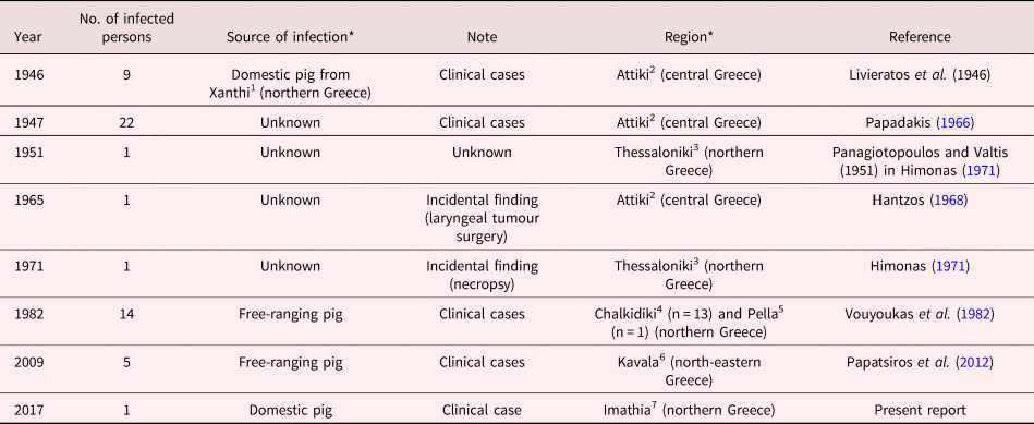
In Greece, the first human trichinellosis infections were documented in the course of an outbreak with nine cases, which occurred in 1946 (Livieratos et al., Reference Livieratos, Danopoulos and Logothetopoulos1948). In total from 1946 to 2017, 54 cases of trichinellosis were documented in Greece, resulting from the consumption of pork from free-ranging pigs or from an unknown source of infection (table 1, fig. 2). In this period of 72 years, the average incidence was of 0.55 cases per 100,000 inhabitants. In domestic pigs, the prevalence of Trichinella spp. infection was quite high (0.12–0.74%) during 1952–1958 (Himonas, Reference Himonas1971; Sotiraki et al., Reference Sotiraki2001). In 1967, a prevalence of 2.2% was detected in pigs slaughtered in Piraeus, Attica (Himonas, Reference Himonas1971). From 1975 to 1978, Trichinella spp. infection was not detected in slaughtered pigs in Greece (Himonas, Reference Himonas1979).
Map of Greece showing the regions where cases of trichinellosis have been recorded. 1: Xanthi (origin of infected meat, see table 1); 2: Attiki; 3: Thessaloniki; 4: Chalkidiki; 5: Pella; 6: Kavala; 7: Imathia.

Trichinellosis in Greece from 1946 to 2017.

*Superscripts (1–7) indicate the region on the map of Greece (fig. 2).
More recently, in a sero-survey performed in wild boars for various pathogens, anti-Trichinella antibodies were detected in 6 (6.4%) of 94 animals (Touloudi et al., Reference Touloudi2015). In the period 2009–2012, T. britovi larvae were detected in 0.29% of free-ranging pigs in northern Greece (Boutsini et al., Reference Boutsini2014). From 1953 to 1968 in Greece, Trichinella spp. infections were documented in red foxes (Vulpes vulpes) and wolves (Canis lupus) (Himonas, Reference Himonas1971). As the one species concept was predominant before the application of biochemical and molecular techniques (Pozio and Murrell, Reference Pozio and Darwin Murrell2006), all Trichinella spp. infections in animals and humans were ascribed to T. spiralis. However, we can speculate that T. britovi could be involved in some, if not in all, of those older cases in Greece, as it is the only species identified in the country by molecular analysis so far (Boutsini et al., Reference Boutsini2014). This is the first time that the etiological agent of human trichinellosis was isolated and identified at the species level in Greece. In the present case, based on the history and eating habits reported by the patient, i.e. consumption of very well cooked wild boar meat and half-cooked meat of domestic pig, the source of infection was suspected to be a domestic pig.
Human trichinellosis may remain asymptomatic when only few infective larvae are ingested, but is often accompanied by various signs and symptoms, including initially gastrointestinal disorders, fever and fatigue, followed by myalgia, arthralgia, dyspnoea and complications such as myocarditis. Some characteristic symptoms of the infection that may occur are periorbital or facial oedema and subungual petechiae. Death due to trichinellosis may be the outcome in a case of untreated, severe infection with complications (Gottstein et al., Reference Gottstein, Pozio and Nöckler2009).
The suspicion of human trichinellosis is based on the clinical and laboratory pattern and history of consumption of raw or semi-raw meat (Pozio and Bruschi, Reference Pozio, Bruschi and Liu2018). Confirmed diagnosis is based on specific laboratory tests, i.e. detection of specific antibodies by serological tests, isolation and identification of Trichinella larvae from muscle biopsies (Gottstein et al., Reference Gottstein, Pozio and Nöckler2009). In a mouse model infected by T. britovi, the collagen capsule around the larvae was observed by light microscope after 30–32 days of infection, whereas the collagen capsule can be detected 16–17 days post infection in T. spiralis infected mice (Pozio et al., Reference Pozio1992). In the present case, the T. britovi larva detected in the muscle biopsy was not encapsulated, suggesting that the date of infected pork consumption was presumably between 25 and 28 November 2017.
The detection of antibodies is a useful tool for the diagnosis of trichinellosis. Patients generally seroconvert 2–5 weeks after infection, but in mild infections seroconversion has also been detected 8 weeks after infection (Pozio and Bruschi, Reference Pozio, Bruschi and Liu2018). Antibodies may persist for months or years, depending on the treatment efficacy (International Commission on Trichinellosis, 2018). In the present case, both IgG and IgM antibodies were detected 25–28 days post infection. The rise of antibody concentration noticed 29 days after the end of treatment is consistent with the antibody kinetics, showing peak of IgG and IgM 60 and 90 days post infection, respectively (Feldmeier et al., Reference Feldmeier, Fischer and Blaumeiser1987), given that the larvae are still an antigenic stimulant after their death. Similarly, the persistence of antibodies 11 months post infection may be attributed to the continuous antigen release from dead larvae.
A prompt and correct diagnosis is of great importance in trichinellosis, as anthelmintic drugs are unable to kill larvae after their encapsulation complications. The most common anthelmintic drugs used for the treatment of trichinellosis in humans are albendazole and mebendazole (benzimidazoles) (Gottstein et al., Reference Gottstein, Pozio and Nöckler2009).
As Trichinella spp. transmission to humans occurs via the consumption of raw or undercooked infected meat, the first line of prevention is the enhancement of prevention awareness among consumers, who should be instructed to eat only well-cooked (internal temperature of 71°C for at least 1 minute) meat of susceptible animals (Gamble et al., Reference Gamble2000). Freezing is also a suggested method to kill Trichinella spp. larvae. For example, pieces of meat up to 15 cm thickness must be frozen at −15°C for three weeks to kill Trichinella larvae. However, some Trichinella taxa, including T. britovi, can survive in frozen muscle tissue at −10 to −15°C for several months (Pozio and Murrell, Reference Pozio and Darwin Murrell2006). In addition, hunters should be instructed to apply proper handling of carcasses and scraps after skinning game animals in the field (Gottstein et al., Reference Gottstein, Pozio and Nöckler2009). Strict veterinary control and good manufacture practices in the pig industry are also crucial in order to prevent human trichinellosis. All potential domestic and wild hosts of the parasite should be tested at the slaughterhouse or after hunting. The European Commission Regulation 2015/1375 requires tests for Trichinella for all slaughtered pigs from holdings that are not officially recognized to apply controlled housing conditions, horses, wild boar and other wild animals intended for human consumption (European Commission, 2015). However, as this regulation is not followed for home-slaughtered pigs nor for hunted animals intended for personal consumption in Greece, consumer education is the only realistic measure to prevent trichinellosis.
In conclusion, although rare, human trichinellosis may occur sporadically in Greece and other European countries, and consequently public health services and physicians should remain vigilant for clinical and laboratory evidence of the disease. Good management practices in farming and hunting, veterinary control of animals susceptible to Trichinella infection, and education and awareness among consumers, farmers and hunters may provide safe meat for human consumption.
Author ORCIDs
A. Diakou, 0000-0001-8347-0769
Conflict of interest
None.