INTRODUCTION
There has been a significant rise in the detection of prostate cancer partly due to Prostate Specific Antigen (PSA) screening.Reference Fonteyne, De Gersem and De Neve 1 Due to the screening of asymptomatic patients, there has been a migration towards a lower stage of prostate cancer at diagnosis.Reference Fonteyne, De Gersem and De Neve 1 However, 35% of newly diagnosed patients in the UK present with locally advanced stage T3 disease. 2 For these stage T3 high-risk patients, the treatment volume required often includes the involved pelvic nodes due to extension beyond the prostate capsule. The challenge with this cohort of patients is the independent motion of the prostate and the lymph nodes (LNs). Historically, around 40% of patients treated radically for clinically localised prostate cancer, suffered from biochemical failure within 5 years.Reference Han, Partin and Zahurak 3 – Reference Kestin, Vicini and Ziaja 5 Fonteyne et al.Reference Fonteyne, De Gersem and De Neve 1 suggest making the assumption that the relationship between higher doses of radiation and better biochemical control in prostate cancer can be applied to LN irradiation also. Hence, a more accurate method of targeting both the prostate and LNs with high doses of radiation could improve the biochemical and clinical control of the disease in a group where local failure is a problem. Two large, randomised, phase III clinical trials have evaluated pelvic nodal radiotherapy for patients with a high risk of microscopic LN metastases. These are RTOG 94-13 from the Radiation Therapy Oncology GroupReference Lawton, DeSilvio and Roach 6 and GETUG-01, from the French Genitourinary Group.Reference Pommier, Chabaud and Lagrange 7 Findings suggested that no benefit is seen in those patients who received nodal radiation. However, it is suggestedReference Bayley, Rosewall and Craig 8 that both trials potentially suffer from flaws due to, under dosing of the LN and the prostate itself and potential geographical miss due to imaging techniques. As discussed by Bayley et al.,Reference Bayley, Rosewall and Craig 8 there is great potential for geographical miss, mainly due to the lack of bladder and rectal protocols and the use of anatomical bone landmarks as a surrogate for prostate position. Current consensus for treating the prostate alone is for fiducial markers to be implanted into the prostate tissue to act as a surrogate for prostate motion.Reference Nederveen, Dehnad and van der Heide 9 – Reference Baker, Fenwick and Mayles 11 Literature suggests suggest that fiducial markers, coupled with effective bladder and rectal protocols, allows for significant reduction in radiotherapy planning margins.Reference Hsu, Pawlicki and Luxton 12 , Reference Eminowicz, Dean and Shoffren 13 When LNs are involved the treatment delivery becomes more challenging, current consensus for prostate only treatment would suggest the need for fiducial markers. However, this does not indicate the position of the LNs where anatomical bone is routinely used as a surrogate. In the clinical centre, from which the patient population is selected, for this study, current practice for these patients is to perform an anatomical bone match on the pelvic bones with a margin of 5 mm around the prostate.
METHODS
Patients
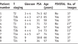
A retrospective analysis of nine patients receiving radiotherapy to either the prostate alone or the prostate and LNs was undertaken. Patient characteristics are detailed in Table 1. This cohort included six patients who were recruited into PIVOTALReference Dearnaley 14 with node negative prostate cancer stage T3b or T4 with a predicted LN risk ≥30%. They had a Gleeson 9-10, or Gleeson 8 and T3 or PSA>20 or Gleeson 7 and T3 or PSA≥30. The remaining three patients were not recruited to the PIVOTAL trial but with node involvement, and fiducial markers implanted, were treated using a departmental protocol. A total of 292 fractions were imaged (mean 32, range 11–37 images) and the images used for analysis.
Patient characteristics

Notes:
a Patients 4, 5, and 6 were not in PIVOTAL and treated using standard department protocol of imaging #1–3 and weekly unless further imaging was necessary.
Abbreviations: PSA, prostate specific antigen.
Patient preparation and immobilisation and treatment
Three gold seed fiducial markers (5×1 mm) were inserted into the prostate trans-rectally, using ultra-sound guidance, ~2 weeks before planning computed tomography (CT). A bladder and rectal protocol was followed before CT planning scan and each treatment fraction, consisting of a micro-enema (RelaxitTM), emptying the urinary bladder, drinking 500 ml of water and waiting for 20 minutes. Patients were positioned supine on an indexed couch top using indexed head, knee and ankle supports. Patients were treated daily using the PIVOTAL dose and fractionation of 74 Gy/37#, with a beam arrangement of either single or double 360° volumetric arc [Volumetric modulated arc therapy (VMAT)] fields.
All CT scans were acquired using Phillips Brilliance CT (© Koninklijke Philips N.V., 2004-2016) with indexed couch top. Images were acquired from the inferior aspect of the L1/L2 vertebral space to the penile urethra (~1 cm below ischial tuberosities). CT data sets were imported into the treatment planning system (Varian Eclipse V.10). For patient receiving irradiation to the prostate and pelvic nodes, I.V contrast was given, where possible, to the patient (Optiray 300 in 75 ml and a delay of 30 seconds between the administration and the CT scan). The target volumes, margins and doses shown in Table 2 are taken from the PIVOTAL planning guidelines.Reference Dearnaley 14
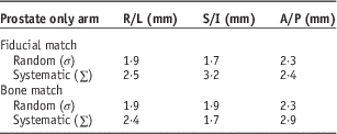
Displacement errors for prostate only arm

Abbreviations: R/L, right-left direction; S/I, superoinferior direction; A/P=anteroposterior direction.
IMRT treatment planning
VMAT ‘RapidARC’ was performed using the treatment planning software (TPS) Eclipse V.10. Either single or double 360° arcs obtained a uniform coverage of the planning target volume (PTV) and satisfied the dose constraints to the OAR. In plans where bowel dose constraints were not met, then the dose to the LN region was decreased from 60 to 55 Gy.
Online set-up strategy
Patients were aligned for treatment using a laser alignment technique, a set isocentre height and shifts from the computer plan. 2D kV Images (15×15 cm) were acquired and assessed online using a Varian On-Board Imager. Pelvic skeletal anatomy was used as a surrogate for the position of the nodal volume and the geometric centre of the three fiducial markers was used as a surrogate for the position of the prostate. The use of fiducial markers as a surrogate for prostate position has previously been shown to give accurate prostate positioning, providing the bladder and bowel protocol is used.Reference Heaton, Mayles and Syndikus 15 The effective dose of a kV image is ~0·01 mSv/image, therefore the concomitant dose is negligible. Assessment of both target volumes was completed daily before treatment. Initially, an anatomical bone match was completed comparing the kV image with a reference digitally reconstructed radiograph obtained from the planning scan. Second, a fiducial marker match was completed. The differences between the matches were calculated in the S/I, A/P and R/L directions. If the difference between the results were <5 mm in each direction the fiducial marker match was applied (due to the 5 mm margin added around the nodes), and the couch was remotely moved. This was performed according to departmental fiducial maker protocol of a 2 mm action level. This includes accuracy of couch repositioning (~1 mm) and 0·5 mm inter-observer variability.Reference Baker, Fenwick and Mayles 11 , Reference Heaton, Mayles and Syndikus 15 If the difference was >5 mm in any one direction then the patient was re-set up and the image process was repeated. If the results demonstrated the same discrepancy, the patient was treated according to the fiducial marker match and a trial planning physicist informed before the next fraction. A Cone Beam CT (CBCT) was then obtained to provide 3D information on both target volumes.
The image acquisition and analysis process was achievable within a standard departmental time slot, aided by the use of VMAT which reduces beam delivery time.Reference Helyer, Stephenson and Heaton 16
Statistical analysis
Data were analysed using Microsoft Excel and Statistical Package for Social Sciences (SPSS) Version 19. Individual and population systematic (∑) and random errors (σ) were calculated from the image analysis results using the formula described by Van-Herk.Reference Van Herk, Remeijer and Rasch 17 The systematic errors found in bone and seed matching were compared using the F-test. Random errors were compared using an unpaired, two-sample t-test. The Van-Herk expression M PTV=2·5Σ+0·7σ was used with the random and systematic errors before online correction. This allowed the margins required for each target volume to be calculated.
RESULTS
Positioning accuracy
The maximum displacement observed between the fiducial match and the bone match was 7, 10 and 4 mm in the A/P, S/I and R/L directions, respectively. The percentage of displacements >5 mm action level was greatest in the S/I direction with 4% >5 mm.
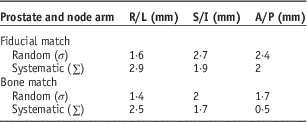
Tables 2 and 3 show the random and systematic errors calculated for each direction (R/L, S/I and A/P) for both the prostate only and prostate and node arms using data before online correction.
Displacement errors for prostate and nodes arm

Abbreviations: R/L, right-left direction; S/I, superoinferior direction; A/P=anteroposterior direction.
Results for prostate only patients and prostate and nodes patients were not combined due to the method of data collection.
When using the 95% confidence interval, the difference in matching to fiducial markers or bone is statistically insignificant in both the S/I and R/L directions. In the A/P direction, the random error is marginally significant (p=0·02); so potentially the coverage of one of the target volumes may be affected by matching to either fiducial markers or pelvic bony anatomy. However, the small sample size (n=9) must be taken into account when considering these results.
PTV margin
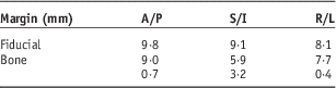
When matching to fiducial markers or anatomical bone, no significant difference was evident, apart from random errors in the A/P direction. These results need to be assessed in terms of the clinical significance and hence the effect this has on the margin required to adequately cover both target volumes. The Van-Herk expression M PTV=2·5Σ+0·7σ was used with the random and systematic errors before online correction. This expression assumes a beam penumbra of 3·2 mm and gives the margin required for 90% of the patients to receive 95% of the dose.Reference Van Herk, Remeijer and Rasch 17
The residual margin is the difference between the two margin calculations, once the online fiducial correction has been applied. The residual margin is what is required around the bone for acceptable coverage.
The similarity of results shown in Table 4 suggests that when treating both prostate and nodes, it may not be necessary to complete the two-step match. Eminowicz et al.Reference Eminowicz, Dean and Shoffren 13 confirmed the safety of fiducial-based image-guided radiotherapy (IGRT), with strict bladder and bowel preparation. They calculated the percentage risk of under dosing the nodal PTV on any given fraction. The likelihood of under dosing in the left, right and anterior directions was <0·25% and <1% in the posterior direction. This confirmed an acceptable reduction of CTV-PTV of the prostate volume and low risk of under dosing of the nodal volume
Margins required for each target volume

Abbreviations: R/L, right-left direction; S/I, superoinferior direction; A/P=anteroposterior direction.
DISCUSSION
Positioning accuracy
The maximum prostate displacement was in the S/I direction, followed by the A/P and smallest in the R/L direction, which is similar to previous findings.Reference Baker, Fenwick and Mayles 11 , Reference Heaton, Mayles and Syndikus 15 , Reference McNair, Keane and Khoo 18 The maximum displacement of 10 mm in the S/I direction is similar to earlier work done at our institution by Baker et al.Reference Baker, Fenwick and Mayles 11 who found prostate displacements of ~11 mm in all three directions. This differs from other studiesReference McNair, Keane and Khoo 18 , Reference Dehnad, Nederveen and van der Heide 19 which found the largest displacement in the A/P direction, this difference is potentially explained by the use of strict bladder and rectal preparation used in this study. The results show that in only 4% of all treatment sessions was there a discrepancy of >5 mm between the prostate position and the position of the nodes.
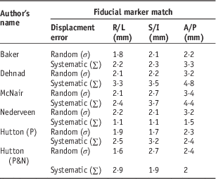
The displacement errors seen for the fiducial marker match in this study are concordant with similar literature.Reference Nederveen, Dehnad and van der Heide 9 , Reference Baker, Fenwick and Mayles 11 , Reference Dehnad, Nederveen and van der Heide 19 Table 5 shows the displacement errors calculated by several authors when using a fiducial marker match.
Fiducial marker match displacement errors from the literature

Abbreviations: R/L, right-left direction; S/I, superoinferior direction; A/P=anteroposterior direction; P, prostate only; P&N, prostate and nodes.
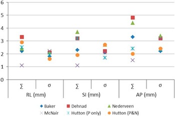
Figure 1 demonstrates the similarity of displacement errors seen in this study and similar recent studies when looking at displacements using fiducial markers. It suggests that the relatively small sample size has not significantly affected the results, compared with other studies.
Displacement errors for fiducial marker matching. Abbreviations: R/L, right-left direction; S/I, superoinferior direction; A/P=anteroposterior direction; ∑, systematic displacement error; σ, random displacement error.

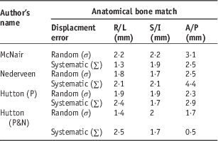
Similarly, Table 6 details the errors calculated by several authors using an anatomical bone match.
Bone match displacement errors from the literature

Abbreviations: R/L, right-left direction; S/I, superoinferior direction; A/P=anteroposterior direction; P, prostate only; P&N, prostate and nodes.
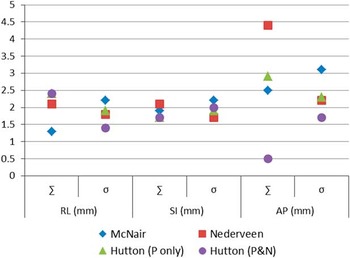
The results seen in Table 6 for an anatomical bone match by other authors are within 5 mm of those found in this study; average random errors were 1·7, 1·9 and 2·0 mm in the R/L, S/I and A/P directions, respectively; average systematic errors were 2·5, 1·7 and 1·7 mm in the R/L, S/I and A/P directions, respectively. The largest discrepancy appears in the A/P direction which is often seen and discussed in the literature.Reference Baker, Fenwick and Mayles 11 , Reference Ten Haken and Randall 20 The most common explanation for this difference is rectal and bladder motion. In this study, a strict bowel and bladder protocol is used before planning CT and each day before treatment. Similarly, McNair et al.Reference Dehnad, Nederveen and van der Heide 19 suggest that patients empty their rectum before treatment, but potentially less reliable than the protocol used in this study; and Nederveen et al.Reference Nederveen, Dehnad and van der Heide 9 fail to discuss any bowel and bladder protocol. This, potentially explains the large systematic error seen in the A/P direction by Nederveen et al.Reference Nederveen, Dehnad and van der Heide 9 (Figure 2), as the bladder volume is known to have a large effect on the position of the prostate.Reference Heaton, Mayles and Syndikus 15 , Reference Greenhalgh 21
Displacement errors for bone matching. Abbreviations: R/L, right-left direction; S/I, superoinferior direction; A/P, anteroposterior direction. ∑, systematic displacement error; σ, random displacement error.

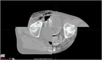
Results from two-step IGRT strategy, coupled with a bladder and bowel protocol, provides accurate coverage of both the prostate volume and the nodal volume. Analysis of imaging data from each fraction of all patients shows the similarity in the prostate and anatomical bone match, it also highlights the random errors seen for one particular patient. This patient had a discrepancy of >5 mm in the S/I direction on 11 out of 37 treatment sessions (30%). Without this patient, the percentage of sessions where a >5 mm discrepancy occurred would have been reduced to 0·8%. It is possible that the pre-treatment preparation of micro-enema and a full bladder was not as effective in this patient, or that this patient had low compliance with the preparation. Evidence of bowel gas was seen on several days using planar 2D images, however, it is difficult to quantify on a 2D image. In a situation where there was a >5 mm discrepancy, a CBCT scan was acquired on the next fraction. Figure 3 shows the transverse view of the CBCT displaying a larger rectum and bladder at planning than on this particular treatment session.
Transverse view of cone beam computed tomography showing larger rectum and bladder.

It is noted that the small sample size used in this study create questions regarding the reliability of the results as F-tests become increasingly unreliable as ‘n’ decreases.Reference Greenhalgh 21 Although the number of image analysis sessions were large, the small patient numbers are a limiting factor when assessing the results, because trends in random and systematic errors are seen for each patient. It is therefore essential to assess the results in relation to those seen in similar studies.
PTV margin
Calculating the margins required on each target volume before any corrections using the formula M PTV=2·5Σ+0·7σ Reference Helyer, Stephenson and Heaton 16 suggested that the margins required on the fiducial markers and bone are very similar. This confirms that whether a fiducial marker match or an anatomical bone match was used, the coverage of both PTV’s is acceptable. This provokes the thought, that one part of the two-step matching process could be eliminated. Results from Eminowicz et al.Reference Eminowicz, Dean and Shoffren 13 confirms this, stating that the risk of under dosing the nodal PTV on any given day is <1%. However, both steps are feasible in a standard 12-minute time slot and are useful in highlighting random errors in the A/P direction. The nodal margin used in the PIVOTAL trialReference Dearnaley 14 is 5 mm in all directions. The residual margin calculated using Van-Herk’s formula gives values of <5 mm and therefore, the IGRT strategy used is appropriate in collaboration with the bladder and bowel protocol.
CONCLUSION
Results suggest that the IGRT strategy of an online correction for both fiducial and bone matches, combined with a strict bladder and bowel protocol and the margins used for the PIVOTAL trial, are acceptable for the treatment of prostate and nodes.
Future work
The protocol used was feasible and reliable and within the quality assurance tolerances of the couch. Our institution is now using a Varian TrueBeam linear accelerator with improved couch repositioning accuracy (0·5 mm) and improved quality CBCT. This will be evaluated with a larger number of patients in order to enhance the statistical reliability of the analytical methods used. Analysis of the next cohort of patients will also include an investigation into rotations as well as translations.
Limitations of study
Patient numbers were limited within the timescale due to the speed of accrual into the PIVOTAL trial. This may have some implications on the results due to one or two patients showing unusual results and, therefore, having a large impact on the findings. It is thought that the findings remain valid, especially when in comparison with other literature, but future work with a larger number of patients would be beneficial.
Acknowledgements
The PIVOTAL Trial Management Group for accepting this project as an IGRT sub-study of the trial. The PIVOTAL trial is funded by Cancer Research UK, CRUK/10/022. The PIVOTAL trial has been scientifically approved by Cancer Research UK’s Clinical Trials Advisory & Awards Committee (CTAAC) and is part of the National Institute for Health Research (NIHR) Clinical Research Network Trial Portfolio. PIVOTAL’s ISRCTN no. (ISRCTN48709247) PIVOTAL was centrally coordinated by the Clinical Trials and Statistics Unit at The Institute of Cancer Research, London.
Disclaimer
The views of the author(s) expressed in this publication are not necessarily those of the NIHR, NHS or the Department of Health.
Conflicts of Interest
None.