Introduction
Temporal lobe surgery for intractable temporal lobe epilepsy (TLE) can result in many patients being rendered seizure free or having a significant reduction in seizure frequency (Choi et al. Reference Choi, Sell, Lenert, Muennig, Goodman, Gilliam and Wong2008). Psychiatric symptoms, however, can develop for the first time following temporal lobe surgery, most commonly depression and anxiety (Foong & Flugel, Reference Foong and Flugel2007; Cleary et al. Reference Cleary, Thompson, Fox and Foong2012). De novo interictal psychosis (Stevens, Reference Stevens1990; Mace & Trimble, Reference Mace and Trimble1991; Christodoulou et al. Reference Christodoulou, Koutroumanidis, Hennessy, Elwes, Polkey and Toone2002; Shaw et al. Reference Shaw, Mellers, Henderson, Polkey, David and Toone2004) following surgery is uncommon, with a mean incidence of 7% (Trimble, Reference Trimble1992; Christodoulou et al. Reference Christodoulou, Koutroumanidis, Hennessy, Elwes, Polkey and Toone2002; Nadkarni et al. Reference Nadkarni, Arnedo and Devinsky2007) but tends to follow a chronic course. The development of psychotic symptoms is not closely related to post-operative seizure outcome (Mace & Trimble, Reference Mace and Trimble1991; Manchanda et al. Reference Manchanda, Miller and McLachlan1993; Leinonen et al. Reference Leinonen, Tuunainen and Lepola1994). De novo interictal psychosis is of particular interest both as a potentially serious complication of the surgery and for the insights it might provide into primary psychosis such as schizophrenia (Shaw et al. Reference Shaw, Mellers, Henderson, Polkey, David and Toone2004).
The underlying pathophysiological mechanisms of psychosis in epilepsy are unclear. Structural and functional neuroimaging studies have implicated left (dominant) temporal lobe pathology with greater reductions in hippocampal volumes (Maier et al. Reference Maier, Mellers, Toone, Trimble and Ron2000), magnetization transfer ratio (Flugel et al. Reference Flugel, Cercignani, Symms, Koepp and Foong2006) and single-photon emission computerized tomography (SPET) perfusion (Marshall et al. Reference Marshall, Syed, Fenwick and Lishman1993; Mellers et al. Reference Mellers, Adachi, Takei, Cluckie, Toone and Lishman1998) in TLE patients with interictal psychosis. Hippocampal sclerosis (HS) is the most common pathology (Roberts et al. Reference Roberts, Done, Bruton and Crow1990; Briellmann et al. Reference Briellmann, Kalnins, Hopwood, Ward, Berkovic and Jackson2000), although low-grade tumours such as dysembryoplastic neuroepithelial tumours have also been reported (Andermann et al. Reference Andermann, Savard, Meencke, McLachlan, Moshe and Andermann1999). Diminished mossy fibre sprouting (Kandratavicius et al. Reference Kandratavicius, Hallak, Young, Assirati, Carlotti and Leite2012), dysplasias of the dentate gyrus (Briellmann et al. Reference Briellmann, Kalnins, Hopwood, Ward, Berkovic and Jackson2000) and excess corpora amylacea (Radhakrishnan et al. Reference Radhakrishnan, Radhakrishnan, Radhakrishnan, Mary, Kesavadas, Alexander and Sarma2007) have been detected in resected tissues of TLE/HS patients with pre-operative interictal psychosis. Detailed histological analysis in patients with de novo interictal psychosis following epilepsy surgery, however, is sparse; one study that explored the density of hippocampal calbindin-immunoreactive neurons in three patients with de novo post-operative and three with pre-operative psychosis compared with controls did not find significant differences (Suckling et al. Reference Suckling, Roberts, Walker, Highley, Fenwick, Oxbury and Esiri2000). The neuropathological changes in de novo interictal psychosis may be similar to primary schizophrenia, given the common clinical symptomatology and pathological findings in the temporal lobe (Perez & Trimble, Reference Perez and Trimble1980; Trimble, Reference Trimble1992; Toone, Reference Toone2000).
In contrast, the neuropathology of schizophrenia has been more widely researched. High-field neuroimaging studies have highlighted structural alterations involving the hippocampal dentate gyrus (Kirov et al. Reference Kirov, Hardy, Matsuda, Messinger, Cankurtaran, Warren, Wiggins, Perry, Babb, Goetz, George, Malaspina and Gonen2013). Hippocampal changes reported in post-mortem studies include altered neuronal densities (Knable et al. Reference Knable, Barci, Webster, Meador-Woodruff and Torrey2004), distribution (Zhang & Reynolds, Reference Zhang and Reynolds2002) and dendritic/synaptic organization (Cotter et al. Reference Cotter, Wilson, Roberts, Kerwin and Everall2000; Rosoklija et al. Reference Rosoklija, Toomayan, Ellis, Keilp, Mann, Latov, Hays and Dwork2000), neuropil abnormalities (Garey, Reference Garey2010) as well as dentate gyrus immaturity (Walton et al. Reference Walton, Zhou, Kogan, Shin, Webster, Gross, Heusner, Chen, Miyake, Tajinda, Tamura, Miyakawa and Matsumoto2012). Altered glutamatergic transmission in the dentate gyrus, including abnormalities of the mossy fibre pathway (Tamminga et al. Reference Tamminga, Stan and Wagner2010) and the endocannabinoid receptor system, have been associated with psychosis (Ujike & Morita, Reference Ujike and Morita2004; Luzi et al. Reference Luzi, Morrison, Powell, di Forti and Murray2008; Fernandez-Espejo et al. Reference Fernandez-Espejo, Viveros, Nunez, Ellenbroek and Rodriguez de Fonseca2009). Post-mortem studies of the neocortex and hippocampal dentate gyrus have also confirmed a reduction in reelin-expressing cells in schizophrenia (Impagnatiello et al. Reference Impagnatiello, Guidotti, Pesold, Dwivedi, Caruncho, Pisu, Uzunov, Smalheiser, Davis, Pandey, Pappas, Tueting, Sharma and Costa1998; Eastwood & Harrison, Reference Eastwood and Harrison2006); reelin has a critical role in the orchestration of normal cortical development as well as ongoing roles in the mature brain, regulating structural plasticity such as axonal growth and synaptogenesis (Lakatosova & Ostatnikova, Reference Lakatosova and Ostatnikova2012; Stranahan et al. Reference Stranahan, Erion and Wosiski-Kuhn2013). Dysregulation of the reelin pathway is implicated in psychosis (Folsom & Fatemi, Reference Folsom and Fatemi2013).
HS is the most common and studied pathology in epilepsy surgical series (Blumcke, Reference Blumcke2009; Blumcke et al. Reference Blumcke, Coras, Miyata and Ozkara2012). In addition to the neuronal loss and accompanying gliosis, frequent epilepsy-specific alterations of the dentate gyrus include granule cell dispersion (GCD) (Blumcke et al. Reference Blumcke, Kistner, Clusmann, Schramm, Becker, Elger, Bien, Merschhemke, Meencke, Lehmann, Buchfelder, Weigel, Buslei, Stefan, Pauli and Hildebrandt2009), mossy fibre sprouting (Sutula et al. Reference Sutula, Cascino, Cavazos, Parada and Ramirez1989), altered calbindin expression (Martinian et al. Reference Martinian, Catarino, Thompson, Sisodiya and Thom2012), and regenerative capacity of granule cells (Thom et al. Reference Thom, Martinian, Sisodiya, Cross, Williams, Stoeber, Harkness and Harding2005a ) and reelin-expressing cell populations (Haas & Frotscher, Reference Haas and Frotscher2010). Some pathological features have been associated with epilepsy co-morbidities, e.g. hippocampal regenerative capacity and calbindin loss have been correlated with memory dysfunction (Coras et al. Reference Coras, Siebzehnrubl, Pauli, Huttner, Njunting, Kobow, Villmann, Hahnen, Neuhuber, Weigel, Buchfelder, Stefan, Beck, Steindler and Blumcke2010; Karadi et al. Reference Karadi, Janszky, Gyimesi, Horvath, Lucza, Doczi, Kallai and Abraham2012). Furthermore, alterations to the endocannabinoid pre-synaptic receptor system have recently been reported in HS. This system modulates both glutamatergic and GABAergic synaptic transmission (Katona & Freund, Reference Katona and Freund2008), and decreases in cannabinoid type 1 receptor (CBR1), CBR1 binding protein mRNA and protein levels have been observed in human HS tissue (Ludanyi et al. Reference Ludanyi, Eross, Czirjak, Vajda, Halasz, Watanabe, Palkovits, Magloczky, Freund and Katona2008).
The aim of this study was to explore relevant cellular alterations gathered from a large series of patients with HS who underwent temporal lobe surgery for epilepsy and to determine whether specific pathological changes in the dentate gyrus could be associated with the development of de novo interictal psychosis.
Method
Subjects
Patients who had developed de novo interictal psychosis following temporal lobe surgery for drug-resistant TLE over a 21-year period (1991 to 2012) were assessed for inclusion in the study. A total of 20 patients who met Diagnostic and Statistical Manual of Mental Disorders, fourth edition, text revision (DSM IV-TR; APA, 2000) diagnosis criteria for a psychotic disorder due to a general medical condition were identified from the hospital records in the neuropsychiatry clinics at the National Hospital for Neurology Neurosurgery (NHNN). Of the 20 patients, 15 had a diagnosis of HS, four patients had low-grade glioneuronal tumours (dysembryoplastic neuroepithelial tumours) and one patient had an epidermoid cyst involving the mesial temporal lobe. Of the 15 patients with HS and psychosis (HS-P), consent was available in 11 for further study. A total of 10 HS patients who also underwent temporal lobe surgery but without either a history of pre- or post-operative psychosis were selected as control cases (HS-NP). They were matched to the HS-P group with respect to age and the year of surgery. The study was approved by the Joint Research and Ethics committee of the Institute of Neurology and NHNN.
Histopathological techniques
Immunohistochemistry
For each patient, histological sections from all specimens from the temporal lobe surgical resection [neocortex (anterior 3–4 cm of temporal lobe and pole), parahippocampal gyrus, amygdala and hippocampus] were reviewed with haematoxylin and eosin and Luxol fast blue stain for myelin. The tissue block from the mid-body of the hippocampus, to include the most complete contour of the dentate gyrus, was selected for an immunohistochemistry panel including: glial fibrillary acidic protein (GFAP) (1:1500; Dako, UK), NeuN (neuronal nuclei; 1:1500; Chemicon International); calbindin D28 K (1:10 000; Swant, Switzerland); reelin (gift from Professor Goffinet, University of Namur, Belgium; dilution. 1:200, microwave pre-treatment); mini-chromosome maintenance protein 2 (MCM2) (1:900; BD Biosciences, USA); dynorphin (1:100; AbD Serotec MorphoSys UK Ltd, UK); CBR1 (1:1000; Abcam, UK); doublecortin (DCX) (1:250; Cell Signaling, UK); nestin (1:1000; Abcam, UK); and GFAP-δ (1:5000; Chemicon International, USA). For this purpose, sections were cut at 7 μm, dewaxed, and immersed in a 3% hydrogen peroxide solution to block endogenous peroxidase activity prior to antibody incubation. Dako EnVision™ diaminobenzidine (DAB) was used as the chromogen and slides were counterstained with haematoxylin.
Qualitative analysis and measurements
On the NeuN sections, the pattern of HS was classified as International League Against Epilepsy (ILAE) type 1, 2 or 3 (Blumcke et al. Reference Blumcke, Thom, Aronica, Armstrong, Bartolomei, Bernasconi, Bernasconi, Bien, Cendes, Coras, Cross, Jacques, Kahane, Mathern, Miyata, Moshe, Oz, Ozkara, Perucca, Sisodiya, Wiebe and Spreafico2013). The presence of neuronal loss in the dentate gyrus, as assessed by the presence of gaps of depletion or discontinuity in the cell layer, was semi-quantified in four grades: <25% (not detectable); 25–50% (mild); 50–75% (moderate); and >75% (severe) neuronal loss. All assessments were made independently by two observers (J.M. and M.T.) with good agreement. The extent of GCD was assessed as in a previous study (Thom et al. Reference Thom, Liagkouras, Elliot, Martinian, Harkness, McEvoy, Caboclo and Sisodiya2010). Using an image analysis program (Image Pro Plus, version 6; Media Cybernetics, UK) linked to a Zeiss microscope (Carl Zeiss, UK), between six and 10 non-overlapping images per case were taken at 10x magnification representing the entire length of the dentate ribbon. For each image frame, the position of the 10 outermost cells in the granule cell layer (GCL) was measured relative to the basal layer in the perpendicular axis providing a maximum of 100 measurements per case. For each case a mean of all these measurements and a mean of the maximum measurement recorded per field were obtained as measures of the overall and maximal severity of GCD. These measurements were repeated by one observer (J.M.) in five cases for reproducibility, with an intra-class correlation coefficient (ICC) of 0.99.
The presence of mossy fibre sprouting (MFS) in the dentate gyrus on dynorphin-labelled sections was graded as previously (Thom et al. Reference Thom, Martinian, Catarino, Yogarajah, Koepp, Caboclo and Sisodiya2009): grade 0 (no MFS); grade 1 (focal MFS in the inner molecular layer; ML); grade 2 (extensive MFS throughout ML). Calbindin expression in the dentate gyrus granule cells was also graded as previously (Martinian et al. Reference Martinian, Catarino, Thompson, Sisodiya and Thom2012): grade 0 (normal expression through the GCL); grade 1 (loss of expression in basal GCL); and grade 2 (extensive loss of expression throughout the GCL).
Quantitative analysis
Reelin
A region of interest (ROI) was drawn at ×2.5 objective magnification using Histometrix image analysis software (Kinetic Imaging, UK) in the hippocampal white matter, with the dentate GCL and the pyramidal cell layer of Cornu ammonis (CA) area CA3–1 as the boundaries. This included the stratum moleculare, lacunosum and radiatum of CA1–3 in addition to the stratum moleculare of the dentate gyrus. The rationale to include all the white matter was that it was not feasible to confidently separate these layers on the reelin-stained sections. All reelin-positive cells within this ROI, regardless of size or morphology (Abraham & Meyer, Reference Abraham and Meyer2003), were counted at ×40 objective magnification by a single observer (M.K.).
MCM2
A ROI was drawn with Histometrix at ×2.5 magnification around the dentate gyrus GCL as in a previous study (Thom et al. Reference Thom, Sisodiya, Beckett, Martinian, Lin, Harkness, Mitchell, Craig, Duncan and Scaravilli2002). Within this ROI (mean area 3.2 × 102 mm2), all fields were examined at ×40 magnification and all positive nuclei counted by a single observer (J.M.) and the mean cell density (per unit area) recorded. These measurements were repeated in eight cases with an ICC of 0.92.
DCX
Sequential images were captured along the entire length of the dentate gyrus at ×40 objective magnification with a Nikon (80i) microscope (average length of dentate gyrus studied per case = 3.6 mm). Using Image Pro Plus analyser (version 6.3), positive cells were manually tagged and the mean cell density (per unit area) recorded.
CBR1
Representative, non-overlapping images from the dentate gyrus (GCL and ML), CA4 and the subiculum were taken at ×20 objective magnification (four to seven images per subfield) with a Nikon 80i microscope. Using RGB thresholding and Image Pro Plus (version 6), the field fraction of synaptic staining (percentage labelling) was quantified for each image. The same thresholds were applied for each image within a case and a mean labelling index for each subfield was obtained. Both the image capture and measurements were carried out by a single observer (J.M.) and repeated in six cases with good inter-observer reproducibility (ICC = 0.96).
Statistical comparisons
Statistical comparisons between HS-P and HS-NP patients were carried out using SPSS for Windows (version 20; IBM, USA) and non-parametric tests, including the Mann–Whitney test and Spearman's correlation.
Results
Clinical characteristics
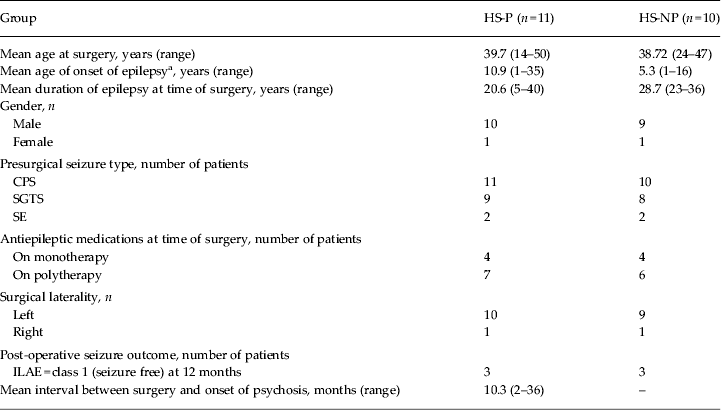
The clinical characteristics of both groups are summarized in Table 1. The two groups did not differ in age, presurgical seizure history or number of antiepileptic medications. Only one patient who was in the HS-P group had a history of cannabis use but no patient was prescribed anti-psychotic medications prior to surgery. There was also no family history of psychosis for any of the subjects. The number of patients who were seizure free (ILAE class 1) at 12 months did not differ between the groups.
Clinical data of HS-P and HS-NP patients

HS-P, hippocampal sclerosis and de novo post-surgical interictal psychosis; HS-NP, hippocampal sclerosis and epilepsy alone with no psychosis (control); CPS, complex partial seizures; SGTS, secondary generalized seizures; SE, episode of status epileptic; ILAE, International League Against Epilepsy.
a Exact age of onset of seizures unknown for three HS-NP cases.
In the HS-P group, the psychotic symptoms were predominantly persecutory delusions and auditory hallucinations, resembling schizophrenia. The onset of psychosis ranged from 2 to 36 months following surgery, with a mean of 10.3 months. The psychosis followed a chronic course (duration ranging from 3 to 20 years) and all patients were maintained on antipsychotic medications at the time of our study.
Histopathological findings
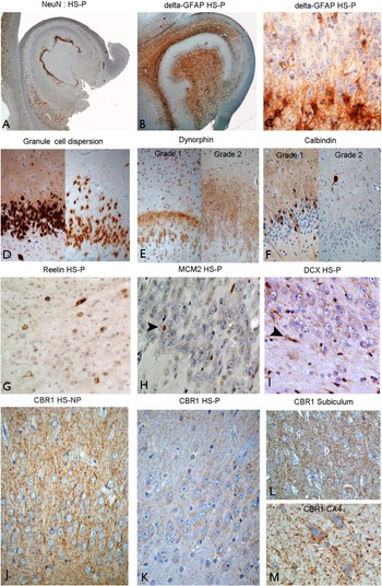
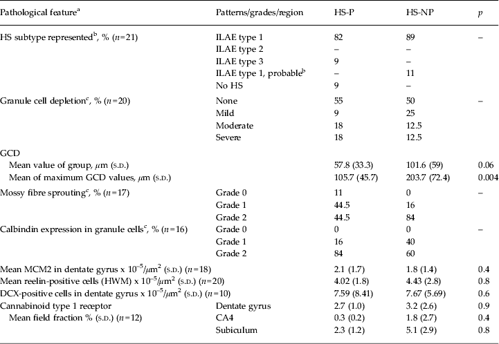
The patterns of HS were similar in the HS-P and HS-NP groups, with HS ILAE type 1 being the commonest pattern in both groups (Table 2 and Fig. 1A). In one of the HS-P patients, gliosis alone was confirmed in the hippocampus and this subject was reclassified as a no-HS case according to the new criteria (Blumcke et al. Reference Blumcke, Thom, Aronica, Armstrong, Bartolomei, Bernasconi, Bernasconi, Bien, Cendes, Coras, Cross, Jacques, Kahane, Mathern, Miyata, Moshe, Oz, Ozkara, Perucca, Sisodiya, Wiebe and Spreafico2013). The severity of granule cell loss, as assessed semi-quantitatively, was also similar between the groups with no perceived loss in around half of the cases in each group. Nestin- and GFAP-δ-positive glial cells were noted in CA4, particularly in the subgranular zone, and CA1 in all HS cases as previously described (Martinian et al. Reference Martinian, Boer, Middeldorp, Hol, Sisodiya, Squier, Aronica and Thom2009), but with no clear discrimination between groups on qualitative evaluation (Fig. 1B, C). GCD was less severe in the HS-P group than in HS-NP cases (Fig. 1D and Fig. 2); statistically significant differences were shown for the maximum GCD values but not for the mean GCD values (Table 2). This significance remained when the no-HS case was excluded from the analysis. Whereas grade 2 mossy fibre sprouting was confirmed in over 80% of HS-NP cases, it was noted in less than half of HS-P cases (Fig. 1E and Table 2). All HS cases showed abnormal calbindin expression in the dentate gyrus, with 84% in the HS-P group and 60% in the HS-NP group showing a marked reduction (grade 2); the remaining cases showed reduced expression restricted to the basal granule cells only (grade 1) (Fig. 1F).
(A) NeuN (neuronal nuclear antigen)-stained section from a patient with hippocampal sclerosis (HS) and post-operative psychosis (HS-P) with the classical pattern of neuronal loss in Cornu ammonis (CA) area CA4 and CA1 with sparing of the CA2 region (International League Against Epilepsy type 1). (B) Glial fibrillary acidic protein (GFAP)-δ labelling confirmed cells with astrocytic morphology in the regions of neuronal loss, particularly in the subgranular zone of the dentate gyrus (C). (D) Varying degrees of granule cell dispersion were measured in the HS-P cases with mild dispersion shown on the left and moderate dispersion on the right. (E) Dynorphin staining for mossy fibre sprouting was graded as grade 1 (mild sprouting into the inner molecular layer of the dentate gyrus) and grade 2 (extensive sprouting throughout the molecular layer). (F) Reduction of calbindin expression in the granule cell layer (GCL) was graded as grade 1 (reduction in basal cells only) and grade 2 (extensive loss of calbindin in granule cells): in these figures interneurons remain labelled, confirming technical efficacy of the immunostaining protocol. (G) Reelin immunohistochemistry reveals distinctively positive cytoplasmic labelling of a population of small round cells and larger cells with horizontal processes in the hippocampal white matter. (H) Mini-chromosome maintenance protein 2 (MCM2) immunostaining highlights positive nuclear labelling of small immature cells (compared with the size of the granule cell neuron); similar small cells also show cytoplasmic labelling for doublecortin (DCX) with bipolar processes (I), through the GCL and subgranular zone (arrowheads in both). (J) Cannabinoid type 1 receptor (CBR1) staining in the dentate gyrus and molecular layer shows diffuse synaptic-like staining surrounding the neurones in a HS case without psychosis (HS-NP); in some cases, and more frequent in HS-P cases, diminished staining was noted, and confirmed with quantitative analysis (K). A similar labelling pattern was noted in the subiculum (L) but with less pronounced labelling in the CA4 region in many HS cases (M). Original magnification objectives in (A) and (B) × 1.6; in (C) and (G–M) × 40; and in (D–F) × 10.

Scaled bar charts of measurements of dentate gyrus (DG) parameters for patients with hippocampal sclerosis (HS) who developed interictal psychosis (HS-P) post-operatively and a matched surgical HS group without psychosis (HS-NP). On the y axis the units for granule cell dispersion (GCD) mean and maximum (Max) are μm × 101. Reelin, mini-chromosome maintenance protein 2 (MCM2) and doublecortin (DCX) are expressed as cells per × 10–5/μm2. The cannabinoid type 1 receptor (CBR1) labelling index is shown as the percentage of immunolabelling over each subfield (DG, Cornu ammonis (CA) area CA4 and the subiculum). Values are means, with standard deviations represented by vertical bars. * Mean values were significantly different (p < 0.05).

Neuropathological findings in HS-P and HS-NP patients

HS-P, hippocampal sclerosis and de novo post-surgical interictal psychosis; HS-NP, hippocampal sclerosis and epilepsy alone with no psychosis (control); HS, hippocampal sclerosis; ILAE, International League Against Epilepsy; GCD, granule cell dispersion; s.d., standard deviation; MCM2, mini-chromosome maintenance protein 2; HWM, hippocampal white matter; DCX, doublecortin; CA4, Cornu ammonis area 4.
a n = Number of cases on which assessment was possible; in some cases assessment was not possible due to technical reasons or insufficient representation of the dentate gyrus on further sections.
b Classification of the HS subtype was based on ILAE system of 2013 (Blumcke et al. Reference Blumcke, Thom, Aronica, Armstrong, Bartolomei, Bernasconi, Bernasconi, Bien, Cendes, Coras, Cross, Jacques, Kahane, Mathern, Miyata, Moshe, Oz, Ozkara, Perucca, Sisodiya, Wiebe and Spreafico2013) with probable HS referring to cases with incomplete subfields available for analysis.
c See text for details of grading schemes for assessment of immunostaining.
The morphology of reelin-positive cells in the hippocampal white matter was similar in both groups and included small round cells with a thin peripheral rim of cytoplasmic labelling in addition to larger cells with neuronal morphology and bipolar orientation, reminiscent of Cajal–Retzius cells (Fig. 1G). Quantitative analysis revealed no significant difference in the density of reelin-labelled cells between the HS-P and HS-NP groups (Table 2). Frequent MCM2-positive nuclei of small immature cells were observed in the GCL in most cases (Fig. 1H); we have previously reported that these cells were mainly negative with mature glial or microglial markers (Thom et al. Reference Thom, Martinian, Williams, Stoeber and Sisodiya2005b ). Cells of similar morphology and distribution in the dentate gyrus, with bipolar or delicate multipolar cytoplasmic process, were highlighted with DCX immunohistochemistry through the dentate gyrus and subgranular zone (Fig. 1I). Quantitative analysis, however, revealed no difference in MCM2 or DCX cell densities in the dentate gyrus between the groups (Fig. 2).
CBR1 immunohistochemistry revealed dense networks of processes in the hippocampus (Fig. 1J–M), with fibres and synaptic-like staining in the neuropil, sometimes observed surrounding residual negatively stained neuronal cell bodies (Fig. 1M). This pattern is in keeping with previous descriptions of CBR1 expression in HS (Magloczky et al. Reference Magloczky, Toth, Karlocai, Nagy, Eross, Czirjak, Vajda, Rasonyi, Kelemen, Juhos, Halasz, Mackie and Freund2010). In all HS samples, there was an impression of reduced CBR1 labelling in the CA4 subfield compared with the dentate gyrus and subiculum. Quantitative analysis confirmed significant differences in CBR1 labelling between the three subfields over all cases (p < 0.01). The mean CBR1 labelling fractions in all three regions (dentate gyrus, CA4 and subiculum), although lower in the HS-P group compared with the HS-NP group (Fig. 1G, H and Fig. 2), were not statistically significantly different (Table 2). There was no significant difference in the CBR1 labelling in the dentate gyrus in relation to the severity of granule cell loss, suggesting that this trend for group differences was independent of the degree of sclerosis.
Clinical–pathological correlations revealed an inverse correlation between the time interval to the development of psychosis post-operatively to both the mean value of GCD (p < 0.05, Spearman's correlation) and CBR1 expression in the subiculum (p = 0.05, Spearman's correlation). There was a negative correlation between the CBR1 index in the CA4 region and age of onset of seizures in the HS-P (p = 0.005, Spearman's correlation) but not in the HS-NP group.
Discussion
Neuropathological studies of resected tissues from patients developing de novo psychosis following temporal lobe surgery for epilepsy are limited. Importantly, they provide a unique opportunity to capture potential cellular responses that may predispose to the development of psychosis without the confounding factor of anti-psychotic treatment-induced changes. The patterns of hippocampal neuronal loss in the HS-P and HS-NP patients in our study were typical of those encountered in epilepsy (82% and 89%, respectively, of patients showing classical or ILAE type 1 HS: neuronal loss and gliosis primarily involving CA1 and CA4), consistent with reports of 60–80% in large epilepsy series (Blumcke et al. Reference Blumcke, Thom, Aronica, Armstrong, Bartolomei, Bernasconi, Bernasconi, Bien, Cendes, Coras, Cross, Jacques, Kahane, Mathern, Miyata, Moshe, Oz, Ozkara, Perucca, Sisodiya, Wiebe and Spreafico2013).
We found less prominent MFS in HS-P patients compared with the HS-NP group. This is in keeping with recent findings of diminished MFS, as visualized with Timms staining, in TLE patients with interictal psychosis compared with patients with HS and epilepsy alone or depression (Kandratavicius et al. Reference Kandratavicius, Hallak, Young, Assirati, Carlotti and Leite2012). The mechanisms promoting axonal sprouting in epilepsy are uncertain; experimental models demonstrate MFS after electrical stimulation but it also occurs in association with loss of hilar neurones and their projection pathways to the dentate gyrus (Nadler, Reference Nadler2003). MFS may therefore represent an aberrant reparative mechanism to restore function, but with evidence that its overall effect is to enhance synchronization and facilitate seizures. Local growth factors and components of the extracellular matrix, as GAP-43 and tenascin-C, may promote MFS (Proper et al. Reference Proper, Jansen, van Veelen, van Rijen, Gispen and de Graan2001; Heck et al. Reference Heck, Garwood, Loeffler, Larmet and Faissner2004). The maturity of granule cells may also govern the extent of MFS (Cameron et al. Reference Cameron, Zhan and Nadler2011; Martinian et al. Reference Martinian, Catarino, Thompson, Sisodiya and Thom2012). Several of these factors may have influenced the diminished axonal sprouting observed in the HS-P patients. It has also been suggested that changes in the dynamics of axonal sprouting or synaptic reorganization in the remaining brain may contribute to the development of psychosis following temporal lobe surgery (Stevens, Reference Stevens1990). It is possible that in our HS-P group, a tendency for diminished axonal sprouting as a reparative response may extend beyond the resected hippocampus or extratemporally and may predispose to the development of psychosis following surgery.
There was no family history of psychosis in our HS-P group but as our sample size was small, the possibility that genetic or epigenetic factors may also contribute to the diminished capacity for axonal sprouting cannot be excluded. For example, the disrupted-in-schizophrenia 1 (DISC1) gene has been reported to be associated with major psychiatric disorders including psychosis and schizophrenia (Enomoto et al. Reference Enomoto, Asai, Namba, Wang, Kato, Tanaka, Tatsumi, Taya, Tsuboi, Kuroda, Kaneko, Sawamoto, Miyamoto, Jijiwa, Murakumo, Sokabe, Seki, Kaibuchi and Takahashi2009). Interestingly, DISC1 has a wide range of regulatory functions, including roles in axonal guidance and neurite extension. Clearly, further research into genetic risk factors in patients who develop psychosis following epilepsy surgery is warranted.
GCD is a well-recognized and relatively specific feature of HS in epilepsy. It is associated with hippocampal (typically hilar) neuronal loss, is observed in around 50% of cases in large surgical series and is associated with early onset or long duration of epilepsy (Blumcke et al. Reference Blumcke, Thom, Aronica, Armstrong, Bartolomei, Bernasconi, Bernasconi, Bien, Cendes, Coras, Cross, Jacques, Kahane, Mathern, Miyata, Moshe, Oz, Ozkara, Perucca, Sisodiya, Wiebe and Spreafico2013). A small series of six patients with TLE and interictal psychosis confirmed GCD in 20% of cases compared with 64% of TLE patients without psychosis, although the differences were not significant (Suckling et al. Reference Suckling, Roberts, Walker, Highley, Fenwick, Oxbury and Esiri2000). We also noted significantly less GCD in the HS-P group, but based on the maximal dispersion measurements only, and this was independent of the pattern of HS. Experimental epilepsy models propose that seizure activity itself induces migration of mature granule cells (Chai et al. Reference Chai, Munzner, Zhao, Tinnes, Kowalski, Haussler, Young, Haas and Frotscher2013) and cell ‘ectopia’ is influenced by γ-aminobutyric acid (GABA) activity following febrile seizures (Koyama et al. Reference Koyama, Tao, Sasaki, Ichikawa, Miyamoto, Muramatsu, Matsuki and Ikegaya2012). Studies in human epilepsy tissues have also proposed that acquired hippocampal reelin deficiency arising in HS has a primary mechanistic role in GCD (Haas & Frotscher, Reference Haas and Frotscher2010). Decreased reelin protein and expressing cells have been shown in HS in mesial MLE patients (Frotscher et al. Reference Frotscher, Haas and Förster2003) and, in experimental studies, diminished reelin follows induction of seizures with the subsequent development of GCD (Haas & Frotscher, Reference Haas and Frotscher2010). In this study, we therefore investigated reelin-expressing cell populations in view of this observation as well as the accumulating evidence also implicating the reelin-signalling pathway in schizophrenia. Variations of the reelin gene have been linked to increased risk of schizophrenia in women (Ben-David & Shifman, Reference Ben-David and Shifman2010; Kuang et al. Reference Kuang, Sun, Zhu and Li2011). Reduced reelin-expressing cells have been shown in multiple brain regions including the frontal cortex and hippocampus in schizophrenia (Impagnatiello et al. Reference Impagnatiello, Guidotti, Pesold, Dwivedi, Caruncho, Pisu, Uzunov, Smalheiser, Davis, Pandey, Pappas, Tueting, Sharma and Costa1998; Fatemi, Reference Fatemi2005; Eastwood & Harrison, Reference Eastwood and Harrison2006, Habl et al. Reference Habl, Schmitt, Zink, von Wilmsdorff, Yeganeh-Doost, Jatzko, Schneider-Axmann, Bauer and Falkai2012) and, in a meta-analysis, this reduction only approached significance in the ML of the dentate gyrus and declined with disease progression (Knable et al. Reference Knable, Barci, Webster, Meador-Woodruff and Torrey2004). Abnormalities in the reelin-signalling pathway could potentially link the association of epilepsy and psychosis. We found lower Cajal–Retzius cell numbers in the HS-P group but the differences were not statistically significant. There was also no association between cell number and the period to the onset of psychosis post-operatively. This lack of significance could be attributed to the relatively small sample size in our study and does not exclude a role for reelin in interictal psychosis.
An ‘immature dentate gyrus’ has been proposed as an underlying template of both experimental and human studies of schizophrenia, characterized by increased numbers of immature neuronal progenitors and a lack of calbindin-positive mature neurons (Walton et al. Reference Walton, Zhou, Kogan, Shin, Webster, Gross, Heusner, Chen, Miyake, Tajinda, Tamura, Miyakawa and Matsumoto2012; Shin et al. Reference Shin, Kobayashi, Hagihara, Kogan, Miyake, Tajinda, Walton, Gross, Heusner, Chen, Tamura, Miyakawa and Matsumoto2013). Studies have identified reduced levels of neural stem cell proliferation in the dentate gyrus as quantified by Ki-67 immunohistochemistry in post-mortem studies in schizophrenia (Reif et al. Reference Reif, Fritzen, Finger, Strobel, Lauer, Schmitt and Lesch2006). In patients with epilepsy and HS, the residual proliferative capacity of the dentate gyrus and isolation of stem cells has been shown to directly correlate with memory function (Coras et al. Reference Coras, Siebzehnrubl, Pauli, Huttner, Njunting, Kobow, Villmann, Hahnen, Neuhuber, Weigel, Buchfelder, Stefan, Beck, Steindler and Blumcke2010). We utilized MCM2 as a marker of regenerative capacity in the dentate gyrus, which labels cells licenced for replication, expressed through the cell cycle, and therefore is a more sensitive marker than Ki-67 in fixed tissue samples (von Bohlen und Halbach, Reference von Bohlen und Halbach2011). It has been used in previous studies of HS (Thom et al. Reference Thom, Martinian, Williams, Stoeber and Sisodiya2005b ; Fahrner et al. Reference Fahrner, Kann, Flubacher, Heinrich, Freiman, Zentner, Frotscher and Haas2007) where small immunopositive nuclei were observed to co-localize with immature nestin-positive cells, and a minority co-localizing with mature glial/microglial markers (Thom et al. Reference Thom, Martinian, Williams, Stoeber and Sisodiya2005b ). We confirmed MCM2-positive cells in both patient groups which were morphologically similar to DCX-positive cells in the dentate gyrus. Similar DCX-positive cells have been recently described in the infant dentate gyrus (Paine et al. Reference Paine, Willsher, Nicholson, Sebire and Jacques2013) but such immature-appearing DCX-positive cells have not previously been reported in the adult human hippocampus or in HS (D'Alessio et al. Reference D'Alessio, Konopka, Lopez, Seoane, Consalvo, Oddo, Kochen and Lopez-Costa2010). There were, however, no group differences in the numbers of DCX or MCM2 cells to suggest altered dentate maturation or neurogenesis in HS-P relative to HS-NP patients. It is possible that seizures and duration of epilepsy have a more over-riding influence on the numbers of DCX-positive cells.
Calbindin expression in the granule cells reaches full maturation by 11 years in humans (Abraham et al. Reference Abraham, Richter, Gyimesi, Horvath, Janszky, Doczi and Seress2011). Loss of calbindin expression in granule cells has been reported in TLE/HS with complete loss varying between 13% and 35% of cases in studies (Magloczky et al. Reference Magloczky, Halasz, Vajda, Czirjak and Freund1997; Arellano et al. Reference Arellano, Munoz, Ballesteros-Yanez, Sola and DeFelipe2004; Martinian et al. Reference Martinian, Catarino, Thompson, Sisodiya and Thom2012); it is also documented in experimental models of epilepsy (Shin et al. Reference Shin, Kobayashi, Hagihara, Kogan, Miyake, Tajinda, Walton, Gross, Heusner, Chen, Tamura, Miyakawa and Matsumoto2013). Loss of calbindin expression in granule cells has been directly correlated with memory impairment (Karadi et al. Reference Karadi, Janszky, Gyimesi, Horvath, Lucza, Doczi, Kallai and Abraham2012) and a reduction noted in patients with schizophrenia and bipolar disorder (Walton et al. Reference Walton, Zhou, Kogan, Shin, Webster, Gross, Heusner, Chen, Miyake, Tajinda, Tamura, Miyakawa and Matsumoto2012). Gene expression studies of dentate granule cells in post-mortem schizophrenic brains have shown significant reductions in calbindin (Altar et al. Reference Altar, Jurata, Charles, Lemire, Liu, Bukhman, Young, Bullard, Yokoe, Webster, Knable and Brockman2005). A previous study of only three patients with psychosis following epilepsy surgery did not identify a significant reduction in calbindin in the dentate granule cells compared with controls (Suckling et al. Reference Suckling, Roberts, Walker, Highley, Fenwick, Oxbury and Esiri2000). In our study, marked loss of calbindin was more common in HS-P than HS-NP patients (84% v. 60%); although this was not statistically analysed this could suggest that loss of calbindin expression may be a risk factor for developing interictal psychosis following epilepsy surgery.
The endocannabinoid system has been implicated in schizophrenia (Fernandez-Espejo et al. Reference Fernandez-Espejo, Viveros, Nunez, Ellenbroek and Rodriguez de Fonseca2009). Cannabis use is associated with acute psychotic episodes and an increased risk of developing schizophrenia in the long term. The main receptor, CBR1, is widely and abundantly expressed in the CNS, primarily at pre-synaptic terminals. Released endocannabinoids act as retrograde signal molecules, mediating glutamatergic and GABAergic neurotransmission, with an overall net effect considered to be inhibitory (Katona & Freund, Reference Katona and Freund2012). Genetic alterations in the CNR1 gene which codes for CBR1 have been shown in hebephrenic schizophrenia (Chavarria-Siles et al. Reference Chavarria-Siles, Contreras-Rojas, Hare, Walss-Bass, Quezada, Dassori, Contreras, Medina, Ramirez, Salazar, Raventos and Escamilla2008) and polymorphisms associated with pharmacological responses to antipsychotic medications in schizophrenia (Hamdani et al. Reference Hamdani, Tabeze, Ramoz, Ades, Hamon, Sarfati, Boni and Gorwood2008). Elevated binding of CBR1 has also been shown in photon emission computerized tomography (PET) imaging in schizophrenic patients (Wong et al. Reference Wong, Kuwabara, Horti, Raymont, Brasic, Guevara, Ye, Dannals, Ravert, Nandi, Rahmim, Ming, Grachev, Roy and Cascella2010). Post-mortem findings in schizophrenia have reported an up-regulation of CBR1 in the frontal (Dean et al. Reference Dean, Sundram, Bradbury, Scarr and Copolov2001) and cingulate cortex (Zavitsanou et al. Reference Zavitsanou, Garrick and Huang2004) using autoradiography, although no tissue studies, to the best of our knowledge, have been carried out on the hippocampus. In post-mortem studies, antipsychotic medications may influence receptor-binding studies (Fernandez-Espejo et al. Reference Fernandez-Espejo, Viveros, Nunez, Ellenbroek and Rodriguez de Fonseca2009) whereas, in contrast, all patients in our study were naive to antipsychotic medications at the time of tissue resection. Furthermore, only one patient in the HS-P group had a history of cannabis use.
In epilepsy, the endocannabinoid system is considered to have both anticonvulsive and neuroprotective effects (Monory et al. Reference Monory, Massa, Egertova, Eder, Blaudzun, Westenbroek, Kelsch, Jacob, Marsch, Ekker, Long, Rubenstein, Goebbels, Nave, During, Klugmann, Wolfel, Dodt, Zieglgansberger, Wotjak, Mackie, Elphick, Marsicano and Lutz2006). PET studies using the ligand [18F]MK-9470 showed an increase in CBR1 receptor availability associated with HS, suggesting an adaptive protective mechanism of neurons against hyperexcitability and seizure activity (Goffin et al. Reference Goffin, Van Paesschen and Van Laere2011). In tissue resections from HS patients there is evidence for an overall reduction in CBR1 (Ludanyi et al. Reference Ludanyi, Eross, Czirjak, Vajda, Halasz, Watanabe, Palkovits, Magloczky, Freund and Katona2008) although relative increases in CBR1 in the dentate gyrus in HS have been shown, primarily associated with GABAergic cell terminals (Magloczky et al. Reference Magloczky, Toth, Karlocai, Nagy, Eross, Czirjak, Vajda, Rasonyi, Kelemen, Juhos, Halasz, Mackie and Freund2010) and confirmed in experimental models (Karlocai et al. Reference Karlocai, Toth, Watanabe, Ledent, Juhasz, Freund and Magloczky2011). These findings therefore support alteration of the endocannabinoid system in HS and epilepsy. In our study, we also noted a similar pattern of alteration of the CBR1 receptor system in all HS cases including significant increases in the dentate gyrus relative to CA4. There was a qualitative impression of a reduction in hippocampal subfield CBR1 in HS-P compared with HS-NP patients although the differences were not statistically significant. Further studies at a molecular–genetic level, as well as receptor affinity studies to explore the role of CBR1 receptor in epilepsy with co-morbid psychosis, are warranted.
The clinical characteristics of our HS-P group are in keeping with previous reports, with the majority being male with left-sided HS and not seizure free following surgery (Flor-Henry, Reference Flor-Henry1969; Lindsay et al. Reference Lindsay, Ounsted and Richards1979; Glosser et al. Reference Glosser, Zwil, Glosser, O'Connor and Sperling2000; Maier et al. Reference Maier, Mellers, Toone, Trimble and Ron2000; Inoue & Mihara, Reference Inoue and Mihara2001; Kanner et al. Reference Kanner, Byrne, Chicharro, Wuu and Frey2009). Our observations therefore support the preponderance of involvement of the dominant hemisphere in psychotic patients with epilepsy which, in the current series, revealed itself only following surgical intervention. We can speculate the existence of lateralized neuronal networks that become altered in TLE/HS, but with different responses following surgery (as has been shown for lateralization of dynorphin levels following traumatic brain injury as an example; Hussain et al. Reference Hussain, Fitting, Watanabe, Usynin, Yakovleva, Knapp, Scheff, Hauser and Bakalkin2012) which ultimately, in these patients, predisposes to the psychotic state. However, we did not identify any clear relationships between the clinical variables, laterality and pathology measurements in the HS-P group.
Several limitations to our study should be considered. The number of cases in our series was small due to the rarity of the condition. A normal (non-epilepsy) hippocampal surgical control group was not available. With quantitative immunohistochemistry, as always, variations in tissue processing and fixation can influence the intensity of immunolabelling; we aimed to minimize this by closely matching our control group for both clinical demographics as well as the year of surgery. Furthermore, pre-operative medications, including anti-epileptic drugs, may have influenced CBR1 expression although both patient groups would have been similarly affected as most patients were on polytherapy.
In summary, we report here the largest published series of patients with HS who developed interictal psychosis following epilepsy surgery. We identified a pattern of reduced mossy fibre axonal sprouting, dispersion of granule cells and calbindin expression as well as qualitative differences in hippocampal CBR1 expression. These findings suggest that subtle differences in the hippocampal circuitry and cellular response to seizures may predispose to the development of psychosis following epilepsy surgery.
Acknowledgements
This work was undertaken at University College London Hospital/University College London, which received a proportion of funding from the Department of Health's National Institute for Health Research (NIHR) Biomedical Research Centre's funding scheme. Neuropathology components of this work and J.L. and C.R. are supported by a grant from the Medical Research Council (MR/J01270X/1).
Declaration of Interest
None.