Introduction
Despite several deficiencies, morphology observation is still a widely used conventional method for embryo selection because of its non-invasiveness and convenience. Both the introduction of sequential culture and the improvement in laboratory technology have led to the successful clinical practice of day 3 (D3) and day 5 (D5) transfers, which have greatly increased both the pregnancy rate (PR) and implantation rate (IR). However, even blastocyst transfer can not ensure successful pregnancy, suggesting the insufficiency of present morphological assessment systems for predicting assisted reproductive technologies (ARTs) outcome. To perform more accurate embryo selection, clinical embryologists have been trying to seek new valuable markers that can reflect more detailed information about the embryos and/or the blastocysts.
During maturation and fertilization, the human oocyte releases the first and second polar body (PB1 and PB2), respectively, via two asymmetric divisions (meiosis). The relationship between PBs and oocytes has attracted much attention because of the homology of their chromatin with the oocytes (Munné et al., Reference Munné, Dailey, Sultan, Grifo and Cohen1995; Ebner et al., Reference Ebner, Moser, Yaman, Feichtinger, Hartl and Tews1999, Reference Ebner, Yaman, Moser, Sommergruber, Feichtinger and Tews2000, Reference Ebner, Moser, Sommergruber, Yaman, Pfleger and Tews2002; Durban et al., Reference Durban, Benet, Boada, Fernandez, Calafell, Lailla, Sanchez-Garcia, Pujol, Egozcue and Navarro2001; Verlinsky Reference Verlinsky2003; Fancsovits et al., Reference Fancsovits, Tothne, Murber, Takacs, Parp and Urbancsek2006; Younis et al., Reference Younis, Radin, Izhaki and Ben-Ami2009; Halvaei et al., Reference Halvaei, Khalili, Soleimani and Razi2011; Reich et al., Reference Reich, Klatsky, Carson and Wessel2011). However, most research has focused on PB1 and controversy still exists (Munné et al., Reference Munné, Dailey, Sultan, Grifo and Cohen1995; Ebner et al., Reference Ebner, Moser, Yaman, Feichtinger, Hartl and Tews1999, Reference Ebner, Yaman, Moser, Sommergruber, Feichtinger and Tews2000, Reference Ebner, Moser, Sommergruber, Yaman, Pfleger and Tews2002; Durban et al., Reference Durban, Benet, Boada, Fernandez, Calafell, Lailla, Sanchez-Garcia, Pujol, Egozcue and Navarro2001; Verlinsky Reference Verlinsky2003; Fancsovits et al., Reference Fancsovits, Tothne, Murber, Takacs, Parp and Urbancsek2006; Younis et al., Reference Younis, Radin, Izhaki and Ben-Ami2009; Halvaei et al., Reference Halvaei, Khalili, Soleimani and Razi2011; Reich et al., Reference Reich, Klatsky, Carson and Wessel2011). Moreover, our knowledge on the relationship of PB2 morphology to oocyte competence, especially in human zygotes, is far from complete. A recent study using animal models has demonstrated that PB2 also contains a representative transcript profile to that of the zygote, and the genes in PB2 reflect the embryo developmental competence (Jiao & Woodruff, Reference Jiao and Woodruff2013). PB1 and PB2 can be observed simultaneously as a sign of fertilization, and assessed at the time of pronuclei (PN) observation on day 1 (D1) (Fabian et al., Reference Fabian, Čikoš, Rehák and Koppel2012). This situation raises two questions: (1) Is it necessary to observe and assess the PBs morphology at this time? and (2) Can PBs be used as an additional marker for predicting embryo quality or pregnancy outcome?
To answer the above questions, we recorded PB morphology at the time of PN observation on D1 and analyzed retrospectively the zygotes’ developmental potential and pregnancy outcome according to PB morphology. We hope that this study will contribute to a better understanding of the physiology of PB and provide new insight towards the improvement of existing morphology assessment in clinical practice.
Materials and methods
Data collection and ethics statements
This study was approved by the Human Research Ethics Committee of Beijing Chao-Yang Hospital; all patients provided their informed consent. The data were collected from 3048 zygotes in 382 cycles with 355 patients who accepted standard in vitro fertilization (IVF) or intracytoplasmic sperm injection (ICSI) treatment after controlled ovarian hyperstimulation (Decapeptyl, Ferring and Gonal-F, Serono) in our centre from January 2014 to September 2014. Embryo quality and pregnancy outcome were analyzed retrospectively according to PB morphology.
In vitro fertilization procedures
Oocyte retrieval was performed at 37–38 h after recombinant human chorionic gonadotropin (hCG) administration (Ovitrelle, Serono). Briefly, the cumulus–oocyte complexes (COCs) were picked up from the follicular fluid and washed twice with G-MOPS and G-IVF (VitroLife, Sweden), respectively. After 2–4 h incubation with G-IVF at 37°C in an atmosphere of 6% CO2 and 20% O2 in air, the COCs were treated with standard IVF or ICSI according to the time of hCG injection.
Semen samples were collected immediately after oocyte retrieval. Density gradient centrifugation and swim-up method were employed to optimize the sperm. Briefly, the liquefied semen were pooled and layered over a Pureception (40 and 80%, SAGE, America) and centrifuged at 300 g for 20 min. The resulting white pellet was collected and washed twice with G-IVF, then transferred carefully to the bottom of a new centrifugal tube containing 2 ml of G-IVF. After swim-up for 15 min, the upper cloudy supernatant containing the optimized motile sperm was collected for the following IVF or ICSI.
Each oocyte or embryo was cultured in a separate droplet of 20–30 μl within a 3001 Petri dish (Becton-Dickinson, Falcon) covered with paraffin oil (VitroLife). G-IVF, G1 and G2 (VitroLife, Sweden) were used for fertilization, cleavage and blastocyst stage culture, respectively. Embryo observation was performed by three professional clinical embryologists and the embryo assessment did not interfere with each other.
Polar body morphology assessment
Assessment of fertilization was performed at 16–18 h after IVF or ICSI. Only normal fertilized zygotes were recruited for PB morphology assessment. Because of the specificity of human samples, it was impracticable to distinguish PB1 and PB2 just by morphology assessment. Thus, the PB morphology was categorized as: intact (both PB1 and PB2 had normal size, a smooth surface and an ovoid shape) or fragmented (PBs with fragmentation, irregular shape or a rugose surface). The zygotes with one fragmented and one intact PB were also categorized as fragmented (Fig. 1).
Different morphology of polar bodies (PBs). (A) PBs were of normal size, a smooth surface and an ovoid shape (intact group). (B, C) Zygotes with one intact and one fragmentized PB (fragmented group). (D) Zygotes with fragmentized PBs (fragmented group). Black arrow indicates the intact PB.

Embryo grading
Assessment of D3 cleavage-stage embryos was based on the Istanbul consensus with slight modifications (Alpha et al., 2011). The grades were as follows: (1) grade 1: embryos with 8–10 blastomeres, had even division and regular morphology and fragments <5%, without multi-nucleation or vacuoles; (2) grade 2: embryos greater than six blastomeres with slightly uneven size and fragments <20%; (3) grade 3: blastomeres with uneven size and fragments were 20–50%; and (4) grade 4: >50% fragments or obviously different cell diameter. In this grading system, embryo development rate was also taken into consideration. Only embryos with 8–10 blastomeres could be categorized as grade 1. The highest score for 6–7 or >10 cells was grade 2, and embryos with blastomeres of less than six cells could only be assessed as grades 3 or 4. Embryos scored as grades 1 and 2 were recorded as high quality.
The Gardner's grading system was applied for blastocyst assessment (Gardner & Schoolcraft, Reference Gardner, Schoolcraft, Jansen and Mortimer1999; Alpha et al., 2011). The scoring systems were as follows: stage 1: with a blastocoel less than half of the volume of embryo; stage 2: with a blastocoel occupying at least half of the volume of embryo; stage 3: with a full blastocoel completely filling the embryo; grade 4: an expanded blastocyst with the volume of the blastocoel larger than the early embryo; grade 5: a hatching blastocyst in which the trophectoderm was escaping through the zona; and grade 6: a completely hatched blastocyst. Blastocysts beyond stage 2 were further classified according to their inner cell mass (ICM) and trophectoderm (TE). For ICM, grade A: a tightly packed ICM with many cells; grade B: loosely grouped ICM with many cells; and grade C: an ICM with very few cells. For TE: grade A: a TE with many cells forming a cohesive epithelium; grade B: a TE with few cells forming a loose epithelium; and grade C: a TE with very few cells. Stage 3–6 blastocysts with ICM and TE scored as or over grade C were defined as good quality.
Selection of embryos for transfer was based on the above established morphological scores and high grade embryos were prior to low grade embryos. For most patients, ET was performed on D3 and the rest of the embryos scored as grade 3 or higher grades and were cultured sequentially to blastocyst stage. For patients with the risk of ovarian hyperstimulation syndrome (OHSS), all D3 embryos equal to or higher than grade 3 were moved directly to G2 medium for blastocyst culture. The decision of ET or cryopreservation on D5 was made according to the clinical factors.
Outcome measurements and statistical analysis
Embryo quality and pregnancy outcome were recorded, including: cleavage rate (CR) = the number of cleaved embryos/that of fertilized embryos × 100%; D3 good quality embryo rate (GER) = the number of embryos of grade 1 and 2 on D3/that of cleaved embryos × 100%; blastocyst formation rate (BFR) = the number of blastocysts/that of cultured embryos for blastocyst formation × 100%; good quality blastocyst rate (GBR) = the number of high quality blastocysts/that of formed blastocyst × 100%; available embryo rate (AR) = the number of transferred and frozen embryos/that of total cleaved embryos; pregnancy rate (PR) = the number of patients with clinical pregnancy/that of transferred patients; and implantation rate (IR) = the number of implanted embryos/that of transferred embryos. The data were analyzed statistically by SPSS 18 and a chi-squared or t-test was used to detect the differences between the two groups. Fisher's exact test was used when n < 40 or a value of <5 was expected. A P-value < 0.05 was considered to be statistically significant.
Results
General information for patients
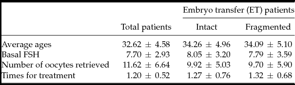
For total patients, the average age, basal follicle-stimulating hormone (FSH), number of retrieved oocytes and times for treatment were 32.62 ± 4.58, 7.70 ± 2.93, 11.62 ± 6.64 and 1.20 ± 0.52 (mean ± standard error of the mean (SEM)), respectively. The average age of total patients was 2 years younger than that of patients in fresh ET cycles, because the fresh ET was cancelled in some young patients with excessive follicles in order to decrease the risk of OHSS. Therefore the average number of retrieved oocytes for total patients was higher than that of fresh ET patients. Moreover, there were no differences in the average age (34.26 ± 4.96 versus 34.09 ± 5.10), basal FSH (8.05 ± 3.20 versus 7.79 ± 3.59), number of retrieved oocytes (9.92 ± 5.03 versus 9.70 ± 5.90) or treatment times (1.27 ± 0.76 versus 1.32 ± 0.68) between the intact and fragmented groups in fresh ET cycles (Table 1).
General clinical information for patients

Intact: zygotes with intact polar bodies (PBs). Fragmented: zygotes with fragmentation, irregular shape or rugose surface PBs. FSH, follicle-stimulating hormone. The results were analyzed by t-test.
Embryo development potential was related to PBs morphology
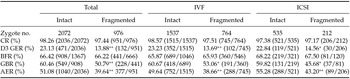
According to PB morphology, fertilized 3048 zygotes from 382 cycles (263 IVF and 119 ICSI cycles) were divided into two groups: intact and fragmented. Several parameters reflecting the embryo quality were compared between the two groups. As shown in Table 2, there was no difference in CR or BFR between the intact and fragmented group. However, the D3 GER (23.13 versus 13.88%, P < 0.01), GBR (60.46 versus 50.79%, P < 0.01) and available embryo rate (AER) (51.08 versus 39.64%, P < 0.01) of the intact group were all significantly higher than that of the fragmented group. Moreover, the zygotes from IVF group showed a similar developmental potential to that of the ICSI group. In Table 2, it is also demonstrated that the influence of PBs morphology on zygotes competence presented the same trend in total, IVF or ICSI groups. In these three groups, the D3 GER, GBR and AER of the fragmented group were all lower than that of the corresponding intact group.
Developmental potential of zygotes according to different PBs morphology

Intact: zygotes with intact polar bodies (PBs). Fragmented: zygotes with fragmentation, irregular shape or rugose surface PBs.
AER: available embryo rate; BFR: blastocyst formation rate; CR: cleavage rate; GBR: good blastocyst rate; GER: good quality embryo rate. ICSI, intracytoplasmic sperm injection; IVF, in vitro fertilization.
The results were analyzed by t-test. *P < 0.05, **P < 0.01.
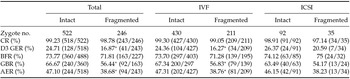
For some patients, ET or/and cryopreservation was conducted on D5, which enabled recording of the outcome of each D3 available embryos (Table 3). In good accordance with the above results, the D3 GER, GBR and AER of the intact group were all significantly higher than that of the fragmented group in total and IVF cycles. Although no difference between each parameter was detected between the intact and fragmented groups in ICSI cycles because of the limitation in sample size, the D3 GER, GBR and AER of the intact group were still higher than that of the fragmented group.
Developmental potential of zygotes in blastocyst culture cycles according to PBs morphology
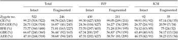
Intact: zygotes with intact polar bodies (PBs). Fragmented: zygotes with fragmentation, irregular shape or rugose surface PBs.
AER: available embryo rate; BFR: blastocyst formation rate; CR: cleavage rate; GBR: good blastocyst rate; GER: good quality embryo rate. ICSI, intracytoplasmic sperm injection; IVF, in vitro fertilization.
The results were analyzed by t-test. *P < 0.05, **P < 0.01.
The relationship between PB morphology and pregnancy outcome
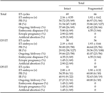
In 238 fresh ET cycles, 69 cycles were transferred with embryos derived from intact PB zygotes, 66 cycles were from fragmented PB zygotes and the rest of the 103 cycles was from a combination of intact and fragmented PBs zygotes. For the accuracy of the results, we only compared the pregnancy outcome between 69 intact and 66 fragmented PB zygote-derived embryo ET cycles. In contrast with the embryo quality results, statistical analysis showed no significant differences in PR or IR between the intact and fragmented groups and the time for ET performance (D3 or D5) showed no influence on these results (Table 4). Moreover, the clinical general information of these patients also showed no obvious differences in age (34.26 ± 4.96 versus 34.09 ± 5.10), basic FSH (8.05 ± 3.20 versus 7.79 ± 3.59), number of retrieved oocytes (9.92 ± 5.03 versus 9.70 ± 5.90), number of ET embryos (2.16 ± 0.55 versus 1.92 ± 0.62) or treatment time (1.27 ± 0.76 versus 1.32 ± 0.68) between the two groups (Table 1).
Pregnancy outcome of fresh transfer cycles according to PB morphology

Intact: zygotes with intact polar bodies (PBs). Fragmented: zygotes with fragmentation, irregular shape or rugose surface PBs.
ET: embryo transfer; IR: implantation rate; PR: pregnancy rate.
The results were analysed by chi-squared test. Fisher's exact test was used when n < 40 or a value of <5 was expected.
Discussion
With the rapid development of genetic diagnosis and embryo/blastocyst biopsy technology, the PB, as a homologous DNA representative of the oocyte, has became an attractable candidate for predicting embryo quality and/or pregnancy outcome. The results, however, are paradoxical (Ciotti et al., Reference Ciotti, Notarangelo, Morselli-Labate, Felletti, Porcu and Venturoli2004; Fancsovits et al., Reference Fancsovits, Tothne, Murber, Takacs, Parp and Urbancsek2006; Navarro et al., Reference Navarro, de Araújo, de Araújo, Rocha, dos Reis and Martins2009; Halvaei et al., Reference Halvaei, Khalili, Soleimani and Razi2011; Reich et al., Reference Reich, Klatsky, Carson and Wessel2011), suggesting that there is still a need to look seriously upon the value of PBs in embryo selection. In the present study, we observed PB morphology at the time of zygotic observation (16–18 h after insemination) and analyzed retrospectively the relationship between PB morphology and embryo quality. The results demonstrated that zygotes from the PBs intact group showed a better developmental potential than that of the fragmented group and that this trend was not affected by insemination manner. To exclude the interference of embryo transfer in which some higher grades embryos had already been selected for transfer before sequential culture, we also analyzed the developmental potential of zygotes from patients in blastocyst culture cycles. It was also shown that the D3 GER, GBR and AER of the intact group were all significantly higher than that of the fragmented group. These results were consistent with other groups’ research (Ebner et al., Reference Ebner, Yaman, Moser, Sommergruber, Feichtinger and Tews2000; Navarro et al., Reference Navarro, de Araújo, de Araújo, Rocha, dos Reis and Martins2009; Younis et al., Reference Younis, Radin, Izhaki and Ben-Ami2009; Reich et al., Reference Reich, Klatsky, Carson and Wessel2011; Jiao & Woodruff Reference Jiao and Woodruff2013). Thus, it is inferred that PB morphology, to some extent, can reflect embryo quality when using the present morphology grading system.
In contrast with the positive relationship of PB morphology to embryo quality, there was no difference in PR and IR between the PB intact and fragmented groups. The method for embryo selection for ET, therefore, might be an affecting factor. For the good of the patient, while ascertaining the clinical predicting value of PB morphology for embryo viability, the embryo selection criteria must be still based on established morphological assessment rather than PB morphology. Furthermore, PRs in fresh ET cycles cannot be fully equal to the AER. In future studies, to better examine the relationship between PBs and recruited oocyte quality in one stimulation cycle, and the accumulated PR needs to be taken into consideration. Moreover, an optimal evaluating indicator should be non-invasive, convenient and easy to handle, and cannot bring too much extra workload to laboratory embryologists who are already working under intense pressure. In the present study, we simultaneously recorded the state of PBs at the time of PN observation and analysed the outcome of zygotes according to PB morphology after the end of all treatments. The implementation of a better-powered prospective randomized controlled study in future research will be more meaningful than a retrospective study.
In addition, the present results showed that some zygotes had two fragmentized PBs, some had one intact and one fragmentized PB, but most had two typical intact PBs. Because of the high price of time-lapse experiments, the observation was only performed at fixed time points (about 16–18 h after insemination); at this observation point, it was difficult to make an accurate distinction between PB1 and PB2. Several studies have demonstrated that PB1 decay usually happened at approximately 4–20 h after its extrusion and PB2 can exist in the perivitelline space for several days after its extrusion (Ortiz et al., Reference Ortiz, Lucero and Coxatto1983; Gardner, Reference Gardner1997; Ciotti et al., Reference Ciotti, Notarangelo, Morselli-Labate, Felletti, Porcu and Venturoli2004). Although there is little comparability in the clinical characteristics of different patients in different studies, these results and our present study indicated that the time for PB1 and PB2 degeneration might be different for each embryo. Moreover, a recent study suggested that there was no difference in the time for PB2 extrusion between the implanted and non-implanted embryos (Aguilar et al., Reference Aguilar, Motato, Escribá, Ojeda, Muñoz and Meseguer2014). According to our results, premature degeneration of PBs seemed to be positively correlated with worse embryo quality. Thus, there must be a time axis and morphological change process of generation and degeneration of PBs in normal embryos. The value of PB status, intact or fragmented, for predicting embryo quality and pregnancy outcome still needs further investigation. In future studies, to distinguish accurately between PB1 and PB2, it would be better to only recruit ICSI cycles, which will allow direct observation of the status of PB1. Moreover, to obtain more accurate information for assessing and selecting ‘good’ embryos, we recommend the use of time-lapse photography to record the whole evolution process of both PB1 and PB2, from appearance to decay by time-lapse video. By comparing the different morphodynamics between PB1 and PB2, we can better comprehend their relationship to embryo quality and pregnancy outcome.
In conclusion, although PB morphology assessed at 16–18 h after insemination had little effect on PR or IR in fresh ET cycles, better embryo quality can be achieved in the PB intact group. The value of PB morphology in embryo selection and prediction of pregnancy outcome in ART treatment still awaits further thorough investigation, especially in a long-term follow-up study on the health of offspring. We are still on the way to improvement of the existing morphology evaluation methods.
Acknowledgements
This work was supported by National Natural Science Foundation of China (grant nos. 81471511, 81270753 and 81370758), Personnel Training Plan of The Health Care System of Beijing (grant nos. 2013-3-021 and 2013-2-009), Cooperation Project of Basic Research, Clinical Application of Capital Medical University (grant no. 13JL11) and Project of Beijing Chao-Yang Hospital for Young Scientists (grant no. YQ2014-12).