Introduction
The developing foetus is completely reliant on its mother for nutrients, so reduced in utero nutrient availability due to a large number of foetuses, reduced maternal feed intake or both in late pregnancy may have a negative impact on lamb birth weight (Louca et al., Reference Louca, Mavrogenis and Lawlor1974; Robinson, Reference Robinson1977; Mellor, Reference Mellor1983). Moreover, lambs’ postnatal growth rates can be negatively affected by reduced milk production (Robinson and Forbes, Reference Robinson and Forbes1968; Peart et al., Reference Peart, Doney and Smith1979; Tygesen et al., Reference Tygesen, Tauson, Ranvig, Nørgaard, Husted and Nielsen2008). It is known from research, mainly in humans and rodents, that specific physiological adaptations may be induced, adaptations that enables the foetus to survive the adverse intrauterine environment (McMillen and Robinson, Reference McMillen and Robinson2005). It has, however, been demonstrated that these physiological adaptations may be persistent (McMillen et al., Reference McMillen, Adams, Ross, Coulter, Simonetta, Owens, Robinson and Edwards2001; Whorwood et al., Reference Whorwood, Firth, Budge and Symonds2001), being associated with permanent alterations in foetal and postnatal homeostatic mechanisms (Harding and Johnston, Reference Harding and Johnston1995; McMillen et al., Reference McMillen, Adams, Ross, Coulter, Simonetta, Owens, Robinson and Edwards2001; Bloomfield et al., Reference Bloomfield, Oliver, Giannoulias, Gluckman, Harding and Challis2003). This phenomenon has been termed ‘foetal metabolic programming’ (Lucas, 1991; McMillen and Robinson, Reference McMillen and Robinson2005). It appears clear that the physiological adaptation and associated programming events in response to a sub-optimal intrauterine environment are crucial for the survival and health of the foetus and the newborn (McMillen et al., Reference McMillen, Adams, Ross, Coulter, Simonetta, Owens, Robinson and Edwards2001). The persistent postnatal manifestations of the programming involve the altered metabolism and function of a number of endocrine systems involved in normal growth and development (McMillen and Robinson, Reference McMillen and Robinson2005). These include the malfunction of the glucose–insulin axis and altered functions of the growth hormone – IGF-1 axis and hypothalamic–pituitary–adrenal cortex (HPA) axis (Fowden and Hill, Reference Fowden and Hill2001; Fowden et al., Reference Fowden, Giussani and Forhead2005 and Reference Fowden, Giussani and Forhead2006). Studies of foetal metabolic programming have been performed on rodents and on a significant number of sheep, however, from a human health perspective. Knowledge of the consequences is lacking for animal production-related parameters such as milk intake, growth rate, and muscle and fat deposition. Furthermore, increased knowledge of the underlying physiological mechanisms involved is essential.
Previous literature has suggested that leptin could be a growth factor in the foetus (Henson and Castracane, Reference Henson and Castracane2000; Sagawa et al., Reference Sagawa, Yura, Itoh, Mise, Kakui, Korita, Takemura, Nuamah, Ogawa, Masuzaki, Nakao and Fujii2002; McMillen et al., Reference McMillen, Muhlhausler, Duffield and Yuen2004) and that leptin may play an important role in determining the consequences of maternal nutrient restriction for offspring’s postnatal metabolism and performance. It has therefore been speculated whether an adverse intrauterine environment alters plasma leptin secretion, i.e. alters the endocrine feedback loop between the adipose tissue and the central and peripheral neuroendocrine system, and whether postnatal growth is influenced by possible changes in plasma leptin concentrations.
The objective of this study was to test two hypotheses (1) that restricting the nutrient intake of ewes in late gestation will have permanent impact on the metabolic and endocrine function of the offspring, impairing the growth and performance of postnatally well-nourished lambs and (2) that leptin may be involved in this adaptation of animal performance to a prenatal nutritional insult.
Genotype also plays a major role in animal performance. The twin-bearing Shropshire ewes used in this study were sired by two different rams, having different breeding indices for carcass lean : fat ratio. This study thus also offers the opportunity to elucidate the extent to which the consequences of adverse in utero nutrient supply on the endocrine and metabolic systems can be affected by different sire genotypes.
Material and methods
Animals
Experimental procedures were approved by and found compliant with the guidelines of the National Committee on Animal Experimentation, Denmark.
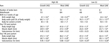
Twenty twin-bearing Shropshire ewes in their second or third parities, born and raised on the Royal Veterinary and Agricultural University’s experimental farm, and having uniform body condition scores and live weights, were included in this study. The ewes were mated with pure-bred Shropshire sires having similar breeding indices for high daily growth rate, but with either a high (‘meat’ (M), index of 117) or a low (‘growth’ (G), index of 93) index for cross-sectional area of m. Longissimus dorsi (LD) combined with minimal subcutaneous fat thickness above LD (lean : fat ratio). A total of 40 lambs were born and all of them were included in the study (Table 1). The ewes were assigned to one of two feeding treatments in the last 6 weeks of gestation: (1) the high-nutrition treatment (H) comprising silage ad libitum (56% dry matter (DM), 6.1% ash, 7.9% crude protein (CP) and 1% fat) + 200 g of whole barley (88.7% DM, 2.2% ash, 10.4% CP and 2.3% fat) + 200 g of protein supplement (89.5% DM, 5.68% ash, 45.4% CP, 5.1% fat; DLG Fårekraft, DLG, Denmark) or (2) the low-nutrition treatment (L) comprising unsupplemented silage with an energy allowance of approximately 50% of the total metabolisable energy intake of the H-ewes. The feed allowance for the L-ewes was adjusted weekly. After lambing, all ewes were fed the H-diet and the supplement was replaced with the one suitable for lactating ewes (88.8% DM, 6.8% ash, 14% CP and 3.2% fat) and gradually increased to 1 kg/day per animal over the first week for H-ewes, while L-ewes were adapted to the supplement over 10 days. The animals had free access to fresh water and a commercial mineral mixture at all times.
Number of lambs included in the experiment, birth weight, body water pool as a fraction of body weight and body fat content at birth

a,b,cValues with different superscripts differ significantly (P < 0.05).
Depth of m. Longissimus dorsi (muscle depth), subcutaneous fat above m. Longissimus dorsi and body weight at 110 and 146 days of age, as affected by high or low maternal feeding level during the last 6 weeks pre partum as well as sire genotype.
Data are LSmean ± s.e. (n = 40).
The lambs were fed silage (56% DM, 6.1% ash, 7.9% CP and 1% fat) and a commercial concentrate (88.5% DM, 7.1% ash, 17.9% CP and 5.2% fat; Woller, Østsjællands Andel, Karise, Denmark) ad libitum from day 21 post partum. They were weaned at 56 days of age and the roughage supply was then gradually changed to ad libitum access to artificially dried grass (88.8% DM, 6.8% ash, 14% CP and 3.2% fat; Dangrønt, Ølgod, Denmark).
Lamb milk intake, body fat content, live weights, growth rates, and measurement of muscle depth and fat thickness by scanning
Lamb milk intake and body water pools (BWPs) were measured using the deuterium (D2O) dilution technique, the calculations are described in detail by Theil et al. (Reference Theil, Nielsen, Kristensen, Labouriau, Danielsen, Lauridsen and Jakobsen2002). In short, milk intake was calculated as the sum of the water turnover rate and potential metabolic water stored, divided by the potential water from the milk. Lamb body fat content at birth was calculated on the basis of their BWP determined using D2O enrichment as described by Tygesen (2005) and was calculated as body weight minus lean body mass, lean body mass being calculated as BWP divided by literature values for the water content in lean mass of young lambs (0.78) (Gardner and Hogue, Reference Gardner and Hogue1964; Searle, Reference Searle1970; Donnelly and Freer, Reference Donnelly and Freer1974; Bocquier et al., Reference Bocquier, Thériez and Brun1991; Benjamin et al., Reference Benjamin, Koenig and Becker1993).
The lambs were weighed and their growth rates were calculated on a weekly basis. Muscle- and backfat thicknesses were measured approximately 110 and 146 days post partum using ultrasound scanning. Backfat thickness is difficult to measure using ultrasound because the border between the skin and superficial fat is poorly recognisable (Trzybìnska, Reference Trzybìnska1997) causing large variations in estimates. In this experiment, an experienced person made the ultrasound measurements without prior knowledge of the treatment groups, and each lamb was measured in duplicate to minimise errors.
Blood sampling and analyses
Blood samples were taken at birth and 3, 7, 10, 14, 21, 28, 70 and 140 days of age. Blood samples of approximately 5 ml were taken in both heparin/fluorine vacuum glasses (Vacutainer™, Becton Dickinson Vacutainer System Europe, Meylan Cadex, France) and ethylenediaminetetraacetic acid (EDTA) vacuum glasses (BD Vacuntainer System, preanalytical Solution Delliver Industrial Estate, Plymouth, UK) by means of venipuncture. The heparin/fluorine-stabilised plasma was analysed for glucose, insulin and leptin, while the EDTA-stabilised plasma was analysed for non-esterified fatty acid (NEFA), β-hydroxybyturate (BOHB) and acetate concentrations.
All plasma samples for leptin analysis were freeze dried and sent to the University of Western Australia, Perth, and analysed in duplicate by means of a double-antibody radioimmunoassay using ovine leptin raised against bovine leptin as described by Blache et al. (Reference Blache, Tellam, Chagas, Blackberry, Vercoe and Martin2000) with modifications described by Tygesen (2005). All samples were processed in a single assay and the limit of detection was 0.08 ng/ml. The assay included six replicates of three control samples containing 0.49, 1.08 and 1.8 ng/ml. The intra-assay coefficients of variation of the three control samples were 4.8%, 3.2% and 5.3%, respectively. Insulin, glucose, NEFA and BOHB were analysed as described by Tygesen (2005).
Statistical analyses
The dependent variables (blood parameters, body weights, growth rates, scanning values of muscle depth and fat thickness above the muscle) were analysed as repeated measurements using the PROC MIXED procedure in SAS version 8.1. Data were analysed according to the following general linear model:

where μ is the general mean, Fi, Sj, Gk and Tl are the main/fixed effects of maternal feeding, sire genotype, lamb gender and time, respectively. (FS)ij, (FG)ik, (SG)jk and (FSG)ijk are interactions between maternal feeding and sire genotype, maternal feeding and lamb gender, sire genotype and lamb gender and maternal feeding, sire genotype and lamb gender, respectively. (TFS)ijl, (TFG)ikl and (TSG)jkl are fixed interaction effects of time, maternal feeding and sire genotype; time, maternal feeding and lamb gender; and time, sire genotype and lamb gender at time l, respectively.
eijkl is the covariation within animals at time l in treatments i, j, k. It is assumed that eijk is normally distributed with zero means and a covariance matrix Σ. A spatial Gaussian autocorrelation structure (SP(gau)) was chosen to model the covariance matrix. pijkl are the variations between animals at time l in treatments i, j, k. It is assumed that pijkl is independent across indices and normally distributed with zero means and variances σijkl 2.
The PROC REG and PROC CORR procedures in SAS were used to analyse linear relationships and calculate the corresponding Pearson correlation coefficients. If not otherwise stated, the results of the full unreduced model are presented as least square means ± standard error of LSmeans (LSmean ± s.e.); P values are given for P ⩽ 0.05, and for P > 0.05, NS is denoted.
The plasma acetate concentrations measured in the first 4 weeks post partum were under the detection limit of the assay, many values are thus missing for this period, so values prior to day 28 have been omitted from the statistical analyses.
Results
Birth weight
Maternal nutrient restriction in late gestation reduced birth weights, as L-lambs weighed 966 ± 180 g less than H-lambs (P < 0.001). Birth weight was also affected by sire genotype as G-lambs (3.8 ± 0.1 kg) weighed more than M-lambs (3.2 ± 0.1 kg, P = 0.002). In addition, there was an interaction between sire genotype and maternal nutrient restriction resulting in LM-lambs (2.6 ± 0.2 kg) weighing significantly less than LG-lambs (3.4 ± 0.2) at birth, while birth weights of H-lambs were unaffected by sire genotype (4.1 ± 0.2 v. 3.8 ± 0.2 kg, for HG- and HM-lambs, respectively). Female lambs weighed less at birth (3.3 ± 0.1 kg) than male lambs (3.7 ± 0.1 kg, P = 0.01); in particular, females born to the M-ram had a lower birth weight (2.7 ± 0.2 kg, P < 0.001) than females born to the G-ram (3.8 ± 0.2 kg).
Body water pool and body fat content in the first 3 weeks post partum
The lambs’ BWP, as a fraction of body weight, did not change in the first 3 weeks post partum and averaged 72.1 ± 0.05%. BWP was unaffected by maternal feeding but was significantly affected by sire genotype, being significantly larger in M-lambs (73.7 ± 0.01%) than in G-lambs (70.2 ± 0.01%, P = 0.02). The lambs had an average body fat content of 8.8 ± 0.9% at birth, which was unaffected by maternal nutrient restriction in late gestation and by sire genotype (Table 1). Male lambs contained significantly more fat (10.7 ± 1.0%) at birth than female lambs (7.0 ± 1.0%).
Body weight, growth rates and conversion rates of milk used for live weight gain
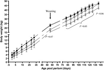
Body weights of lambs, in the two maternal feeding groups, reflected the differences in birth weight throughout the growth period investigated, with L-lambs weighing significantly less than H-lambs . Throughout the experimental period, the effects of maternal feeding were greatest in M-lambs; LM-lambs had the lowest body weights at all times, while LG-lambs reached live weights similar to those of H-lambs at 145 days of age (Figure 1). The live weights of male lambs were higher than those of female lambs from 82 days of age (P < 0.002).
Average live weight of lambs’ post partum as affected by high and low maternal feeding level during the last 6 weeks pre partum and sire genotype (HG-lambs (▪); LG-lambs (□); HM-lambs (▴) and LM-lambs (▵), respectively). Data are LSmean ± s.e. (n = 40). P values are for comparison between LM- and LG-lambs.

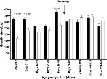
Daily growth rates were significantly higher in H-lambs than in L-lambs in the first 2 weeks post partum and from day 28 until day 56 post partum, thereafter, L-lambs grew at the same rate (g/day) as H-lambs (Figure 2). Overall, sire genotype had no impact on lambs’ daily growth rates, though the average growth was higher in male (306 ± 9 g/day) than in female lambs (269 ± 10 g/day, P = 0.009).
Average daily growth rate of lambs post partum as affected by high (▪) and low (□) maternal feeding level during the last 6 weeks pre partum. Data are LSmean ± s.e. (n = 40).

The lambs’ daily growth rates for the first 3 weeks post partum was related to milk intake as follows: growth rate (g/day) = 67.8 (±23.6) + 0.12 (±0.01) × milk intake (g/day), P < 0.001, R 2 = 0.56, CV = 18.6, n = 58). The conversion rate (CR) of milk/g daily live weight gain increased significantly in H-lambs throughout the first 3 weeks post partum (4.9 ± 0.3 to 6.9 ± 0.3 in week 1 to week 3 post partum, respectively), whereas it remained stable in L-lambs (5.6 ± 0.3 to 5.9 ± 0.3 in week 1 to week 3 post partum, respectively), indicating that H-lambs became less efficient in converting milk to live weight gain. CR was unaffected by sire genotype and lamb gender.
Muscle and backfat depth
Maternal nutrient restriction in the last 6 weeks of gestation reduced the cross-sectional area of LD (muscle depth) in L-lambs (24.0 ± 0.7 mm) as compared with H-lambs (26.1 ± 0.6 mm, P = 0.03) at day 110 post partum, and this also tended to be the case on day 146 post partum (P = 0.07). Muscle depth was unaffected by sire genotype on day 110, while on day 146 post partum M-lambs had a greater muscle depth (30.4 ± 0.7) than G-lambs (28.2 ± 0.7 mm, P = 0.02). Maternal nutrient restriction in late gestation had no effect on subcutaneous fat thickness above LD at day 110; however, on day 146 post partum it was higher in H-lambs (4.8 ± 0.1 mm) than in L-lambs (4.2 ± 0.2 mm, P = 0.02). Sire genotype had no effect on subcutaneous fat thickness above LD either by day 110 or by day 146 post partum (Table 1).
Glucose
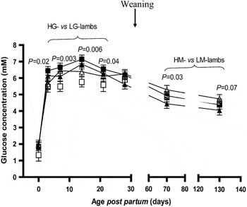
Plasma glucose concentrations increased markedly from a very low level immediately after birth, peaking on day 14 post partum (P < 0.001) after which concentrations decreased from an average of 6.5 ± 0.14 mM on day 14 to an average of 4.4 ± 0.14 mM on day 130 post partum (P < 0.001) (Figure 3).
Postnatal plasma glucose concentrations in lambs as affected by high and low maternal feeding level during the last 6 weeks pre partum and sire genotype (HG-lambs (▪); LG-lambs (□); HM-lambs (▴) and LM-lambs (▵), respectively). Data are LSmean ± s.e. (n = 40).

A significant interaction effect between maternal feeding and sire genotype on plasma glucose concentrations was found, indicating that LM-lambs, which had the lowest growth rates, had lower plasma glucose concentrations than HM-lambs (P < 0.01) from day 3 until day 21 post partum, after which no differences were found. LG-lambs had higher plasma glucose concentrations than HG-lambs (P = 0.03) on day 70, and this difference tended to persist until day 130 post partum (P = 0.07, Figure 3). Plasma glucose concentrations were unaffected by lamb gender.
Non-esterified fatty acids (NEFA) and β-hydroxybyturate (BOHB)
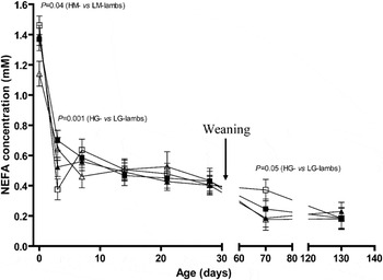
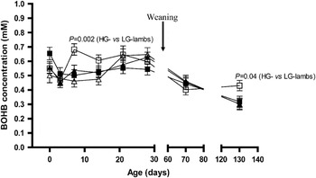
Maternal nutrient restriction, sire genotype and lamb gender had no significant effect on plasma NEFA or BOHB concentrations. Plasma NEFA concentrations were markedly elevated at birth and fell significantly from birth until day 3 post partum (Figure 4), coinciding with increases in plasma glucose concentrations. After day 3, plasma NEFA concentrations decreased slightly throughout the rest of the experimental period. An interaction between maternal nutrient restriction in late gestation and sire genotype was found, with sire genotype affecting concentrations only in L-lambs. This interaction, however, was not systematic. LG-lambs had significantly higher NEFA concentrations than did LM-lambs at birth and at day 70 post partum, while on day 3 post partum LM-lambs had higher plasma NEFA concentrations than LG-lambs. As of the last measurement on day 130 post partum, no differences in NEFA concentrations were found between LG- and LM-lambs. LG-lambs had higher plasma BOHB concentrations than LM-lambs on days 7, 14 and 130 post partum, while the remaining measurements were similar (Figure 5). Plasma NEFA and BOHB concentrations in H-lambs were similar throughout the experimental period, regardless of sire genotype.
Plasma non-esterified fatty acid concentrations in lambs as affected by high and low maternal feeding level during the last 6 weeks pre partum and sire genotype (HG-lambs (▪), LG-lambs (□), HM-lambs (▴) and LM-lambs (▵), respectively). Data are LSmean ± s.e. (n = 40).

Plasma β-hydroxybyturate concentrations in lambs as affected by high and low maternal feeding level during the last 6 weeks pre partum and sire genotype (HG-lambs (▪); LG-lambs (□); HM-lambs (▴) and LM-lambs (▵), respectively). Data are LSmeans ± s.e. (n = 40).

Acetate
Plasma acetate concentrations, generally, were undetectable until day 28 post partum and increased from day 30 to day 70 post partum (0.2 ± 0.02 v. 0.7 ± 0.02 mM, respectively; P < 0.001), coinciding with the lambs developing from primarily being monogastric to becoming ruminants. Maternal nutrient restriction, sire genotype and lamb gender had no significant effects, though maternal nutrient restriction in late gestation reduced L-lambs’ plasma acetate concentration as of day 70 post partum compared with that of H-lambs (0.6 v. 0.8 ± 0.03 mM, respectively; P = 0.02). From day 70 to day 130 post partum, plasma acetate concentrations decreased markedly in H-lambs (from 0.8 to 0.4 ± 0.03 mM, respectively; P < 0.001), while only a small decrease was recorded in L-lambs (from 0.6 to 0.5 ± 0.3 mM, respectively; P = 0.02), resulting in H-lambs having a lower plasma acetate concentration than L-lambs on day 130 post partum (0.4 v. 0.5 ± 0.03 mM, respectively; P = 0.02). Maternal nutrient restriction and sire genotype interacted, resulting in plasma acetate concentrations being lower in LG-lambs (0.5 ± 0.05 mM) than in LM- (0.7 ± 0.06 mM), HG- (0.7 ± 0.05 mM) and HM-lambs (0.8 ± 0.05 mM; P < 0.008) on day 70 post partum. Plasma acetate concentrations were on average 23% higher (0.48 ± 0.2 mM) in male lambs than in female lambs (0.39 ± 0.3 mM; P = 0.01).
Insulin
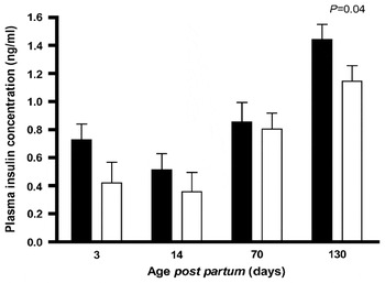
Maternal feeding level, sire genotype and lamb gender had no significant effect on lamb plasma insulin concentrations. However, L-lambs had lower overall insulin concentrations than H-lambs (0.5 ± 0.07 v. 0.8 ± 0.06 ng/ml, respectively; P = 0.004). Plasma insulin concentrations decreased from day 3 to day 14 (0.6 to 0.4 ± 0.08, respectively; P < 0.001) after which they increased from day 14 to day 70 (0.4 to 0.8 ± 0.08, respectively; P < 0.001) and again until day 130 post partum (1.3 ± 0.07 ng/ml) (Figure 6).
Plasma insulin concentrations in lambs from birth to 130 days of age as affected by high (▪) and low (□) maternal feeding level during the last 6 weeks pre partum. Data are LSmean ± s.e. (n = 40).

Leptin
Plasma leptin, which varied greatly between animals, was not significantly affected by maternal nutrient restriction, sire genotype or lamb gender. The average plasma leptin concentration was 0.82 ± 0.34 ng/ml and did not change significantly in the first 28 days post partum. Plasma leptin was not significantly correlated to lambs’ birth weight, body fat content or live weight later in the experiment. Leptin concentrations in milk were significantly higher than those in lambs plasma concentrations for the first 5 days post partum. Leptin concentrations in milk was not correlated to plasma leptin concentrations. After day 5 post partum, leptin concentrations in milk and lamb plasma were similar in magnitude until the last measurement on day 28 post partum. For further information on milk leptin see Tygesen et al. (Reference Tygesen, Tauson, Ranvig, Nørgaard, Husted and Nielsen2008).
Discussion
Impact of foetal nutrition on performance and metabolic/endocrine status of the offspring
From literature it appears that the effect of maternal nutrient restriction in late gestation on lamb birth weight is dependent on the duration and severity of the nutritional insult (Gardner et al., Reference Gardner, Buttery, Daniel and Symonds2007). This study found that a 50% restriction of maternal nutrient supply in the last 6 weeks of gestation reduced lambs’ birth weight by 25%. Measurements of the lambs’ BWP did not indicate, however, that maternal feed restriction had any impact on the lean : fat ratio of the newborn lambs. The estimated average body fat content of 8.8% we found is substantially higher than the literature values for young lambs (Jagusch et al., Reference Jagusch, Norton and Walker1970 (2.7%); Sykes and Field, Reference Sykes and Field1972 (2%); Chiou and Jordan, Reference Chiou and Jordan1973 (3.5%)), which may be because the body fat contents reported here are indirect estimates and not actually chemically determined.
The impaired in utero environment and decreased nutrient availability in early life improved the efficiency with which milk was converted to live weight gain in L-lambs, the difference being significant by the third week of life. This may relate to L-lambs being lighter than H-lambs; the maintenance requirements could thus be expected to be relatively lower, leading to increased efficiency of milk conversion of milk to live weight gain.
The live weights of L-lambs were on average 5 kg lower than those of H-lambs at the time of weaning at 56 days of age. In the post-weaning period, all lambs were fed ad libitum and the growth rates of the L-lambs increased, but only to the same level as observed in the H-lambs, so the body weight difference between the L- and H-lambs remained until the end of the experiment. Farm animal species have the capacity to exhibit compensatory growth when a high plane of nutrition follows a period of restricted feeding and growth (Wilson and Osbourn, Reference Wilson and Osbourn1960; Hornick et al., Reference Hornick, Van Eenaeme, Gérard, Dufrasne and Istasse2000). Low birth weights of pigs (Poore and Fowden, Reference Poore and Fowden2004) and lambs (Greenwood et al., Reference Greenwood, Hunt, Hermanson and Bell1998) born to non-feed-restricted dams have been associated with an increased body fat content later in life, ascribed to a postnatal compensatory growth. The results of the present study indicated that L-lambs had a different partitioning of muscle and fat deposition than H-lambs. L-lambs generally remained smaller than H-lambs throughout the study period, but at slaughter at 146 days of age, the cross-sectional of area of LD was the same in L- and H-lambs (within sire), whereas the layer of subcutaneaous fat above the LD was significantly thinner in L-lambs than in H-lambs. Possibly, the altered partitioning in the direction of a higher muscle : fat deposition ratio in L-lambs was equivalent to what occurs early in a compensatory growth phase (Hornick et al., Reference Hornick, Van Eenaeme, Gérard, Dufrasne and Istasse2000), when the animal would be expected to have an improved feed CR and daily fractional growth rate, similar to our findings in the L-lambs post-weaning. Therefore, we cannot exclude that prolonging the postnatal ad libitum feeding period would have increased L-lambs’ growth rates and fat deposition in excess of those of the H-lambs.
It has been suggested that an adverse foetal environment, in which foetal nutrient demands exceed supply, leads to permanent alterations in foetal and postnatal homeostatic mechanisms (McMillen et al., Reference McMillen, Adams, Ross, Coulter, Simonetta, Owens, Robinson and Edwards2001; Bloomfield et al., Reference Bloomfield, Oliver, Giannoulias, Gluckman, Harding and Challis2003). Indeed, insulin concentrations were significantly lower in L-lambs than in H-lambs during both the pre-weaning period and the period of ad libitum feeding post-weaning. Glucose concentrations were not systematically affected by maternal feeding, but the highest glucose concentrations post-weaning were observed in L-lambs born to the G-sire. It has been reported that maternal undernutrition in late gestation impairs the insulin secretory capacity of the pancreas in young and adolescent lambs, and insulin sensitivity in target tissues may be increased in compensation (Husted et al., Reference Husted, Nielsen, Tygesen, Kiani, Blache and Ingvartsen2007). Our results concur with such a modification in the glucose–insulin axis, and this may explain how the growth rates of L-lambs increased when they were transferred to ad libitum feeding post-weaning; however, higher priority was given to muscle than to fat deposition in comparison with the H-lambs growing at a similar rate. We found no systematic differences in the plasma levels of any of the other studied metabolites or hormones that could be related to undernutrition in late foetal life. It has previously been suggested that plasma leptin in growth-restricted infants is low at birth and that either excess or poor nutrition in utero can result in increased circulating leptin concentrations in later life in humans (Sagawa et al., Reference Sagawa, Yura, Itoh, Mise, Kakui, Korita, Takemura, Nuamah, Ogawa, Masuzaki, Nakao and Fujii2002). In this experiment, reducing the maternal nutrient supply by 50% in the last 6 weeks of gestation had no significant effect on the lambs’ subsequent plasma leptin concentrations, and no significant changes in the first 4 weeks post partum were recorded. This is in line with previous research into growing lambs (Tokuda et al., Reference Tokuda, Kimura and Fujihara2001; McFadin et al., Reference McFadin, Morrison, Buff, Whitley and Keisler2002; Ehrhardt et al., Reference Ehrhardt, Greenwood, Bell and Boisclair2003) demonstrating that irrespective of growth rates, leptin concentrations remained the same. This supports the theory that leptin synthesis in the adipose tissue only increases when a threshold level of available energy is reached (Ehrhardt et al., Reference Ehrhardt, Greenwood, Bell and Boisclair2003). Thus, leptin does not appear to be involved in programming growth and metabolism in postnatal life in response to nutrient deprivation in late foetal life in sheep unlike what has been found in rats (Vickers et al., Reference Vickers, Gluckman, Coveny, Hofman, Cutfield, Gertler, Breier and Harris2005).
Sire genotype impacts the consequences of late foetal life nutrition for postnatal performance
The sheep used in this experiment were sired by two different rams of the same breed. The only apparent difference between them was that one ram (M) had a higher index for lean : fat deposition in the body than the other ram (G), so the genotype differences between the two rams must be considered relatively modest. The interaction between reduced late foetal life nutrition and sire genotype demonstrates that the genotype of the sire did not influence any of the measured traits in adequately nourished lambs, whereas differences were manifested in lambs that had been nutritionally challenged in late foetal life. A negative impact of foetal nutrient deprivation was observed in lambs sired by both rams, but it was particularly pronounced in the M-offspring that were born with an average birth weight of 2.6 kg and thus were small for gestational age. The data thus suggest that in situations of limited nutrient availability, sire genotype may play a more dominant than usual role in determining birth weight and postnatal performance, and some genotypes may be more susceptible than others to undergoing metabolic programming in response to in utero nutrient deprivation.
Conclusions
A 50% reduction of maternal nutrient supply in the last 6 weeks of gestation reduced lambs’ birth weight. Postnatal growth rates were determined by current level of nutrition, being reduced in L-lambs pre-weaning due to lower milk production by the dam, but restored to normal when an adequate post-weaning diet was provided. Prenatal undernutrition lowered plasma insulin in postnatal life, resulting in an improved capacity to convert ingested feed into lean rather than fat growth. We propose that the differences observed in fractional growth rates, feed CRs and growth pattern are likely to be expressions of a metabolic programming induced in L-lambs by nutrient restriction in late foetal life, a programming that has permanently altered the metabolic priorities and endocrine function of the body. Prenatal undernutrition may programme postnatal metabolism and the glucose–insulin axis differentially depending on sire genotype, thus affecting the ability of the ad libitum-fed lamb to exhibit compensatory growth and achieve a given body weight by a certain age.
Acknowledgements
The project was financially supported by the Research School of Animal Production and Health at the Royal Veterinary and Agricultural University, Denmark (RVAU), and the Danish Research Council for Development Research, Denmark. Ruth Jensen and Vibeke G. Christensen are thanked for analysing plasma NEFA, BOHB, acetate, insulin and glucose concentrations, and Margaret Blackberry is thanked for assisting with the leptin analysis at the University of Western Australia, Crawley, Australia. Mette Ostwald Nielsen and the technical staff at RVAU’s experimental animal facilities are thanked for assisting with the practical handling of the animals.