Introduction
Frontal sinus surgery continues to challenge even the most experienced endoscopic sinus surgeon. The anatomical constrictions of the frontal recess and frontal ostium are unforgiving of any mucosal injury or inflammation resulting from frontal sinus surgery. Revision frontal sinus surgery is even more challenging, because of the presence of previous mucosal trauma and the heightened predisposition to further scarring and stenosis.Reference Orlandi and Knight 1
Anatomical studies have demonstrated that the underlying problem in chronic frontal sinusitis is not the sinus itself but rather its drainage pathway, the frontal recess. The frontal recess is an inverted, funnel-like area that connects the frontal ostium to the anterior ethmoid sinus. It is usually pneumatised by a variety of frontal recess cells, which may cause anatomical frontal recess obstruction and be the primary cause of chronic frontal sinusitis. Consequently, function-restoring frontal recess procedures have yielded improved long-term results. However, the complex anatomy and its anterosuperior location render endoscopic frontal recess visualisation and dissection difficult, thereby predisposing to surgical failure.Reference Chan, Melroy, Kuhn, Kuhn, Daniel and Kuhn 2
Restenosis of a surgically enlarged opening of the frontal sinus can occur in three partly related ways. First, restenosis can occur as a result of a persistent obstruction of the opening by blood and fibrin during the immediate post-operative phase. Fibroblasts migrate into the fibrin mesh and form granulation tissue. Collagen is deposited, and the opening can become occluded by scar tissue. Second, marked swelling begins during the third post-operative week and can lead to contact zones at the frontal sinus opening. Contact between areas of granulation tissue can result in occlusion by scar tissue. Third, realignment of collagen fibres during the remodelling phase can lead to a concentric narrowing of ring-shaped openings.Reference Weber, Mai, Hosemann, Draf and Toffel 3
Endoscopic ethmoidectomy can result in scarring of the frontal sinus outflow tract, and this may be a factor in the observed increased incidence of frontal sinus disease in these patients. The occurrence of reocclusion is associated with: the diameter of the frontal neo-ostium; polyposis; excessive denuded bone; remnants of osteitic bone in the frontal recess; severe mucosal disease; lateralisation of the middle turbinate; and excessive removal of the middle turbinate.Reference Hunter, Silva, Youngs, Saeed and Varadarajan 4
The use of stents has long been described, as an attempt to decrease the rate of synechiae and stenosis. The primary purpose of a stent is to separate the edges of a wound surface so as to prevent synechial band formation and stenosis. Stents also serve to take up space that would otherwise be filled with blood, fibrin and/or mucus. Accordingly, stents can decrease the time and discomfort of post-operative debridement as there is simply less clot and debris to remove. Stents may also provide a matrix for epithelial migration, especially in areas of denuded bone, as the stent's surface gives subepithelial scarring a chance to stabilise. Finally, stents serve as an occlusive dressing which has a positive influence on wound healing.Reference Bednarskim and Kuhn 5
Two examples of prefabricated, semi-rigid, silicone frontal sinus stents are the Freeman stent (InHealth Technologies, Carpinteria, California, USA), a biflanged tube, and the Rains stent (Smith & Nephew ENT, Memphis, Tennessee, USA), which has a collapsible, tapered bulb tip.Reference Freeman and Blom 6 , Reference Rains 7
In this study, double J stents were used as frontal sinus stents in patients undergoing revision frontal sinusotomy.
Method
Patients in this study presented to the ear, nose and throat clinic in Fakeeh and United Doctors Hospitals in Jeddah, Saudi Arabia, between January 2007 and December 2010.
The inclusion criteria were frontal sinusitis and a history of previous functional endoscopic sinus surgery (FESS).
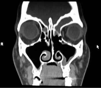
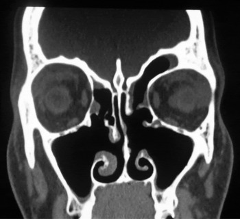
Five patients were included in the study, three males and two females, aged from 17 to 54 years. Revision frontal sinus surgery was indicated in all five. The patients’ main complaint was frontal headache. Their computed tomography (CT) and endoscopic findings were suggestive of fibrosis and/or new bone formation in the frontal recess area (Figure 1). Three of the patients had unilateral frontal sinusitis and two had bilateral disease. One female patient presented with a fistula discharging pus from the left eyelid; her CT scan showed total opacity of the left frontal sinus and erosion of the infero-lateral wall of the left frontal sinus.
Coronal computed tomography scan of a patient with left frontal sinusitis following previous functional endoscopic sinus surgery.

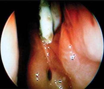
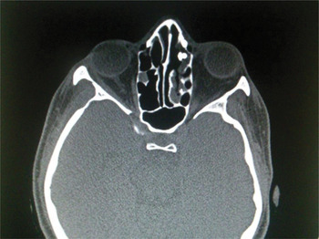
All the patients underwent frontal sinusotomy. The frontal recess was cleared of any ethmoid cells obstructing the outflow tract, especially the agger cells, bulla ethmoidalis and frontal cells if they had not been dealt with during previous surgery. Any scar tissue or new bone in the frontal recess was also excised. At the end of the procedure, a double J stent was introduced using the wire supplied with the stent (which straightened the coil at the end of the stent). The wire, with the overlying stent, was introduced into the frontal sinus, under direct vision using a 30° Hopkins telescope. After successful placement, the wire was withdrawn. The end of the stent, now situated inside the frontal sinus, regained its coil shape. The stent was cut with scissors so that the free end in the nasal cavity appeared just beyond the middle meatus (Figures 2 and 3).
Endoscopic view using a 30° telescope, showing the nasal end of the stent in place.

Axial computed tomography scan showing the stent in place.

Patients were given an oral antibiotic (levofloxacin 500 mg once daily) for 10 days, together with nasal irrigation using a saline nasal spray. Patients also underwent suction of debris and residual blood one and two weeks post-operatively. They were then followed up monthly for six months, when their stent was removed. The duration of post-operative follow up ranged from 10 to 36 months.
Results
Five patients were included in the study: three males and two females. Three patients had unilateral frontal sinusitis and two had bilateral frontal sinusitis.
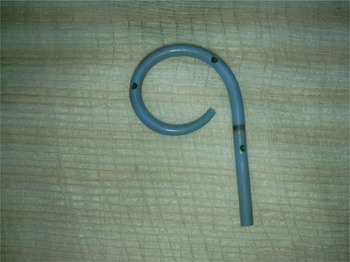
A total of seven frontal sinus stents were applied. The stents were well tolerated in all five patients and were left in place for six months. The stents were self-retained, and there were no cases of migration or extrusion. Mild crustation was cleaned by gentle suction using a 30° telescope in the out-patient clinic, with mild patient discomfort. Six months post-operatively, the stent was easily removed in the out-patient clinic by simply pulling the free tip of the stent which lay within the nasal cavity (Figure 4). The stent was easily removed as its soft, coiling end (within the frontal sinus cavity) straightened during extraction.
The double J stent after removal.

The follow-up period ranged from 10 to 36 months. During this time, four patients (six sinuses) were observed in the out-patient clinic to have patent frontal ostia, either seen or felt with a frontal probe (Figures 5 to 7). One patient who had originally had unilateral frontal sinusitis suffered recurrence, with restenosis of the frontal ostium and recurrent frontal headache and purulent discharge from the pinpoint ostium. He was referred for a Draf III procedure.
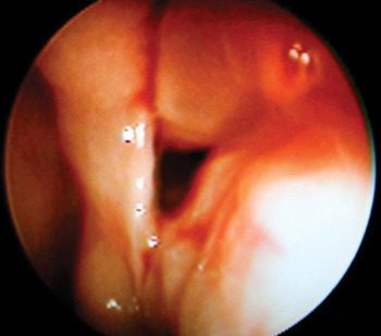
Post-operative endoscopic view using a 30° telescope, showing a patent frontal sinus outflow tract immediately after stent removal.

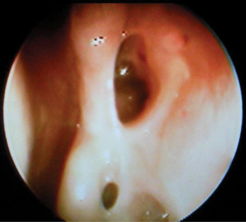
Endoscopic view of the same patient shown in Figure 5, one month later.

Coronal computed tomography scan of the same patient shown in Figure 1, after stent removal.

Discussion
Frontal sinus stenting offers a method of draining the frontal sinus and maintaining the frontal outflow tract without radical surgery. The stent prevents scar tissue formation across the frontal sinus opening, and instead allows epithelialisation to occur along the surface of the stent.Reference Hunter, Silva, Youngs, Saeed and Varadarajan 4
Other authors have reported the use of different stents, including the Rains stent, the Freeman stent and a rolled silicone sheet. Hughes and Rowe-Jones have advocated the use of a ureteric pigtail (double J) stent as a self-retaining frontal sinus stent, following its use in one patient.Reference Hughes and Rowe-Jones 8
In the current study, double J stents were used in five patients (seven sinuses) undergoing revision frontal sinus surgery following previous FESS. These patients had stenosed or obliterated frontal sinus outflow tracts. We used frontal sinus stents in these patients to avoid more radical surgery. The stents were left in place for six months and were well tolerated. It is not known whether this time period is ideal, and it is possible that the patient who suffered restenosis required a longer duration of stenting. Wound healing in the paranasal sinuses following major surgery takes up to three months.Reference Hoseman, Schindler, Wiegrebe and Gopferich 9
Electron microscopy has demonstrated that, when mucosa is only partially removed, ciliated cells regenerate fully by six months post-operatively. However, if the mucosa is completely removed, epithelium forms after a period of six months to one year and contains only scattered ciliated cells.Reference Bednarskim and Kuhn 5
In a study by Weber et al., all stents were removed after six months.Reference Weber, Mai, Hosemann, Draf and Toffel 3 However, in Orlandi and Knight's study stents were left for 11 to 73 months, with a mean of 31.6 months.Reference Orlandi and Knight 1
In a study by Freeman and Blom, stents placed to prevent post-operative stenosis were removed four weeks after surgery. However, if surgery was indicated to correct stenosis, then the stents were left for 6 to 12 months.Reference Freeman and Blom 6
Regarding the indication for frontal sinus stenting, Hunter et al. propose a number of potential factors that surgeons should consider: (1) the size of the frontal sinus outflow tract; (2) extensive polyposis (e.g. as found in allergic fungal sinusitis); (3) circumferentially exposed bone; (4) revision frontal sinus surgery; (5) osteitic bone in the frontal recess; (6) a flail middle turbinate; and (7) traumatic fracture of the frontal sinus outflow tract.Reference Hunter, Silva, Youngs, Saeed and Varadarajan 4
According to Orlandi and Knight, patients with 360° circumferential mucosal defects, or subtotal mucosal defects but long and narrow outflow tracts, following frontal sinus exploration, are candidates for stenting.Reference Orlandi and Knight 1 These authors also proposed that patients with an intra-operative ostial diameter of less than 5 mm are stent candidates.
In the present study, all the patients had previous FESS with frontal recess fibrosis and/or new bone formation. No patient had a tumour or traumatic fracture. Our success rate of 86 per cent (six out of seven sinuses) is difficult to interpret in view of the small number of patients.
Schaefer and Close used a silicone catheter for six weeks following endonasal frontal sinus surgery. They reported that 32 of 36 patients (89 per cent) experienced an overall improvement or resolution of symptoms after an average of 16.4 months post-operatively.Reference Schaefer and Close 10 In a study by Weber et al., 80 per cent of frontal sinus ostia were open 12 to 16 months after long-term stenting, compared with only 33 per cent of frontal sinuses that were not stented.Reference Weber, Hosemann, Draf, Keerl, Schick and Schinzel 11
Orlandi and Knight used Rains frontal sinus stents in nine patients.Reference Orlandi and Knight 1 At the end of the study, seven patients had a stent in place and two patients had a patent frontal sinus outflow tract.
Our study shows that double J stents are well tolerated as frontal stents, and are easily applied and truly self-retained, with no need for sutures. They have a thin wall and a smooth surface which may cause less scar tissue formation. The length of these stents can be controlled by cutting the nasal end as desired. If the stent is too short, granulations can overgrow it and embed it within scar tissue. If the stent is too long, persistent crusting of the nasal end will occur, which can disturb the patient and can cause an unpleasant odour.Reference Weber, Mai, Hosemann, Draf and Toffel 3
-
• Frontal sinus stents can prevent frontal sinus outflow tract restenosis
-
• Double J (ureteric pigtail) stents can be used as frontal sinus stents
-
• They are self-retaining, easy to insert and well tolerated
-
• Revision cases with new bone formation need at least six months' stenting
-
• Frontal sinus stenting may obviate the need for more aggressive procedures
If a large diameter stent is required, both ends can be applied together, as the largest double J stent is an 8 French gauge.
The current study was limited by its small number of patients and lack of a control group. However, it was intended only to demonstrate the tolerability, ease of application, self-retaining properties and modifiable nature (length-wise) of the double J stent.
Conclusion
Double J stents can be used as frontal sinus stents. They are well tolerated by the patient, easily applied and self-retaining, with no need for sutures. The length of the stent can be altered according to the patient's anatomy and pathology.