Implications
This is the first study linking pig 3-hydroxy-3-methylglutaryl-CoA reductase (HMGCR) expression levels with carcass and meat quality traits. This gene, which codes for the limiting enzyme in the de novo synthesis of cholesterol, regulates plasma lipid levels and cholesterol uptake in humans. Our study suggests that it also affects local lipids uptake and deposition in pig peripheral tissues, as muscle HMGCR expression correlates with carcass lean percentage, muscle intramuscular fat (IMF) content and individual fatty acid composition. Moreover, we describe a polymorphism in the coding region of this gene significantly associated with plasma lipid levels and IMF content.
Introduction
The rate limiting step of the de novo biosynthesis of cholesterol occurs at the 3-hydroxy-3-methylglutaryl-CoA reductase (HMGCR) catalysed step, which converts HMG-CoA into mevalonate (Friesen and Rodwell, Reference Friesen and Rodwell2004). Expression of this gene represses the expression of low-density lipoprotein (LDL) receptors in liver, altering the serum cholesterol levels (reviewed in Kajinami et al., Reference Kajinami, Takekoshi, Brousseau and Schaefer2004). In agreement with this, polymorphisms on the human HMGCR gene have been associated with changes in the plasma cholesterol and triglyceride (TG) levels (Tong et al., Reference Tong, Zhang, Li, Su, Kong, Liu, Xiao, Sun and Shi2004). Also, mutations resulting in alternative splicing of human exon 13 are a source of serum LDL-cholesterol variation (Burkhardt et al., Reference Burkhardt, Kenny, Lowe, Birkeland, Josowitz, Noel, Salit, Maller, Pe’er, Daly, Altshuler, Stoffel, Friedman and Breslow2008). Moreover, differences in liver HMGCR activity were reported between pigs selected for thin or thick backfat thickness at 80 Kg body weight for more than 14 generations (Pond and Mersmann, Reference Pond and Mersmann1996).
This gene has been extensively studied in humans as HMGCR inhibitors, known as statins, are the standard treatment for hypercholesterolemic patients. Its sequence and promoter transactivation has also been characterised in model animals such as rat (Osborne et al., Reference Osborne, Pollock, Lagor and Ness2004), mouse (Datta et al., Reference Datta, Wang, Moore and Osborne2006) and opossum (Chan et al., Reference Chan, Donalson, Kushwaha, Ferdinandusse, VandeBerg and VandeBerg2008) with a view to studying the genetics of serum cholesterol and TG variation and their consequences over atherosclerosis. The pig has also been used as a model for atherogenesis, obesity and lipoprotein and cholesterol metabolism due to anatomic and pathogenic similarities with humans (reviewed in Pond and Mersmann, Reference Pond and Mersmann1996). HMGCR activity is regulated at both post-transcriptional and post-translational levels. Regarding the pig, hepatic HMGCR enzyme activity seems to be regulated throughout development but inhibition of hepatic reductase activity by dietary cholesterol is particularly evident in young pigs (McWhinney et al., Reference McWhinney, Pond and Mersmann1996).
In addition to the pathological consequences, high-serum lipid concentrations can also contribute to increased fat deposition in adipose tissue and muscle. A classical example of this is type-2 diabetes mellitus-related obesity (Wilding, Reference Wilding2007). However, to our knowledge the relationship between HMGCR polymorphism, expression levels and fat deposition has not yet been attempted in humans or in any other species.
Following years of intensive selection for leaner pigs, in the last decade the market has shifted to an increasing demand for better quality pork. Two parameters, intramuscular fat (IMF) and fatty acid composition, have a marked effect over several meat quality traits such as tenderness, water holding capacity, juiciness and flavour (Olsson and Pickova, Reference Olsson and Pickova2005). A number of genes with a physiological role on lipid transport and metabolism have been proposed as candidate genes for fat deposition in pigs (Davoli and Braglia, Reference Davoli and Braglia2007) and some of these have been integrated in pig industry’s selection schemes (van der Steen et al., Reference van der Steen, Prall and Plastow2005). With all the above in mind and in order to contribute to the knowledge of genes that control cholesterol and lipid deposition in pigs, we have focussed the present work on the HMGCR gene as a source of genetic variation for traits related to serum lipid levels and fat deposition in pigs. With this aim, we have analysed the level of HMGCR expression in muscle and liver in two groups of commercial Duroc pigs displaying extreme values for carcass fat deposition and serum and muscle lipid traits. We have also characterised the pig HMGCR coding region, where we have described two polymorphisms. Subsequently, we have studied the relationship between liver and muscle expression data, HMGCR genotype and several serum and muscle lipid parameters.
Material and methods
Animal material and phenotypic data
Animals came from a high-IMF commercial Duroc line used in the production of fine quality cured ham. An experimental population was generated on the basis of a half-sib design, by mating five parental boars with 385 females and taking only one male offspring per litter to a total of 385 animals as described before (Gallardo et al., Reference Gallardo, Pena, Amills, Varona, Ramirez, Reixach, Diaz, Tibau, Soler, Prat-Cuffi, Noguera and Quintanilla2008). These animals were castrated and kept under normal intensive conditions, being all subjected to the same management procedures at IRTA-CCP control station.
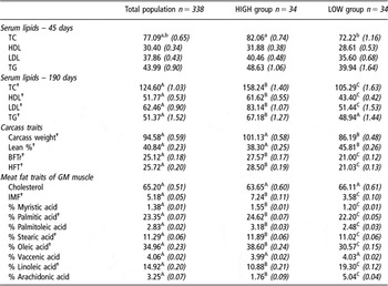
Two blood samples were taken at approximately 45 and 190 days of age to measure total serum cholesterol (TC), LDL, high-density lipoprotein (HDL) and TG concentrations (Gallardo et al., Reference Gallardo, Pena, Amills, Varona, Ramirez, Reixach, Diaz, Tibau, Soler, Prat-Cuffi, Noguera and Quintanilla2008). Animals were slaughtered according to a commercial protocol around 190 days of age (approximately 122 kg of live weight). Samples of 200 g gluteus medius muscle were collected immediately after slaughter for meat analyses carried out at the Centre of Meat Technology (CTC-IRTA). Analyses of gluteus medius lipid components included the determination of percentage of IMF, cholesterol content and fatty acid composition (in the C12 to C22 interval) were performed as follows: IMF content was determined by Near Infrared Transmittance (NIT; Infratec® 1625, Tecator AB, Hoganas, Sweden) (Plastow et al., Reference Plastow, Carrión, Gil, GarcIa-Regueiro, i Furnols, Gispert, Oliver, Velarde, Guàrdia, Hortós, Rius, Sárraga, Diaz, Valero, Sosnicki, Klont, Dornan, Wilkinson, Evans, Sargent, Davey, Connolly, Houeix, Maltin, Hayes, Anandavijayan, Foury, Geverink, Cairns, Tilley, Mormède and Blott2005, Pinhu et al., Reference Pinhu, Park, Yao and Griffiths2008) fatty acid composition was analysed by gas chromatography of methyl esters as described in (Mach et al., Reference Mach, Devant, Diaz, Font-Furnols, Oliver, Garcia and Bach2006), and cholesterol content was measured following (Cayuela et al., Reference Cayuela, Garrido, Banon and Ros2003). The most relevant phenotypic traits analysed in this study, along with their abbreviations and statistics in the studied Duroc population, are shown in Table 1 (complete data set was finally obtained from 338 animals). Differences between group means were computed by a two-tailed t-test. All experimental procedures were approved by the Ethics Committee for Animal Experimentation of IRTA.
Mean (s.e.) of phenotypic records in blood, carcass and muscle samples for the whole Duroc pig population and for the two extreme groups of animals selected for HIGH and LOW values for cholesterol and lipid metabolism traits

TC = total serum cholesterol; LDL = low-density lipoprotein; HDL = high-density lipoprotein; TG = serum triglyceride levels; BFTr = backfat thickness measured between the 3rd and 4th ribs; HFT = ham fat thickness; GM = muscle gluteus medius; IMF = intramuscular fat.
†Traits used in the principal component analysis to select animals for the HIGH and LOW groups. Different superscripts denote significant differences between group means (uppercase: P < 0.001; lowercase: P < 0.05).
Isolation of DNA and RNA samples
Liver and gluteus medius muscle samples were collected at slaughter, snap frozen in liquid nitrogen and stored at −80°C for subsequent RNA analysis. To isolate total RNA, samples were ground with mortar and pestle in liquid nitrogen and homogenised with a mechanical rotor. RNA was isolated by the acid phenol method (Chomczynski and Sacchi, Reference Chomczynski and Sacchi1987) using the RiboPure kit (Ambion, Austin, TX, USA). DNA was isolated from blood samples using standard procedures (Sambrook and Russell, Reference Sambrook and Russell2001).
Characterisation of coding region and polymorphisms
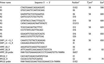
Primer design was carried out with the Primer Express software (Applied Biosystems, Foster City, CA, USA) using pig sequences as a template when available or human cDNA sequences that displayed high homology with other available mammalian sequences. For the amplification of the coding region, a total of five primer sets were designed (Table 2). Total RNA (0.5 to 1 μg) was retrotranscribed to cDNA using an Oligo(dT) primer and MMuLV RT enzyme (Fermentas, Glen Burnie, MD, USA) at 37°C for 1 h (Sambrook and Russell, Reference Sambrook and Russell2001). PCR reactions were carried out in a PTC-100 thermocycler (MJ Research, Waltham, MA, USA) in a volume of 25 μl with 1 × buffer, 200 μM dNTP mix, 2.0 mM MgCl2, 400 nM of each primer, 1U of Taq polymerase (ECOGEN SL, Barcelona, Spain) and 0.5 μl of cDNA. Thermal profile was as follows: denaturation 5 min at 95°C; 35 cycles of 15 s at 95°C, annealing 30 s at temperature indicated in Table 2, 40 s at 72°C; final extension 5 min at 72°C. PCR products were directly sequenced using the BigDye Terminator Sequencing kit v3.1 (Applied Biosystems) in an ABI-3100 capillary electrophoresis system (Applied Biosystems). Sequences obtained were edited using the Sequencing software (Applied Biosystems) and aligned with the ClustalW program (Chenna et al., Reference Chenna, Sugawara, Koike, Lopez, Gibson, Higgins and Thompson2003).
Primers used for the characterisation and genotyping of the pig HMGCR gene

†Position according to exon/intron distribution in human gene.
‡Annealing To used in the PCR reactions.
§Size in base pair of the amplified product.
Polymorphism genotyping
To genotype the HMGCR:c.807A > G polymorphism, primers were designed on exon eight and intron eight of the HMGCR gene (SNP1_A > G_F and SNP1_A > G_R; Table 2). Genotyping was performed by restriction enzyme cleavage of the 650 bp-long PCR product with HhaI (Fermentas). The G allele (two fragments of 450 and 200 bp) but not the A allele was cleaved and the fragments resolved in a 1.5% agarose gel.
Microarray hybridisation design
A differential expression study comparing animals with extreme characteristics for the lipid metabolism traits was carried out in our commercial Duroc population by means of hybridisation in oligo-based microarrays. With this purpose, from the complete set of phenotypic data available for the 385 Duroc pigs, the most important phenotypes (13 traits; see Table 1) were selected to perform a principal component analysis by means of PRINCOMP procedure of SAS (SAS Institute Inc., Cary, NC, USA). The first principal component, which accumulated 30.7% of total phenotypic variance, was defined by several fatness and serum cholesterol measures, together with IMF, monounsaturated and saturated fatty acid content in gluteus medius, and, in opposite direction, by gluteus medius polyunsaturated fatty acid content and carcass lean percentage. We used the values from the first principal component as a selection index to rank all studied individuals. Animals at both extremes of the ranking were selected to form the HIGH and LOW groups (34 animals per group).
Thus, RNA from 68 samples of gluteus medius muscle was individually hybridised on GeneChip Porcine Genome arrays (Affymetrix, Boston, MA, USA). Expression data were normalised using the Robust Multichip Average (RMA) algorithm. Expression values corresponding to the Ssc.16088.1.S1 probe (representing the porcine HMGCR gene) were extracted and analysed as explained below.
Quantitative PCR analysis (qPCR)
In order to study the expression of HMGCR gene in liver, and also to validate the muscle HMGCR microarray expression data in a subset of muscle samples, we carried out real-time qPCR essays in an ABI-7500 device (Applied Biosystems) in a final volume of 5 μl. Primers and TaqMan probes were designed using the Primer Express v2.0 software (Applied Biosystems; Table 2). The HMGCR cDNA was amplified in a PCR reaction containing 200 nM primers (F3 and R2; Table 2) and 1×SYBR Green Master mix (Applied Biosystems). For the two endogenous controls (HPRT and RPL32), PCR reaction contained 300 nM primers, 200 nM TaqMan probe and 1× Universal Taqman Master Mix (Applied Biosystems). The following thermal profile was used for all reactions: 10 min at 95°C, 40 cycles of 15 s at 93°C, 1 min at 60°C and 30 s at 72°C, followed by a quick denaturation at 95°C × 5 min plus a slow ramp to 30°C to generate a dissociation curve to control the specificity of the amplified product. In order to quantify and normalise the expression of the samples we used the ΔΔCt method (Yuan et al., Reference Yuan, Reed, Chen and Stewart2006).
Statistical analyses
Association analysis of polymorphisms detected in the HMGCR gene
Association studies of polymorphism detected in the HMGCR gene with the complete set of available phenotypic data from the experimental Duroc population (n = 338; traits detailed in Table 1) were carried out by means of the GLM procedure of SAS (SAS Institute Inc.). The model used for these analyses was:

where yijklm and yijlm represents phenotypic observation for each individual; μ is the overall mean; fi is the systematic effect farm of origin, with three levels (only for serum lipid concentrations at 45 days); pj is the effect batch of fattening, with four levels; qk is the effect of belonging to the j-th half-sib family, with five levels; gl is the effect of individual genotype (three levels, the two homozygous and the heterozygous); eijklm and eijlm are the residual effects.
In all analysed traits, least-squares (LS) means for the three genotypes were computed and compared between them.
Finally, an adjustment for multiple testing was carried out by Bonferroni correction i.e. as a solution to Pexp = 1−(1−P) N, where Pexp is the experiment-wide P value and N is the number of independent analyses. Nevertheless, an important correlation between performed association tests is expected due to correlation between analysed traits. The fact that Bonferroni correction is overly conservative under these circumstances must be taken into account when interpreting data.
HMGCR differential expression and association with HMGCR genotypes
HMGCR expression data (n = 68) from both muscle (microarray data) and liver (qPCR results) were used to investigate the relationship between the HMGCR expression level and genotype.
An initial differential expression analysis was performed with the following model:

where yjklm represents the individual HMGCR expression in muscle or liver; tm represents the effect of belonging to one of two groups (HIGH- and LOW-lipid metabolism) and pj, qk, gl and ejklmn are the same effects as in the association analysis for the HMGCR polymorphisms. LSmeans for the group and genotype effects were computed.
Correlation study between HMGCR gene expression and phenotypic data
In order to further investigate the relationship between lipid metabolism and HMGCR expression, we performed a correlation study between the phenotypes analysed (Table 1) and HMGCR expression levels both in liver and muscle. The correlation study was performed using the CORR procedure of SAS (SAS Institute Inc.) after adjusting both, phenotypes and expression levels, for the environmental significant effects considered in previous analyses. In order to test possible differences between groups, two correlation analyses were carried out: 1) a conjoint correlation analysis considering all data set (n = 68 for each correlation) after correcting for all environmental factors including group, and 2) a within-group (HIGH and LOW lipid metabolism) correlation analyses (n = 34 for each correlation) after correcting for environmental factors.
Results
Identification and genotyping of polymorphisms in the porcine HMGCR coding region
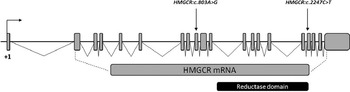
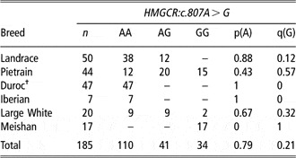
We successfully amplified 2691 bp of the pig HMGCR cDNA, which included the entire coding region, in five partially overlapping PCR fragments (submitted to GenBank with accession number EU726797). This sequence is identical to the reference sequence for porcine HMGCR cDNA (NM_001122988.1) but for two synonymous polymorphisms we identified in our commercial Duroc animals: HMGCR:c.807A > G and HMGCR:c.2247C > T, situated in putative exons 9 and 17, respectively (Figure 1). For the first polymorphism, the studied Duroc population exhibited an intermediate allele frequency for allele G (q = 0.33). Moreover, three of the five founder sires were heterozygous for this polymorphism. We also genotyped this single nucleotide polymorphism (SNP) in a number of animals from different breeds (Table 3). Allele frequencies varied widely between populations: white pig breeds displayed low (Landrace) and intermediate (Large White, Pietrain) allelic frequencies for allele G, while it did not segregate in three other populations where either the A allele (Iberian and a Duroc line of another origin) or the G allele (Meishan pigs) was fixed. The second polymorphism could not be genotyped in a larger set of animals as the lack of information on the pig genomic sequence around the mutation prevented setting up a genotyping protocol on genomic DNA.
Graphical representation and localisation of two new single nucleotide polymorphism in the coding region of the pig HMGCR gene. Exon/intron distribution is taken from the human gene.

Allelic frequency for the HMGCR:c.807A > G polymorphism in several porcine breeds

†Population of different origin from the analysed Duroc population.
Association between HMGCR genotype and lipid metabolism traits
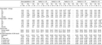
Table 4 shows results corresponding to the association study between the HMGCR:c.807A > G genotype and several serum and meat lipid composition traits, at both population and within-family levels. Concerning the serum lipid concentrations, the A allele was related to higher TC and LDL levels at 45 days in family 3, being these differences significant at experiment-wide level. A similar tendency, although not statistically significant (0.5 < P < 0.1; not indicated in tables), for LDL at 190 days was observed in family 3. Also an experiment-wide significant association of the A allele with higher LDL at 190 days was found in family 5, affecting also the TC levels and the ratio of HDL v. LDL cholesterol (in these two cases at nominal level). At nominal level of significance, the AA genotype showed higher serum TG concentrations at 45 days in the whole-population analysis. Except for family 4, this effect was not significant when a within-family analysis was conducted, although a similar tendency was observed in most cases.
Least-squares means for association study of HMGCR:c.807A > G genotype with serum and carcass and meat lipid traits

TC = total serum cholesterol; TG = serum triglyceride levels; LDL = low-density lipoprotein; HDL = high-density lipoprotein; IMF = intramuscular fat; GM = muscle gluteus medius.
§Means of serum lipid traits with log transformed data. Different superscripts denote significant (P < 0.05) differences at nominal level. Superscripts in uppercase letter denote significant differences at experiment-wide level (P exp<0.05), corresponding to a nominal P < 0.006.
As regards the lipid content of gluteus medius muscle, no experiment-wise significant associations were obtained, but at population level, we observed (nominal) significantly higher IMF in individuals carrying the A allele. The same trend was observed in all within-family analysis except in family 1, but these results did not reach the significance level. Concerning the fatty acid composition of gluteus medius muscle, the A allele was also significantly (P < 0.05) associated with higher oleic content and suggestively (P = 0.06; not indicated in tables) with lower linoleic percentage in family 4.
Association between HMGCR genotype and expression levels
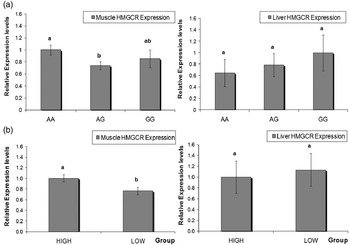
Results from the association analyses between HMGCR:c.807A > G genotype and HMGCR mRNA levels in muscle and liver are shown in Figure 2a. These results revealed significant differences in muscle HMGCR expression levels between animals with genotypes AA and AG (P < 0.05), whereas individuals with genotypes GG (only six animals) showed intermediate expression levels and no differences from the other genotypes. In contrast, liver expression levels were uniform amongst genotypes (Figure 2a). The different behaviour of this gene in these two tissues was also reflected by a lack of correlation between liver and muscle HMGCR mRNA levels (data not shown).
Graphical display of relationship of pig HMGCR muscle and liver expression levels with (a) HMGCR:c.807A > G genotype (P < 0.05; n(AA) = 29, n(AG) = 33, n(GG) = 6) and (b) lipid metabolism phenotypic group (P < 0.001; n(HIGH) = 34; n(LOW) = 34). Mean values are represented. Error bars indicate s.e.

HMGCR gene expression in animals with extreme lipid metabolism and its relationship with phenotypic measures
Figure 2b presents the comparison of HMGCR muscle and liver expression levels in two groups of Duroc pigs corresponding to the most extreme animals for 13 traits related to growth, fattening and cholesterol metabolism traits (HIGH and LOW groups; Table 1). Results indicate that HMGCR expression varied according both to the tissue type and to the group of analysis. In this sense, muscle HMGCR expression was 1.31-fold higher in animals from the HIGH than from the LOW group (P < 0.001). These differences were, however, specific of muscle, as liver expression levels did not differ between groups. We validated the microarray results of muscle HMGCR gene by qPCR of a subset of ten muscle samples from each group. The expression ratios between HIGH and LOW groups remained in the same sense in both assays (microarray and qPCR) obtaining identical significance level (P < 0.001) with both datasets.
In addition, we studied the within-group correlation coefficients between gluteus medius and liver HMGCR expression levels and serum, carcass and muscle phenotypes (Table 5). With some punctual exceptions, we obtained no significant differences between both sets of within-group (HIGH and LOW) correlation coefficients, and neither with the global correlation coefficients obtained with pooled data set (results not shown). In all cases, muscle HMGCR expression was positively correlated with serum TC and HDL levels, and also with IMF, oleic and palmitic fatty acid content in gluteus medius, and negatively correlated with linoleic and stearic fatty acid content, and with lean percentage. In contrast, a positive correlation between serum LDL levels and muscle expression levels was detected only in the HIGH group.
Correlations between muscle and liver HMGCR expression values and serum lipid concentrations, carcass traits and muscle fat content and composition

Ms = muscle; Lv = liver; TC = total serum cholesterol; TG = serum triglyceride levels; Chol GM = cholesterol content in muscle gluteus medius; IMF = intramuscular fat; H = HIGH group; L = LOW group.
*Significant results (P<0.05) are reported in bold.
As far as HMGCR expression in liver is concerned, it shared with muscle expression the negative correlation with stearic acid content and lean carcass percentage, but, in contrast, a negative interaction with serum HDL levels was also detected. Liver HMGCR mRNA levels displayed significant correlation with no other traits analysed.
Discussion
Despite the crucial role of the microsomal membrane-bound glycoprotein HMGCR enzyme in determining serum cholesterol levels in humans (reviewed in Schmitz and Langmann, Reference Schmitz and Langmann2006), the information regarding this gene in pigs is sparse. Previous studies have focused on the regulation of HMGCR hepatic activity by dietary fat indicating that this enzyme is particularly inhibited by dietary cholesterol reductase activity in young pigs (McWhinney et al., Reference McWhinney, Pond and Mersmann1996; Pond and Mersmann, Reference Pond and Mersmann1996) and that the modulation of hepatic reductase activity is related, at least in guinea pigs, to the ratio of saturation to insaturation of the dietary fat (Fernandez and McNamara, Reference Fernandez and McNamara1991). Regarding the porcine HMGCR gene, restriction fragment length polymorphism (RFLP) polymorphisms in the porcine gene have been previously described by Southern blotting (Davis et al., Reference Davis, White and Wheeler1995). Therefore, the position of these mutations was unassigned. Moreover, no study on the relationship between this gene and livestock production traits has yet been reported.
We report here the complete coding region for the pig HMGCR cDNA and describe two synonymous polymorphisms located at position +807 and +2247 of the coding region. The former of these polymorphisms (HMGCR:c.807A > C) was segregating in commercial pig breeds (Table 3) and showed significant whole-population associations, although at nominal level, with gluteus medius IMF content and serum TG at 45 days of age (Table 4). Similar effects have been described in humans, where analyses of an 830A/C polymorphism in human HMGCR gene resulted in higher levels of circulating low-density lipoprotein and TG in patients with the AA genotype (Tong et al., Reference Tong, Zhang, Li, Su, Kong, Liu, Xiao, Sun and Shi2004). Considering the correction for multiple testing, the association tests with LDL-bound cholesterol reached the significance at experiment-wide level (P < 0.006) in family 3 (45 days) and family 5 (190 days), and also with TC in family 3 (45 days). The fact that Bonferroni correction is overly conservative when correlated association tests are performed must, however, be taken into account. In fact, other associations at nominal level were also observed but they were not consistent across all within-family analyses. These inconsistencies are probably caused by the limited sample size of some families, and the corresponding loss of power, but could also be due to family differences in the linkage disequilibrium phase of HMGCR:c.807A > C with the mutation responsible for these differences. More powerful experiments with a larger number of animals would be worthy to disentangle these inconsistencies, but the considerable management and economic difficulties involved in performing this kind of studies in commercial pig populations constitute an obstacle not easy to overcome.
We have also carried out a functional study on the pig HMGCR gene, investigating the relationship of HMGCR expression in liver and gluteus medius muscle with serum lipid levels and fat muscle content. Correlations obtained in this study suggested associations between HMGCR expression levels and phenotypic variability of several fatness traits and serum and meat lipid content, but differences were not always consistent across tissues. Thus, carcass lean and stearic fatty acid percentages in gluteus medius were negatively correlated with both liver and muscle HMGCR expression, whereas HDL levels were negatively correlated with liver expression levels, possibly reflecting the role of the liver in the excretion of HDL-cholesterol particles, but exhibited a positive correlation with muscle HMGCR mRNA levels. In contrast, only muscle HMGCR expression showed a significant relationship with serum TC and LDL levels, IMF and other fatty acids (oleic, linoleic and palmitic) content in muscle. The positive correlation of cholesterol serum concentrations (TC, HDL and LDL) with muscle expression levels allow to hypothesise a relationship between changes in lipoprotein transport levels and muscle gene transactivation, as a results of the lipids uptaken from circulating lipoproteins (Nagao and Yanagita, Reference Nagao and Yanagita2008). The liver plays a critical role in orchestrating cholesterol biosynthesis and transport to and from peripheral tissues. In addition to this established role, we anticipated that local activity of this enzyme in peripheral tissues would potentially regulate local levels of lipid uptake and storing. In this context, results shown in this study suggest that high-serum lipid concentrations can also contribute to enhance fat deposition in adipose and muscle tissue.
The association analysis between structural and functional variations in HMGCR gene (Figure 2a) revealed a relevant association between HMGCR expression levels in gluteus medius muscle and HMGCR:c.807A>G genotype. In contrast, no significant differences for liver expression were found between genotypes. In humans, although a number of intronic SNP have been reported to lower the response to statins (Chasman et al., Reference Chasman, Posada, Subrahmanyan, Cook, Stanton and Ridker2004), structural mutations of this gene have not been associated to changes at liver expression level. Moreover, HMGCR mRNA levels are unaffected by changes in intronic splicing events (Medina et al., Reference Medina, Gao, Ruan, Rotter and Krauss2008). Consistent to this, the slight decrease in liver HMGCR mRNA levels of hemizygous HMGCR mice resulted in no differences in cholesterol or TG content in liver or blood (Ohashi et al., Reference Ohashi, Osuga, Tozawa, Kitamine, Yagyu, Sekiya, Tomita, Okazaki, Tamura, Yahagi, Iizuka, Harada, Gotoda, Shimano, Yamada and Ishibashi2003). In this same line, divergent selection on pigs for serum total cholesterol (seven generations) was not associated with differences in hepatic or jejunum HMGCR activity or with HMGCR RFLP polymorphisms, at least in young animals, but rather with CYP7 polymorphisms (Schoknecht et al., Reference Schoknecht, Ebner, Pond, Zhang, McWhinney, Wong, Klein, Dudley, Goddard-Finegold and Mersmann1994). In contrast with these results, in our Duroc pig population a 1.3-fold change in the muscular expression of this gene is associated with plasma and muscle lipid traits (Figure 2b), such as those defining the HIGH and LOW groups (Table 1). With respect to the LOW group, animals from the HIGH group had overall higher serum lipid concentrations, backfat thickness, IMF and monounsaturated fatty acid content (oleic acid), together with lower lean content and polyunsaturated fatty acid (linoleic and arachidonic acid) content. This is in agreement with a higher frequency of AA animals in the HIGH (p(AA) = 0.53) v. LOW (p(AA) = 0.32) group, and conversely for the lower frequency of AG animals (p(AG) = 0.38 in HIGH v. p(AG) = 0.59 in LOW). The most probable reason is that serum plasma lipids determine the amount of intracellular lipids in muscle and, therefore, the activation of steroids and other signalling molecules affecting the transactivation of the HMGCR promoter, which in turn affects the number of active LDLR (low-density lipoprotein receptor) and, subsequently, the uptake and accumulation of lipids in muscle (Telford et al., Reference Telford, Edwards, Lipson, Sutherland, Barrett, Burnett, Krul, Keller and Huff2003). Indeed, steroids (including cholesterol and mevalonate) promote a negative feed-back over the transcription of HMGCR and a positive enhancement of enzyme ubiquination and degradation (Ness and Chambers, Reference Ness and Chambers2000).
Conclusion
In summary, in this study, we have characterised the coding region and two synonymous polymorphisms for the cholesterol metabolism-related HMGCR porcine gene, one of them showing significant associations with serum LDL concentrations. Moreover, we have for the first time described relevant correlations between the HMGCR expression and fat deposition and meat composition traits, such as carcass lean percentage, IMF or meat fatty acid composition. Also, we have established a relationship between structural variations in this gene and expression levels in the gluteus medius muscle. As a whole, results obtained in this study allow us to conclude that the HMGCR gene displays a relationship with not only lipid serum traits but also with commercially important pig meat quality and production traits, and can be shown as an interesting candidate gene for gene assisted selection in pigs.
Acknowledgements
The authors thank Selección Batallé S.A. for providing the animal material and for their cooperation in the experimental protocol and D. Almuzara for technical support. A. Cánovas received a predoctoral scholarship from INIA. RN Pena received a contractual grant from INIA. This project has been financed by the Spanish Ministry of Education and Science (projects GEN2003-20658-C05-05/CICYT, AGL2002-04271-C03-02 and AGL2007-66707-C02-01).