Introduction
Thérèsemagnanite was originally described as a new cobalt–zinc–nickel sulfate from the Cap Garonne mine, Var, Provence-Alpes-Côte d'Azur, France by Sarp (Reference Sarp1993) with the formula (Co,Zn,Ni)6(SO4)(OH,Cl)10·8H2O, but was redefined recently by Kasatkin et al. (Reference Kasatkin, Plášil, Škoda, Belakovskiy, Marty, Meisser and Pekov2018) as NaCo4(SO4)(OH)6Cl·6H2O; the Co-analogue of gordaite. Unfortunately, Kasatkin et al. (Reference Kasatkin, Plášil, Škoda, Belakovskiy, Marty, Meisser and Pekov2018) could not identify any crystals suitable for single-crystal analysis, however, during a systematic study looking for crystals of guarinoite and thérèsemagnanite in the North mine at Cap Garonne, we were able to find single crystals suitable for study. Here we report the crystal structure of Ni-rich gordaite–thérèsemagnanite near the triline, in order to shed light on the structure of thérèsemagnanite and the as yet unreported Ni-analogue of thérèsemagnanite and gordaite.
Occurrence
The gordaite–thérèsemagnanite specimen studied was collected at pillar 80 in the North mine, Cap Garonne, Var, France (43°4'53''N, 6°1'55''E). Pillar 80 is a long and straight mine wall exhibiting unusual mineralogy, dominated by copper, zinc, cobalt and nickel. The end of the wall includes a large cavity where 5 out of the 14 new species known at Cap Garonne have been found: geminite (Sarp and Perroud, Reference Sarp and Perroud1990), guarinoite (Sarp, Reference Sarp1993), mahnertite (Sarp, Reference Sarp1996), pushcharovskite (Sarp and Sanz-Gysler, Reference Sarp and Sanz-Gysler1997) and thérèsemagnanite (Sarp, Reference Sarp1993). This specimen was collected as part of the systematic study of the mine mineralogy by Association des Amis de la Mine de Cap Garonne (Favreau and Galea-Clolus, Reference Favreau and Galea-Clolus2014; Mills et al., Reference Mills, Christy, Schnyder, Favreau and Price2014, Reference Mills, Christy, Colombo and Price2015, Reference Mills, Christy, Favreau and Galea-Clolus2017, Reference Mills, Christy and Favreau2018; Plášil et al., Reference Plášil, Petříček, Mills, Favreau and Galea-Clolus2018).
There are two different environments that co-exist in the cavity. The lower part of the cavity wall is comprised of a very compact quartz conglomerate, rich in tennantite, where acidic conditions may have existed for the formation of geminite and pushcharovskite. Other copper minerals are found in this environment, such as lavendulan, mahnertite, zdenĕkite, atacamite, brochantite and olivenite. The upper part of the wall and the roof are made of a less compact conglomerate with malachite and cerussite, and the Zn−Co–Ni sulfates ktenasite, guarinoite and thérèsemagnanite. A thick, solid, fine-grained galena vein is located between the lower and upper part, with rare cavities hosting covellite, native sulphur (Kösters et al., Reference Kösters, Galéa-Clolus, Clolus, Heying and Pöttgen2019) and anglesite. The specimen studied was found in a fracture of this galena vein.
Gordaite–thérèsemagnanite occurs as pale pink, vitreous hexagonal plates up to 0.2 mm in diameter, usually stacked as textbooks or organised as rosettes. The unusual thickness and size of the crystals in this specimen allowed for single-crystal studies to be conducted. The specimen studied has been deposited in the collections of Museums Victoria, registration number M54547.
Experimental
Chemistry
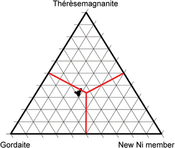
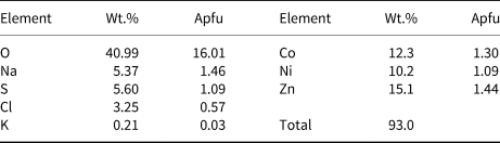
Semi-quantitative chemical analyses of a crystal cluster from the specimen were performed on a JEOL 7001F FEG-SEM (energy-dispersive spectroscopy (EDS) mode, 20 kV, 3 nA and 6 µm beam diameter) at the Monash Centre for Electron Microscopy, Monash University, Australia. The scanning electron microscopy EDS method was used for semi-quantitative chemical analysis due to the hydrated nature of the mineral and its susceptibility to beam damage. Even gentle conditions in an electron microprobe caused burning of the sample. The standards used were: quartz (O and Si), albite (Na), pyrite (S), synthetic NaCl (Cl), synthetic KBr (K), Co metal (Co), Ni metal (Ni) and Zn metal (Zn). Results (Table 1) are recorded to only one decimal place for all elements, while H cannot be detected by EDS. Sodium is overestimated in the analyses due to the overlap between NaKα and ZnKα. The analyses for Co, Ni and Zn (Fig. 1) show a spread of values similar to those presented in fig. 6 in Kasatkin et al. (Reference Kasatkin, Plášil, Škoda, Belakovskiy, Marty, Meisser and Pekov2018), where some points are Zn-dominant (18 points) and some Co-dominant (5 points), but with the average calculated Co:Ni:Zn ratio of 0.34:0.28:0.38. However, the Co:Zn average ratio is within error of each other given the uncertainties of the analytical method. The specimen studied shows more Ni than the samples of Kasatkin et al. (Reference Kasatkin, Plášil, Škoda, Belakovskiy, Marty, Meisser and Pekov2018), suggesting that a Ni-dominant member of the gordaite group could also be found at Cap Garonne, probably at pillar 80.
Triangular diagram showing the Co:Zn:Ni proportions analysed in the gordaite–thérèsemagnanite crystals (23 analyses). Plotted using Graham and Midgley (Reference Graham and Midgley2000).

The average composition of gordaite–thérèsemagnanite (23 analyses) determined by SEM–EDS*.

* H is not included. Apfu is atoms per formula unit, based on 23 non-H atoms.
X-ray diffraction
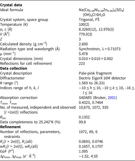
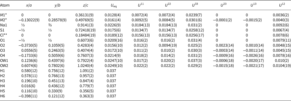
The single crystal X-ray diffraction experiment was carried out on the micro-focus macromolecular MX2 beamline at the Australian Synchrotron, part of ANSTO. A 10 µm × 10 µm × 2 µm pale-pink single crystal (a fragment of a larger hexagonal plate) was selected from specimen M54547. Data were collected at 100 K by a Dectris EigerX 16M detector and monochromatic radiation with a wavelength of 0.71073 Å. After processing the data using XDS (Kabsch, Reference Kabsch2010), XPREP (Bruker, Reference Bruker2001) and SADABS (Bruker, Reference Bruker2001), 10,670 reflections were integrated with a R int of 0.1352. Structure solution in P  $\bar{3}$ was carried out by direct methods using SHELXT (Sheldrick, Reference Sheldrick2015a) and located all atoms. Structure refinement by full-matrix least-squares was implemented by SHELXL (Sheldrick, Reference Sheldrick2015b), using neutral atomic-scattering factors. Three reflections with calculated F 0/F C errors >5.00 were omitted from the final refinement. All hydrogen atoms were located via calculation of the bond valence sums on the oxygen atoms, consideration of non-bonded O···O distances which might indicate the presence of hydrogen bonds, and careful inspection of the difference-Fourier maps. All hydrogen atoms were successfully located in this way. Hydrogen atoms were constrained softly to a distance of 0.90(2) Å and a U eq 1.5 times that of its corresponding oxygen atom. The Cl site refined to a final occupancy of 93% Cl and 7% OH. A difference peak located at (0, 0, 0.112, site occupancy factor = ⅓), 0.918 Å from the Cl/OH site may also indicate the presence of a partially occupied H atom. In the final stage of the refinement the Co:Ni:Zn ratio was fixed at 0.34:0.28:0.38 on the basis of the average chemical analyses. Details concerning data collection and structure refinement are provided in Table 2. Fractional coordinates and atomic displacement parameters are provided in Tables 3 and 4, respectively. A bond-valence analysis is provided in Table 5, using the parameters of Gagné and Hawthorne (Reference Gagné and Hawthorne2015) for all ion–oxygen bonds except Na (Wood and Palenik, Reference Wood and Palenik1999) and H (Brown, Reference Brown2002). The M1–Cl bond valence was calculated using the parameters of Brese and O'Keefe (Reference Brese and O'Keeffe1991).
Summary of information relating to data collection and structure refinement of gordaite–thérèsemagnanite.

Atom coordinates, site occupancies and atomic displacement parameters (Å2) for gordaite–thérèsemagnanite.

* Occupancies for M sites fixed to 34% Co, 28% Ni and 38% Zn.
** Occupancy refined to Cl 0.93(3) and O (as OH) 0.07(3)
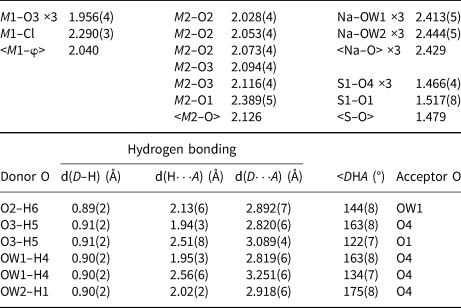
Bond lengths (Å) for gordaite–thérèsemagnanite*.

* Occupancies for M sites fixed to 34% Co, 28% Ni and 38% Zn; occupancy for Cl site refined to 93% Cl and 7% OH.
Bond-valence sums (in valence units) for gordaite–thérèsemagnanite, (A) indicates an accepted hydrogen bond.

* 34% Co, 28% Ni and 38% Zn; ** 93% Cl and 7% OH
Structure description
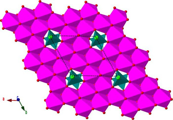
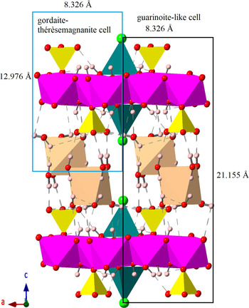
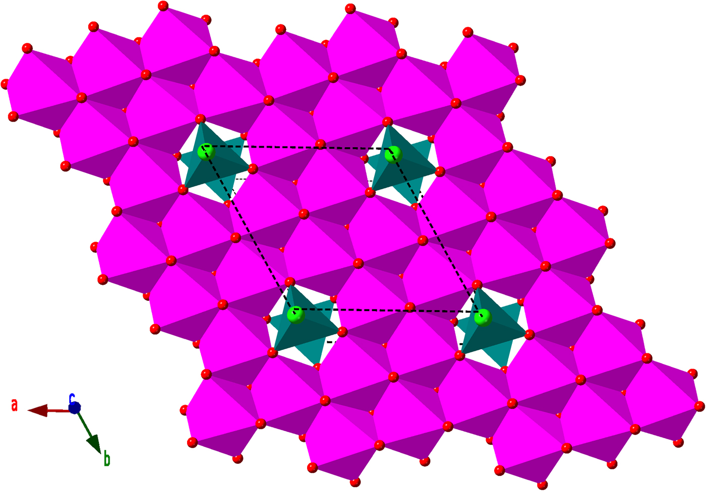
The structure of the gordaite–thérèsemagnanite from Cap Garonne (Figs 2 and 3) is very similar to those reported for gordaite (Adiwidjaja et al., Reference Adiwidjaja, Friese, Klaska and Schlüter1997; Zhu et al., Reference Zhu, Seff, Witzke and Nasdala1997 – although only the former study located H atoms on the H2O group oxygen atoms), with the only main difference noted in the hydrogen bonding. The structure is layered parallel to {001}, with a brucite-like sheet formed from edge-sharing [M2(OH)5O]3– octahedra. Each octahedron is surrounded by six others, but one-seventh of the octahedral sites are vacant, providing three corner-linking O3 sites for two [M1(OH)3Cl]2– tetrahedra to be bonded to the layer, one above and one below the sheet. SO4 tetrahedra are corner-linked to one-third of the [M2(OH)5O]3– octahedra, also pointing both above and below the sheet. To compensate for the overall negative charge of the [M1(OH)3Cl]2– tetrahedra, [M2(OH)5O]3– octahedra and SO4 tetrahedra, isolated Na(H2O)6 octahedra are found between the layers. Hydrogen bonding is integral in linking the layers together along [001].
One sheet of gordaite–thérèsemagnanite viewed down [001]. [M2(OH)5O]3– octahedra are in magenta, [M1(OH)3Cl]2– tetrahedra in dark cyan, O atoms in red, Cl atoms in bright green. H atoms are not shown for clarity.
The structure of gordaite–thérèsemagnanite viewed down [010]. Colour scheme is as above, with SO4 tetrahedra in yellow, Na(H2O)6 octahedra in pale orange and H atoms in pale pink. H bonds are shown as dashed lines.

The hydrogen bonding network is similar to that described by Adiwidjaja et al. (Reference Adiwidjaja, Friese, Klaska and Schlüter1997). Identical H bonds are seen connecting the channel H2O groups to SO4 oxygen atoms, and from sheet OH groups to channel H2O oxygen atoms. The main difference is that no H bonds in our structure are oriented towards the Cl anions or between adjacent Na(H2O)6 octahedra. Instead, the H bonds are more typically oriented towards other H2O groups (Fig. 2).
The structure has a superficial resemblance to the sulfate members of the hydrotalcite supergroup (Mills et al., Reference Mills, Christy, Génin, Kameda and Colombo2012), however, in hydrotalcites SO4 tetrahedra do not bond directly onto the brucite layer. Therefore, the gordaite–thérèsemagnanite structure is more closely related to minerals such as spangolite, Cu6Al(OH)12(SO4)Cl·3H2O (Hawthorne et al., Reference Hawthorne, Kimata and Eby1993) and jamborite, Ni2+1−xCo3+x(OH)2−x(SO4)x·nH2O (Bindi et al., Reference Bindi, Christy, Mills, Ciriotti and Bittarello2015) where the SO4 tetrahedra bond directly to the layer.
Comment on relationship to guarinoite
Sarp (Reference Sarp1993) described guarinoite as ideally (Zn,Co,Ni)6(SO4)(OH,Cl)10·5H2O and thérèsemagnanite as (Co,Zn,Ni)6(SO4)(OH,Cl)10·8H2O, with the unit cells a = 8.344(4) and c = 21.59(2) Å and a = 8.363(8) and c = 26.18(17) Å for guarinoite and thérèsemagnanite, respectively. We note that the a axis and chemistry reported by Sarp are very similar.
The correct unit cell for thérèsemagnanite is actually half of that originally reported: a = 8.3260(12) and c = 12.976(3) Å. Given that the crystal structure is similar to hydrotalcite supergroup minerals, it is reasonable to suggest that the gordaite crystal structure may also be polytypic. If we consider the gordaite–thérèsemagnanite crystal structure has one layer (i.e. 1T), and then we take two layers and draw the unit cell from Cl atom to Cl atom we get the parameters a ≈ 8.34 and c ≈ 21.16 Å (Fig. 3). This unit cell would then match that reported by Sarp (Reference Sarp1993) and would be considered the 2H polytype. It is also possible that there could be a loss of ~H2O in the structure, corresponding to a ~1 Å decrease in c. If this is correct, then the guarinoite formula reported by Sarp (Reference Sarp1993) is also likely to be incorrect and is more likely to be Na(Zn,Co)4(SO4)(OH)6Cl·~5–6H2O. That would, therefore, mean guarinoite is equivalent to Co-rich gordaite-2H and would not be a distinct mineral species.
We were not able to find a suitable crystal for single-crystal analysis on the type specimen of guarinoite (Natural History Museum of Geneva, specimen 435/85). Any analyses with Co > Zn and the unit cell parameters a ≈ 8.34 and c ≈ 21.16 Å would be considered thérèsemagnanite-2H.
Acknowledgements
This research was undertaken in part using the MX2 beamline at the Australian Synchrotron, part of ANSTO, and made use of the Australian Cancer Research Foundation (ACRF) detector. The authors acknowledge use of the facilities and the assistance of Nick McDougall and Xi-Ya Fang at the Monash Centre for Electron Microscopy. Support funding has also been provided to OPM by an Australian Government Research Training Program (RTP) Scholarship. We would like to thank Jakub Plášil, Peter Leverett and Nikita Chukanov for providing helpful comments on the manuscript.
Supplementary material
To view supplementary material for this article, please visit https://doi.org/10.1180/mgm.2019.6