INTRODUCTION
Radiocarbon (14C) dating of bones is frequently used in archaeological research because they are often the only evidence remaining of human presence. However, because of contact with soil, water or sediments, bones can suffer diagenetic processes or absorb contaminants from burial sites, modifying their original composition.
Efforts have been made to find bone fractions with enough isotopic integrity to provide reliable information for archaeological research. The methods developed for the extraction of collagen from ancient bones seek to remove organic molecules incorporated postmortem from the environment. For severely degraded bones, recovery methods aim to restore collagen or fragments which retain the original carbon in order to obtain reliable dating (DeNiro Reference DeNiro1985; Brock et al. Reference Brock, Wood, Higham, Ditchfield, Bayliss and Ramsey2012).
The Mexican AMS laboratory (LEMA), established in 2013 at the Institute of Physics of the National Autonomous University of Mexico (UNAM), began a joint research program with the National Institute of Anthropology and History (INAH) for the purpose of carrying out interdisciplinary studies from Mexican archaeological zones. Absolute AMS 14C dating was applied to some bones to complement the reconstruction of the chronology of Mexican history.
Archaeological sites in Mexico have diverse environmental conditions, including dry caves in arid zones, warm and moist tropical areas, and submerged caves. In addition, the micro-environmental conditions in which individual bones are buried make each of them unique, since their preservation is strongly influenced by the local micro-environment. For example, bones from submerged caves have been seldom studied, and there seems to be evidence of a strong ion exchange in the mineral part of the bone and an important loss of collagen due perhaps to warm water temperatures (~20°C) and changing conditions of salinity (Gonzalez et al. Reference González, Terrazas, Stinnesbeck, Benavente, Avilés, Rojas, Padilla, Velásquez, Acevez and Frey2013). Under unfavorable conditions, bones can be severely chemically or physically degraded and become susceptible to biological attack. As a result, part of the original collagen associated with the bones is often lost or replaced by other compounds (Collins et al. Reference Collins, Nielsen-Marsh, Hiller, Smith, Roberts, Prigodich and Turner-Walker2002). Under these conditions, traditional collagen-extraction methods often do not yield sufficient amounts of high-quality collagen to permit 14C analysis.
Elemental composition and collagen-extraction yields are among the best indicators of bone collagen quality. Each of these measures is useful to determine which samples are suitable to be dated, thus avoiding the unnecessary destruction of important archaeological material. For example, the C/N ratio of bone can be obtained by elemental analysis, and for well-preserved materials suitable for dating, this ratio has been found to be in the 2.9–3.6 range (DeNiro Reference DeNiro1985). At C/N ratios below 2.9, bone collagen tends to be degraded, and at ratios above 3.6, the samples are usually contaminated with exogenous carbon (DeNiro Reference DeNiro1985). However, C/N analysis can be misleading, since there have been cases in which the raw bones with a C/N value of 11, have been found suitable for dating (Kim et al. Reference Kim, Hong, Park, Woo, Hodgins, Jull and Kim2011).
The purpose of our study was to search for new ways of dating collagen when its integrity is compromised. Our goal is to revisit the process of extraction of collagen in order to analyze each step of the extraction process and to attempt to date the samples usually discarded due to insufficient levels of high-quality collagen. An approach was developed to characterize bones from different regions of Mexico in order to create a database indicating how parameters such as %N, %C, and the C/N ratio are related to bone sample preservation state measured through the collagen yield. Samples were classified into four groups based on the microclimate of the particular site from which bones were recovered: arid, humid, tropical, and submerged caves. In order to date bones, we adopted the method of extraction of collagen used in Zurich (Hajdas Reference Hajdas2008), based on a modified Longin method (Longin Reference Longin1971) that included ultrafiltration. Besides or instead the ultrafiltered collagen 14C analyses, were performed on two other collagen fractions: total and insoluble, in order to detect if other fractions could provide a reliable 14C dating in case no ultrafiltered collagen was obtained. Infrared spectra of raw bone, total collagen, and insoluble collagen were obtained and analyzed at the molecular level and compared with the infrared spectra for ultrafiltered collagen.
MATERIALS AND METHODS
The bone samples analyzed in this study included modern bones from forensic science and bones from pre-Columbian and Preceramic archeological sites with a variety of environmental conditions (Figure 1 and Table 1).
Map showing the location of bone samples. Geometric shapes represent different climatic zones.

Classification of samples shown in Figure 1 according to their climatic context and state of origin.

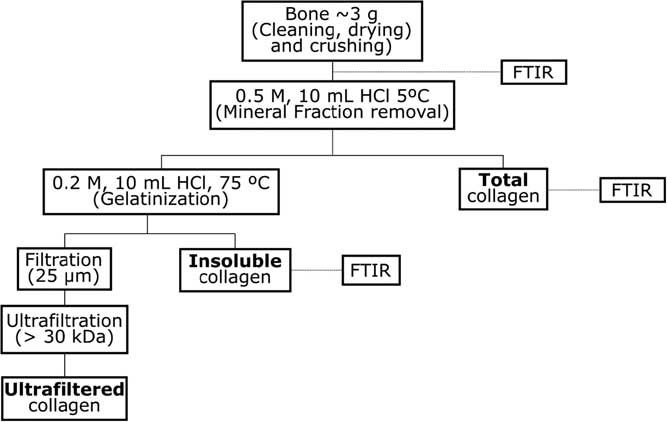
Bones were cleaned by removing their surface and were crushed in a mill. Three fractions were obtained as shown in Figure 2. The cleaned bones underwent a demineralization process with 10 mL HCl 0.5M at 5°C for 24 hr to remove contaminants. This demineralized portion was the total collagen fraction, from which the soluble and insoluble collagen fractions were obtained. If the samples were still bubbling after 24 hr with the addition of more HCl 0.5 M, they were left for a longer time at 5°C in order to demineralize them further. A low temperature was chosen to minimize the destruction of collagen. The alkali treatment step was avoided, to recover as much collagen as possible and because previous studies show that more accurate ages are obtained when only HCl pretreatment is applied (Fülöp et al. Reference Fülöp, Heinze, John and Rethemeyer2013). The gelatinization of the total collagen fraction was carried out adding 10 mL of 0.2M HCl at 75°C for 10–12 hr. In most of the samples, this treatment resulted in the separation of the acidified solution into two parts: at the top, a glue-like transparent colloidal mixture and a solid residue at the bottom.
Collagen-extraction process.
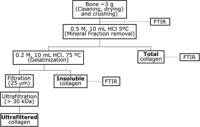
Then, the colloidal mixture was filtered through a coarse filter (20μm Millex) and an ultrafiltration step was carried out (Millipore Amicon) to remove the low molecular weight contaminants and degraded collagen. Finally, the sample was lyophilized. Both filters were cleaned before use. The procedure for cleaning the Amicon ultrafilters began with sonication in ultrapure water for 15 min. The filter was later filled with ultrapure water and centrifuged for 30 min. The previous process was repeated three times. Ultrafilters were kept in ultrapure water until the moment they were used. Millex filters were also cleaned three times by sonication in ultrapure water and kept wet in ultrapure water before use. The solid fraction (at the bottom of the container) with bone residue from the separation step described above had the insoluble collagen (Figure 2) was rinsed and received no further treatment.
For AMS 14C analyses, the carbon was converted to graphite in automatized graphitization equipment AGEIII (Ion Plus). Oxalic acid II (NIST SRM 4990C) was used as a primary standard, and phtallic acid (with no 14C) was used as a blank. The measured 14C/12C isotopic ratios were corrected for isotopic fractionation using the 13C/12C isotopic ratios measured in the accelerator. 14C age was expressed as Fm and calculation was performed as described in Solis et al. (Reference Solís, Chávez-Lomelí, Ortiz, Huerta, Andrade and Barrios2014). Calibration of the measured 14C ages was performed by OxCal v4.2 (Ramsey and Lee Reference Ramsey and Lee2013) using the IntCal13 calibration curve (Reimer et al. Reference Reimer, Bard, Bayliss, Beck, Blackwell, Bronk Ramsey, Buck, Cheng, Edwards, Friedrich, Grootes, Guilderson, Haflidason, Hajdas, Hatté, Heaton, Hoffmann, Hogg, Hughen, Kaiser, Kromer, Manning, Niu, Reimer, Richards, Scott, Southon, Staff, Turney and van der Plicht2013).
Infrared spectra were taken on a Nicolet 6700 FTIR spectrometer from Thermo Scientific using a single reflection ATR with a diamond crystal, at a resolution of 4 cm–1 and an accumulation of 100 scans in the spectral range of 4000 to 400 cm–1. Infrared spectra of raw bone and the three collagen fractions described in Figure 2 were measured for most of the samples.
RESULTS
The performance of the 14C ultrafiltered collagen preparation was tested through the analysis of the Fifth International Radiocarbon Intercomparison reference materials (VIRI F and VIRI H) supplied by Marian Scott from the University of Glasgow. The mean value and its estimates of uncertainty for samples prepared at LEMA and consensus values are shown in Table 2 for each standard. The results agree with the expected values.
Average values of Fm for collagen extracted from VIRI F bones (n = 22) and VIRI H bones (N = 15).

The collagen from the 54 Mexican bone samples was extracted using the method depicted in the upper part of Figure 2 and carried out to the last step whenever possible (ultrafiltered collagen). Afterwards, the ultrafiltered collagen was weighted to obtain the collagen yield in order to relate this factor to the %C, %N, and C/N values of each sample. The results are shown in Figure 3. For nitrogen content, it was observed that for collagen yields higher than 2%, nitrogen values stabilized at approximate values between 12% and 14%. The behavior of carbon was similar to that of nitrogen; for yields higher than 2%, carbon values became stable in the interval from 35% to 45%.
Scatter plot of wt% ultrafiltered collagen yield versus (a) %N, (b) %C, and (c) C/N ratio of raw bone; (d) scatter plot showing C/N ratio of raw bone versus C/N ratio of ultrafiltered collagen.

Figure 3c shows ultrafiltered collagen yield versus C/N ratio. The C/N ratio showed a wide range of values for collagen yields lower than 1%, but for higher yield values, C/N ratio was stabilized at around 2.9. From Figures 3a–3c it can be observed that samples from arid zones (black squares) had in generally higher collagen yields as compared to samples from the other zones.
Figure 3d shows the values of C/N ratios of 37 samples before (raw bone) and after ultrafiltered collagen extraction. Although initially some C/N ratio values were very high, the ultrafiltration process resulted in a decrease in these values until most of them were located within the 2.9–3.6 ratio limits (dotted line). This indicates that the ultrafiltration step releases external contamination by carbonates and that bones tend to recover the C/N ratio of a fresh bone. Short chain collagen and proteins were removed as well. From the results shown in graphs from Figure 3, we estimated that 84% of the samples had an ultrafiltered collagen yield lower than 3% and most of those samples (~74%) did not reach 1%. When collagen was obtained, the gelatinization and ultrafiltration step resulted in the elimination of some organic material. This led to a marked improvement in the C/N ratio, regardless of the C/N of the raw bone, but also in a decrease in the collagen yield.
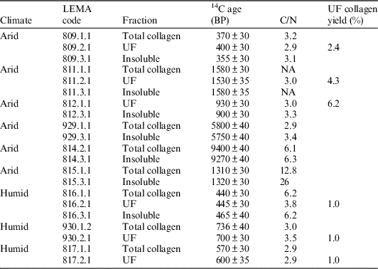
The ages obtained for either two or three of the collagen fractions from a set of samples were compared and are shown in Table 3. Most of them had a low C/N ratio for ultrafiltered collagen with a collagen recovery yield higher than 1%. For this set, the ages obtained were consistent and reproducible for all samples. Although the conventional approach is to use the ultrafiltered collagen, these results show that all fractions yielded similar ages.
Comparison of 14C ages for two or three different collagen fractions from two climate contexts. The C/N ratios and the UF collagen yield are also included.

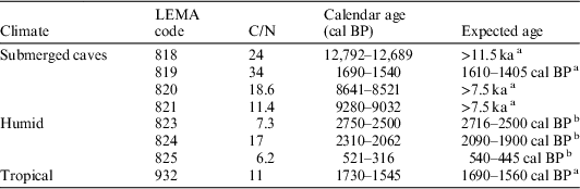
Bones with high C/N ratios are expected to have very low ultrafiltered collagen yields and they are normally not dated. However, based on the results shown above, samples from Table 3 could have been dated using either the total or insoluble collagen fraction. In Table 4 ages from a set of bones with high C/N are shown, and the collagen yield for them was lower than 1%. Dating was based only on the insoluble collagen fraction. In the case of bones analyzed from submerged caves, there are no previous 14C dates but their age could be estimated from the depth of their recovery site. Most of the remains were deposited in dry caves which became flooded afterwards. It is well established from hydrogeological data that the flooding occurred later because sea levels rose. Based on these facts it is possible to infer a limit for the expected age considering the dates of the floods or increases in sea levels (Blanchon and Saw Reference Blanchon and Shaw1995). In the case of bones from humid climates, ages were obtained through other collagen fractions.
Comparison between the 14C ages of insoluble collagen fraction of bones from humid, tropical and submerged caves and their expected ages.

a Origin of reference value by archaeological interpretation (stratigraphy or associated material).
b Origin of reference value from total collagen fraction.
Although it was not possible to obtain ultrafiltered collagen for the cases shown in Table 4, the ages obtained from the insoluble collagen fraction were consistent with those expected from archaeological criteria or with those obtained from total collagen fraction. This can be explained considering that for bones from flooded caves, collagen was degraded but not contaminated, or that the steps taken to obtain the insoluble fraction resulted in the removal of contamination by humic acids.
It is interesting to note that 14C dating of the insoluble collagen fraction of bones found in submerged caves, shown in Table 4, allowed for discrimination between samples belonging to different temporalities; sample LEMA 819 is clearly a pre-Columbian bone, while the others are more than 8000 yr old. The results show that preservation of bones does not depend on the time of deposit. Sample LEMA 819 is the youngest bone in Table 4 and it had the highest C/N ratio (C/N = 34). High C/N ratios (as expected) are reflecting the degradation state of bones and not necessarily the age of the bones.
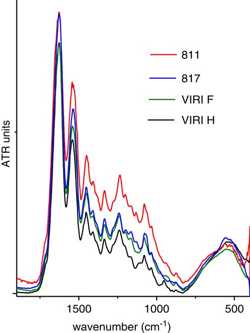
Infrared spectra were taken for most of the samples listed in Tables 3 and 4. The ultrafiltered fraction of collagen could not be obtained for all the samples analyzed, but was obtained for the standards VIRI F and VIRI H, as well as for the samples listed in Table 3. The infrared spectra of ultrafiltered collagen for both standards and samples 811 and 817 are shown in Figure 4. The spectra shown in Figure 4 are the typical spectrum of pure collagen, with no remnants of mineral moieties (Payne and Veis Reference Payne and Veis1988; Petibois et al. Reference Petibois, Gouspillou, Wehbe, Delage and Déléris2006).
Infrared spectra of ultrafiltered collagen from the standards VIRI F, VIRI H, samples 811 and 817 from 1900 to 400 cm–1.

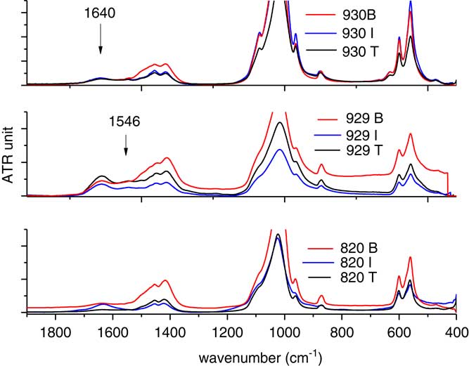
Representative spectra for the three fractions analyzed are displayed in Figure 5: raw bone (B), total collagen (T), and insoluble (I) fractions. The infrared data corroborated the efficiency of the demineralization process through the marked decrease in intensity of the mineral components of bone in the T and I fractions as compared to the raw bone spectrum. The spectra shown in Figure 5 belong to samples selected from three different microclimates: a submerged cave (820), an arid zone (929), and a humid area (930). Infrared results confirmed the presence of collagen in the D and R fractions, as evidenced by the amide I band at approximately 1640 cm–1, which is easily observed in all fractions and the amide II band at 1546 cm–1, which is much weaker in these spectra and sometimes not observed (Yizhaq Reference Yizhaq, Mintz, Cohen, Khalaily, Weiner and Boaretto2005). Thus, the presence of collagen can be assessed in all fractions analyzed: raw bone, total collagen and insoluble fractions. All infrared spectra displayed the characteristic bands of bone: ν3 bands for B-type carbonate substitution (CO3) in the hydroxyapatite matrix, at 1455 and 1416 cm–1 and phosphate (PO4 3−) modes, ν1 around 1020 cm–1 and ν4 and ν3 at 600 and 560 cm–1, respectively (Pucéat Reference Pucéat, Reynard and Lécuyer2004; Lebon Reference Lebon, Reiche, Bahain, Chadefaux, Moigne, Fröhlich, Sémah, Schwarcz and Falguères2010; Hollund Reference Hollund, Ariese, Fernandes, Jans and Kars2013). Thus, the low molecular weight collagen of the fractions not usually dated by 14C was used as dating material.
Infrared spectra from 1900 to 400 cm–1 of three bone fractions: raw bone (B), total collagen (T), and insoluble collagen (I), for representative samples from a humid zone (930), an arid zone (929), and a submerged cave (820).

DISCUSSION AND CONCLUSION
In this work, we performed 14C analyses of three collagen fractions extracted from bones. The Fm values obtained for VIRI F and VIRI H standards showed a good agreement with the expected values. This method was therefore applied to several bones from different archaeological sites of Mexico and processed at LEMA.
The obtained results showed that there is a relationship between the measured parameters and the burial place. As expected, bones from caves in arid zones were the best-preserved ones with the highest collagen yields and adequate C/N ratios. Moving toward the tropical areas with higher temperatures and humidity, as well as submerged caves, collagen degradation increased. In most cases, no collagen was obtained using the ultrafiltration process. When dating other collagen fractions, we found that these results were close to dates predicted with archaeological and hydrogeological data. Therefore, we consider that when no other more suitable datable fraction is present, these fractions can be used as an additional information source to establish sample age estimates, providing valuable information to archaeologists who otherwise would not have samples available for dating.
Although this work demonstrates that ultrafiltration significantly reduces the C/N ratio, in many cases the obtained ages do not differ significantly from those obtained using the other fractions, total or insoluble collagen.
In the case of severely degraded bones, such as those coming from the submerged caves analyzed in this study, very little collagen was present in the original bone, apparently consisting of low molecular weight collagen, so no ultrafiltered collagen could be obtained. On the other hand, if additional contextual information is carefully considered, we have found that if there is enough collagen in the insoluble collagen fraction, it can be dated. This was the case for bones from submerged caves, as their obtained ages were consistent with expected ages from archaeological and hydrogeological evidence. Therefore, the insoluble fraction is well-suited for the absolute dating of bones even though there was no ultrafiltered collagen for conventional 14C dating.
The standard collagen extraction method is not applicable to highly degraded bones, such as those found in submerged caves. The analysis of the total collagen and insoluble fractions obtained during the collagen extraction process seems to indicate that even though collagen integrity parameters are out of range, it is possible to characterize the fractions to decide if they can be used in the dating process. This approach can be useful for unique bones like those found in submerged caves and other archaeological Mexican sites. However, more studies on collagen degradation and diagenesis on these bones are necessary before a final statement. A bigger number of samples as well as more information about the burial conditions, will help to understand more about the factors that determine bone degradation in Mexican archaeological zones.
ACKNOWLEDGMENTS
We thank A. Huerta for operation of the AMS system and Sergio Martínez for technical support. We acknowledge the many archaeologists from INAH who provided the samples. We also acknowledge John D. Powers and two anonymous reviewers who helped us to improve the manuscript with their helpful comments. This work received partial support from projects PAPIIT IG100216, CONACYT through projects 232718, 264245, 271802, 205317, and 280769.