Levoatriocardinal vein is a rare vascular abnormality generally encountered in left-sided obstructive lesions, and is often associated with partial anomalous pulmonary venous drainage.Reference Bernstein, Moore, Stanger and Silverman 1 This association is a real diagnostic challenge. We report two cases where non-invasive imaging was decisive for the definitive diagnosis and for orienting to the best therapeutic solution.
Case 1
A full-term newborn weighing 3 kg, prenatally diagnosed with hypoplastic left heart syndrome, was transferred to our department immediately after birth on prostaglandin infusion for further management. At clinical evaluation, transcapillary oxygen saturation was 94% and mild tachypnoea was present, suggesting imbalance of the pulmonary/systemic flow ratio with pulmonary overflow. Diffuse radiological signs of pulmonary engorgement were also present during chest radiography.
Surprisingly, echocardiography showed evidence of mitral and aortic atresia with intact interatrial septum and the presence of a tortuous levoatriocardinal vein, decompressing the left atrium and draining into the innominate vein with a mild aliasing in its middle tract observed during the color flow Doppler examination (Supplementary movie 1). The ascending aorta was severely hypoplastic, the right brachiocephalic artery and the left carotid artery arose from the horizontal aortic arch, and the left subclavian artery arose from the descending aorta distally to the large patent arterial duct; however, the drainage of the pulmonary veins was poorly visualised.
The newborn rapidly developed metabolic acidosis and multiorgan system dysfunction, despite usual pharmacological and ventilatory strategies. Therefore, in order to elucidate the pulmonary vein drainage and the levoatriocardinal vein course, as cardiac catheterisation and MRI were regarded as hazardous in this critically ill patient, the patient underwent non-gated cardiac CT with the following scan parameters – rotation time 400 ms, pitch 0.984, slice thickness 32×0.625 mm, voltage 80 kV, and current varied during acquisition – and the following injection parameters – 2 ml/kg of contrast medium (Iopamerol 300 mg/ml; Schering SA, Berlin, Germany), followed by 5 ml saline solution at a flow rate of 1 ml/second. The acquisition lasted 1.4 seconds and the examination lasted ~15 minutes in total. The effective irradiation dose was estimated to be 1.6 mSv.
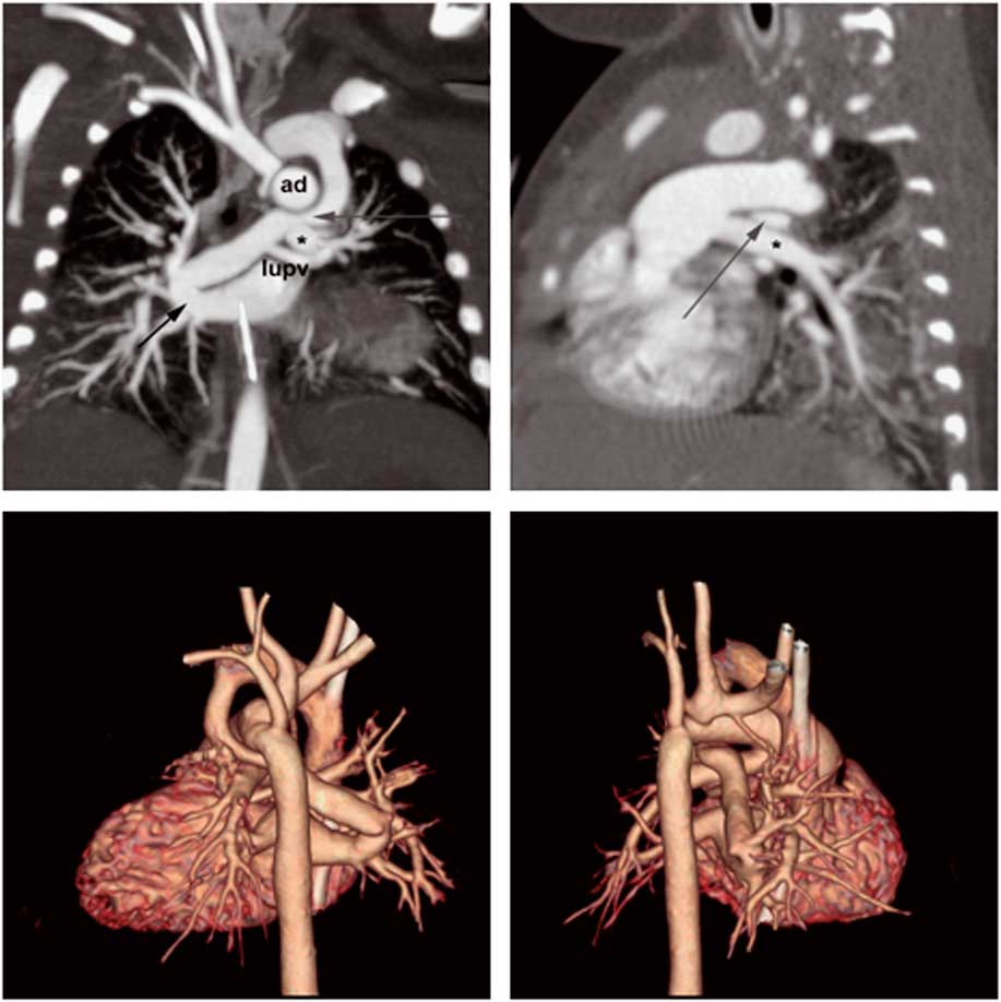
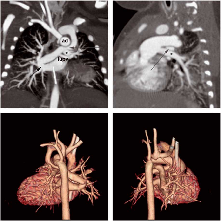
The cardiac CT confirmed the diagnosis of an anomalous tortuous levoatriocardinal vein emerging from the right lower pulmonary vein, ascending into the mediastinum, passing anterior to the left main bronchus, and mildly compressed between the arterial duct and the left pulmonary artery to finally drain into the innominate vein. The left upper pulmonary vein drained directly into the levoatriocardinal vein. The ascending aorta was severely hypoplastic. The horizontal tract of the aortic arch supplying the right brachiocephalic trunk and the left carotid artery seemed to rise directly from the arterial duct. The left subclavian artery arose from the descending aorta distally to the large patent arterial duct (Fig 1; Supplementary movie 2).
Anterior (left upper panel) and sagittal (right upper panel) views of the CT scan: the short black arrow indicates the origin of the levoatriocardinal vein emerging from the left atrium and receiving the drainage of the left upper pulmonary vein (lupv). The vessel was mildly compressed (long grey arrow) between the arterial duct (ad) and the left pulmonary artery (asterisk); three-dimensional volume rendering from a posterior view (left inferior panel) and sagittal view (right inferior panel).
As the high transcapillary oxygen saturation (94%) was suggestive of pulmonary overflow and adequate blood mixing through the levoatriocardinal vein, percutaneous blade and balloon atrial septostomy was considered ineffective in this clinical setting. We opted for a rapid 2-stage strategyReference Gomide, Furci and Mimic 2 consisting of immediate bilateral pulmonary banding associated with prostaglandin infusion to maintain the patency of the arterial duct to re-balance the pulmonary/systemic flow ratio and improve end-organ perfusion without the need to implant a stent. This first step served as a bridge to the Norwood operation, which was performed after 2 weeks, associated with interatrial septectomy and closure of the levoatriocardinal vein distal to the left upper pulmonary vein drainage. The postoperative period was unremarkable.
Case 2
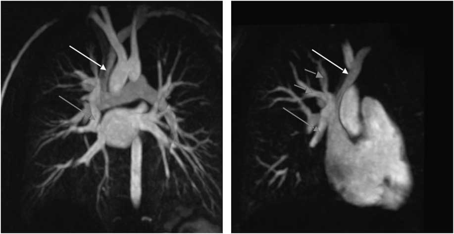
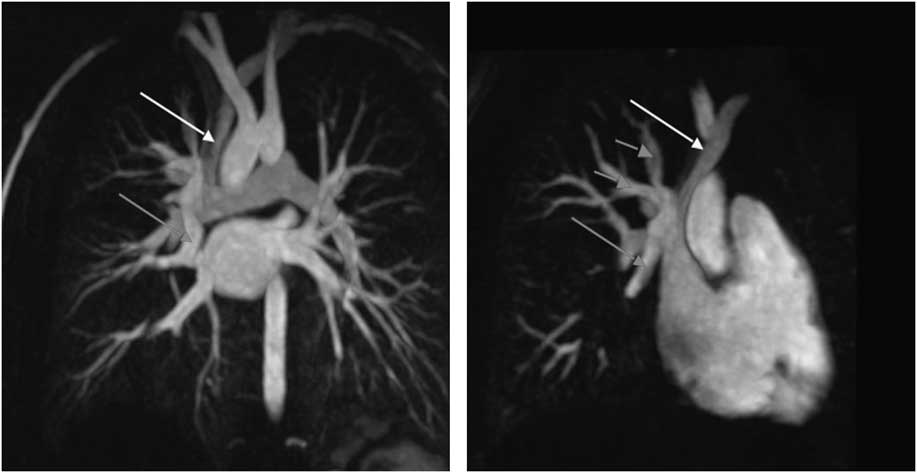
An 8-year-old boy was referred to our department because of progressing right heart dilatation during echocardiography evaluation. In his past, the patient had undergone aortic de-coarctation associated with ventricular and atrial septal defects closure in the neonatal period. Cardiac magnetic resonance angiography – repetition time/echo time 4.5/2 ms; image matrix size from 128 to 190×256; field of view of 180–400 mm and 1–3 mm for slice thickness, peripheral injection of 0.1 mmol/kg of gadolinium – revealed the presence of a large levoatriocardinal vein connecting the middle segment of the middle right pulmonary vein with the right superior caval vein (Fig 2). Moreover, two small pulmonary veins drained the pulmonary blood return of the right upper pulmonary lobe directly into the right superior caval vein distal to the levoatriocardinal vein drainage. Indexed right end-diastolic volume was 123 ml/m2. Cardiac catheterisation confirmed the anatomical diagnosis, finding a pulmonary/systemic flow ratio of 2.7. The patient underwent surgical closure of the levoatriocardinal vein re-directing the right upper pulmonary return to the left atrium using a pericardial patch. The postoperative course was uneventful, and 1 month after surgery the patient was on oral anticoagulant treatment.
Gadolinium-enhanced angiography: the red arrows indicate the levoatriocardinal vein emerging from the middle segment of the middle pulmonary vein and draining into the right superior caval vein (white arrow). Note the less-intense signal of the right superior caval vein compared with the signal of the left atrium. Short red arrows indicate two pulmonary veins originating from the right superior pulmonary lobe and draining into the right superior caval vein distal to the levoatriocardinal vein drainage.
Discussion
The levoatriocardinal vein is an abnormal residual connection between the pulmonary venous system and the cardinal (systemic) venous system through the splanchnic plexus of the embryonic foregut. This abnormality is supposed to serve as a decompression pathway of the pulmonary venous return in case of left-sided obstructions.Reference Bernstein, Moore, Stanger and Silverman 1 In fact, it is mostly found associated with cor triatriatum, mitral and aortic atresia, and aortic coarctation; however, the levoatriocardinal vein may also be an isolated finding.Reference Cullen, Breen, Rihal, Simari and Ammash 3
A wide spectrum of origin and drainage of the levoatriocardinal vein has been previously observed.Reference Bernstein, Moore, Stanger and Silverman 1 In this report, we describe a levoatriocardinal vein emerging from the right lower pulmonary vein crossing the mediastinum to drain into the innominate vein in a newborn with hypoplastic left heart syndrome with intact interatrial septum; and a levoatriocardinal vein egressing from the right middle pulmonary vein in a patient operated for aortic coarctation in the neonatal period. To the best of our knowledge, these two anatomical courses of the levoatriocardinal veins have not been previously described.
Color flow Doppler mapping and two-dimensional echocardiography are the first-level examinations to recognise and describe the levoatriocardinal vein. This may masquerade as the vertical vein that is encountered in the supracardiac total anomalous pulmonary venous drainage. When the latter is suspected, left-sided obstructive lesions should always be excluded during the echocardiographic scan. Conversely, the diagnosis of left-sided obstructive lesions should always prompt the search for the levoatriocardinal vein, as this abnormality may be a cause of misdiagnosed left-to-right shunt, as occurred in Case 2. Moreover, when a levoatriocardinal vein is recognised, the anatomical course of the pulmonary veins should always be examined, because partial anomalous pulmonary vein drainage is frequently associated.Reference Bernstein, Moore, Stanger and Silverman 1 , Reference Cullen, Breen, Rihal, Simari and Ammash 3
As occurred in Case 2, in order to elucidate pulmonary and systemic vein abnormalities, we normally prefer cardiac MRI; however, as occurred in Case 1, we recur to CT imaging in critical patients because the scanning time is lesser. Moreover, CT has higher spatial resolution compared with magnetic resonance and this might be crucial in newborns. In both our cases, the second-level non-invasive imaging, based on the clinical conditions of the patients, allowed to assert the definitive diagnosis, providing important information for clinical management.
In patients with hypoplastic left heart syndrome, we normally prefer the hybrid approach as the first choice for Stage 1 palliation; however, in Case 1, we speculated that arterial duct stenting would have exacerbated the levoatriocardinal vein compression and that the risk of retrograde coarctation would not have been negligible in the follow-up when considering the aortic anatomy revealed by the CT scan.Reference Vance 4 Therefore, we opted for a rapid 2-stage Norwood strategy, associated with interatrial septectomy and section of the levoatriocardinal vein distal to the left upper pulmonary vein drainage.
In isolated levoatriocardinal veins, percutaneous embolisation of the vein using Amplatzer devices is a widely adopted solution;Reference Cullen, Breen, Rihal, Simari and Ammash 3 however, in Case 2, the percutaneous occlusion of the levoatriocardinal vein emerging from the medium right pulmonary vein would have left the superior right pulmonary veins draining into the superior caval vein. For this reason, we preferred the surgical solution.
In conclusion, these two cases highlight that in patients with left-sided obstructive lesions the existence of the levoatriocardinal vein should always be kept in mind, that the levoatriocardinal veins are often associated with abnormal pulmonary venous drainage, and that cardiac CT and MRI are a pivotal resource to elucidate this rare and complex association, both able to provide anatomical information useful to tailor the best therapeutic strategy.
Acknowledgement
The authors thank Damien Kenny (Rush University, Chicago, Illinois, United States of America) for providing English revision.
Financial Support
This research received no specific grant from any funding agency, commercial, or not-for-profit sectors.
Conflicts of Interest
None.
Supplementary Material
For supplementary material referred to in this article, please visit http://dx.doi.org/10.1017/S1047951115002425