Introduction
Benign tumours affecting the external nose encompass a diverse range of pathologies, presenting diagnostic and therapeutic challenges in clinical practice. These benign nasal tumours pose difficulties due to their various locations, size and adverse cosmetic effects on nasal aesthetics. Unlike other sinonasal pathologies, most external nasal tumours primarily manifest as visible mass lesions on the nasal tip and the dorsum. Therefore, the surgeon needs to simultaneously achieve better nasal aesthetics and tumour removal. These goals may be challenging in cases where tumours exhibit unclear boundaries or involve extensive surrounding tissues, occasionally necessitating the intentional retention of residual tumour components to achieve better cosmetic outcomes.
Due to the scarcity and diverse nature of benign tumours in the external nasal region, previous case reports are mostly anecdotal. Furthermore, to the best of our knowledge, no cohort studies have conducted a comprehensive analysis of benign tumours of the external nose. A comprehensive understanding of benign external tumours of the nose is difficult due to the relative paucity of literature. Therefore, this retrospective cohort study presents our 18 years of surgical experience in treating benign external tumours affecting the nasal region to provide useful information for diagnosing and managing such tumours. In the present study, the spectrum of these lesions is described, the surgical methods used for excision and subsequent reconstruction are outlined, treatment outcomes are evaluated and factors correlated with tumour recurrence are analysed.
Materials and methods
Patients selection and data collection
This retrospective study includes 32 patients who underwent surgical intervention for benign tumours of the external nose at Asan Medical Center between November 2006 and March 2023. A single senior surgeon (Y.J.J.) performed all surgeries for complete or partial resection with cosmetic maintenance. Patients were excluded from the analysis if the tumours simultaneously involved other areas of the external face, such as the cheek (two cases), if they lacked final pathology results (one case), or if the post-operative pathological report demonstrated malignancy (three cases). A benign inflammatory lesion (two cases) was not categorised as a benign tumour. A total of 24 patients were included in final analysis. The Institutional Review Board of Asan Medical Center approved this study (No. S2023-2738-0001).
Demographic information including patient age and gender, and tumour characteristics such as location and size, were extracted from electronic medical records. Slides of all surgical specimens were stained with haematoxylin and eosin (H&E) and underwent comprehensive immunohistochemical review to assess morphology according to established diagnostic criteria for the pathology. Details regarding the surgical procedures performed, including the approach used for mass removal, the extent of excision, reconstruction techniques and any intentional retention of residual tumours, were analysed.
Surgical techniques
Surgeries were performed under general or local anesthesia. The incisional approach was tailored to the tumour site and size, with the open rhinoplasty approach being the most common. Although the primary aim was complete resection, in cases where clear demarcation of the tumour was challenging, partial resection was performed using curettage or debridement using a microdebrider. Intentional retention of residual tumours was occasionally unavoidable for aesthetic considerations. In addition, some partial skin excisions were performed to manage skin redundancy overlying the tumour. Auricular conchal cartilage grafting was used for alar reconstruction when necessary, and diced septal cartilage was utilised to camouflage palpable deformities of the nasal dorsum.
Follow-up and outcome assessment
Post-operative patient data were collected to assess treatment outcomes, detect tumour recurrence and monitor potential complications. Recurrence was evaluated through physical examination via inspection or palpation and imaging studies, particularly paranasal sinus MRI scans. Any instances of tumour recurrence and the consistency of surgical approaches with the primary procedures were systematically recorded and analysed.
Statistical analysis
Descriptive statistics were used to summarise patient demographics, tumour characteristics, surgical interventions and treatment outcomes. Continuous variables are presented as mean ± standard deviation (SD), while categorical variables are presented as frequency. Statistical analyses were performed using SPSS 27.0 (SPSS Inc., Chicago, IL, USA). Associations between categorical variables were assessed using the chi-squared test. All tests were two-sided, and statistical significance was defined as p less than 0.05.
Results and analysis
Patient characteristics
In this retrospective 18-year study focusing on the surgical management of benign external nasal tumours, 24 patients were included. The cohort consisted of 10 males and 14 females, with a mean age of 32.71 ± 19.18 years (range, 3 months to 65 years). Tumour distribution showed comparable incidence on the right side (7 cases), left side (9 cases) and midline (8 cases). The most common tumour location was on the nasal dorsum (11 cases), followed by the nasal root (4 cases), nasal ala (4 cases), nasal tip (3 cases) and nostril (2 cases) (Figure 1). Patients self-reported an average duration of 3.67 ± 3.06 months between identification of the mass and their visit to the clinic. Five individuals reported delays of more than 4 years, extending up to 10 years before seeking medical attention.
Pre-operative external photographs showing various regions of the external nose, including nasal root and dorsum (A), nasal columella (B), nasal ala (C) and nostril (D).

Surgical procedures and tumour characteristics
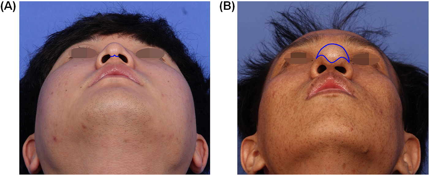
Mass removal in most cases (17 patients, 70.8 per cent) involved a conventional open rhinoplasty procedure, utilising trans-columellar inverted V and marginal incisions (Figure 2A). The other seven patients, in whom the tumour affected the nasal tip, nostril or nasal ala, underwent tumour excision through direct incision over the lesion (Figure 2B). In three patients where extensive skin redundancy coincided with the underlying mass and in one case with a mass in the alar area requiring treatment for nostril symmetry, partial skin excision was performed.
Representative pre-operative external photographs displaying the conventional open rhinoplasty approach (A) and direct incision on the lesion (B), both marked by a blue incision line in the pre-operative basal view.

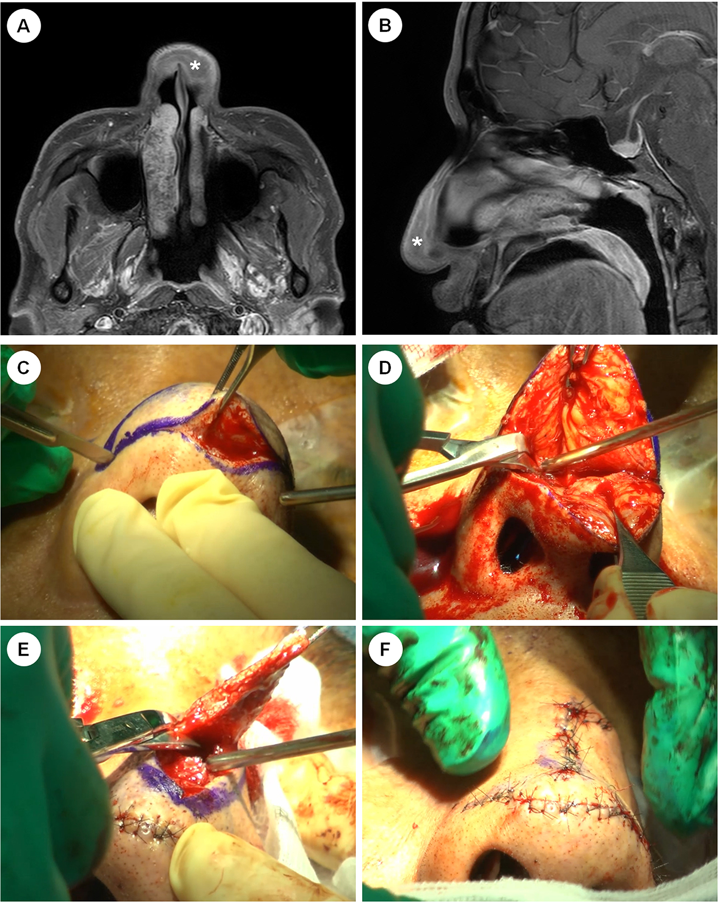
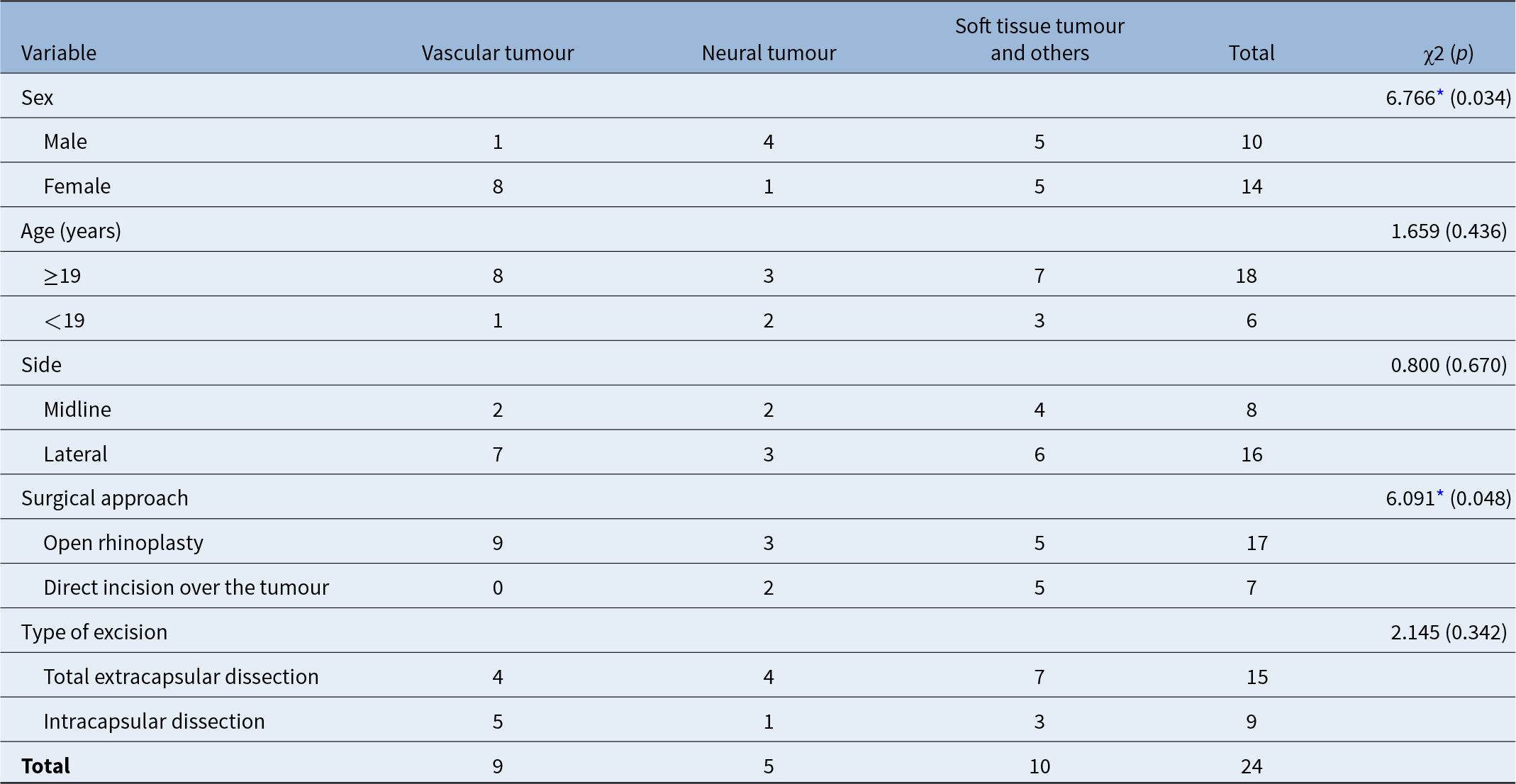
Figure 3 shows intra-operative findings from a representative case involving direct incision over the tumour and partial skin excision. Partial excision of the skin was performed to control the redundancy of the overlying skin and subsequent deformity resulting from the tissue-expander-like effect of the tumour. Furthermore, four patients were treated with deliberate partial tumour resection because of ill-defined borders and to maintain aesthetic outcomes. Alar reconstruction was performed using auricular conchal cartilage in three patients. Crushed septal cartilage was used as a dorsal onlay graft to camouflage nasal bone defects in three other patients, resulting in enhanced cosmetic appearance. Multiple different tumour pathologies were identified, with vascular tumours (nine cases) and neural tumours (five cases, Table 1) the most common types. Patients with vascular tumours were more likely to be female and be managed through an open rhinoplasty approach (Table 2).
Pre-operative axial (A) and sagittal (B) images on T1-enhanced paranasal MRI, with the asterisk indicating the tumoir location. Intra-operative photographs depict the following stages: (C) direct incision on the lesion, (D) meticulous dissection for complete tumour excision, (E) partial excision of redundant skin caused by the tissue-expander-like effect of the tumour and (F) primary skin suture performed at the end of the operation.

Clinical characteristics and surgical details of patients (n = 24)

Abbreviations: CT, computed tomography.
Associations between patient characteristics and tumour pathological types along with surgical technique

Note: Values are presented as number.
* p<0.05
Treatment outcomes and recurrence
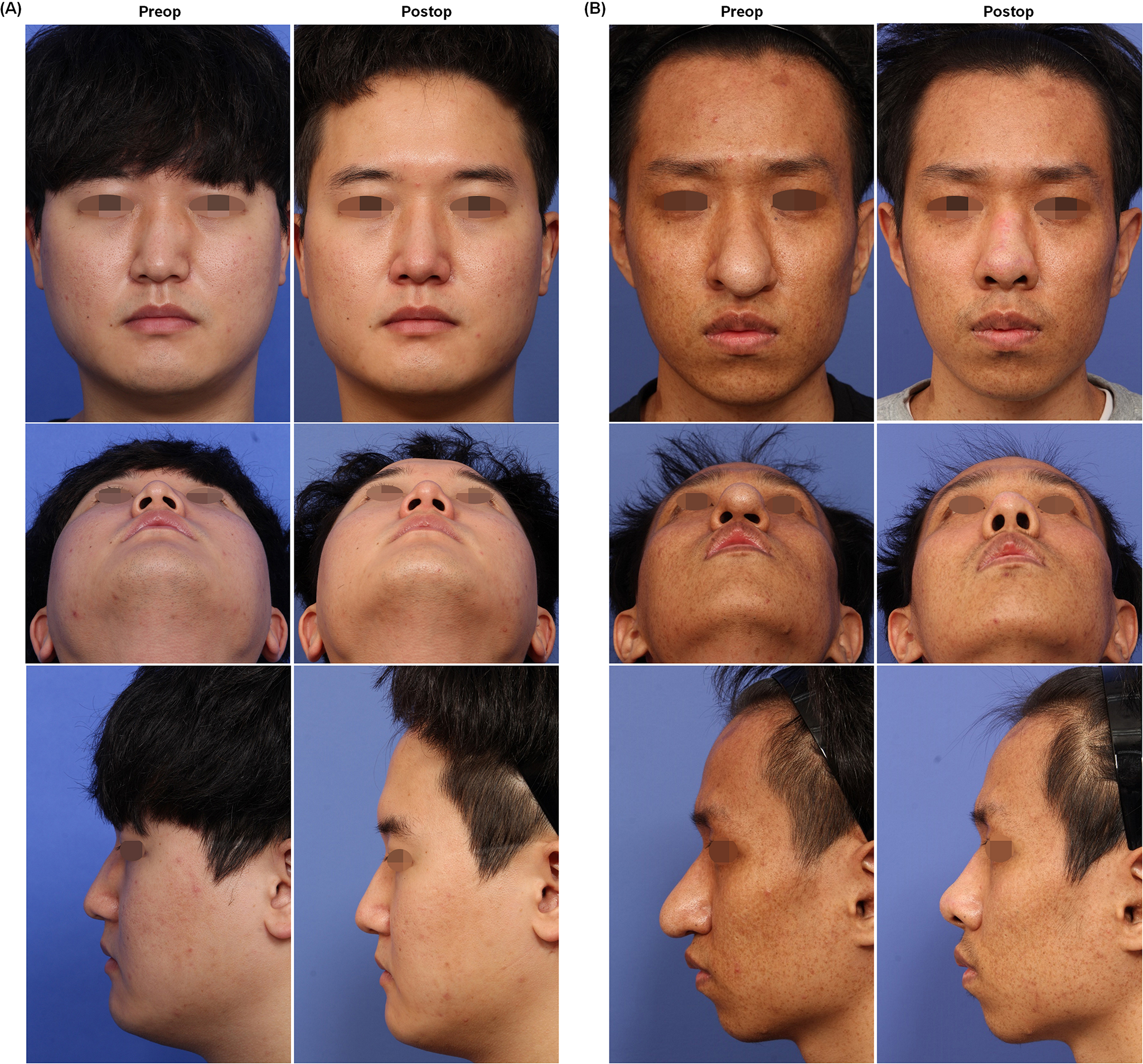
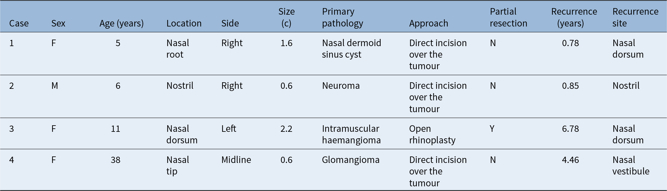
The average follow-up time was 2.19 ± 4.02 years after the index resection. Twenty patients (83.3 per cent) showed no recurrence or major complications. Importantly, these patients expressed subjective satisfaction with the aesthetic outcomes (Figure 4). However, tumour recurrence was detected in four patients, identified via physical examination as a visible mass in three cases and through post-operative paranasal sinus MRI in one case. Recurrence occurred at an average interval of 3.21 ± 2.93 years following initial surgery (Table 3). Among the recurrences, three cases occurred in paediatric patients and, only one case in an adult patient. All three paediatric patients were diagnosed with the lesion at birth or within their first year of life. The histologic diagnoses for these patients were haemangioma, nasal dermoid sinus cyst and neuroma. Among the four recurrences, only one was initially treated with intentional partial excision. Complete excision was attempted for the other three patients. Revision surgeries were performed using the same approach, open rhinoplasty versus direct incision over the tumour, as the primary surgery in all cases.
Representative photographs exhibiting post-operative nasal contour enhancements at one year. (A) Open rhinoplasty approach. (B) Direct incision over the tumour.

Discussion
Benign tumours of the external nose are a rare and challenging condition. The primary objective in managing this uncommon clinical entity is to effectively remove the tumour while preserving both the functional aspects and aesthetic appearance of the nose. The present study provides a comprehensive overview of various histologic diagnoses associated with benign tumours of the external nose, covering a spectrum of types including vascular (haemangioma, vascular malformation), neural (neurofibroma, schwannoma), dermoid cysts (nasal dermoid sinus cyst), soft tissue variants (nodular fasciitis, granuloma, angioleiomyoma trichofolliculoma) and other benign tumours (glomangioma, xanthogranuloma). The nasal dorsum was the most commonly affected anatomical site, accounting for 45.8 per cent of the 24 cases, although tumours can arise at different locations on the external nose such as the nasal root, tip, alar, nostril or columella.Reference Jeon, Kim, Lee and Lim1 In the present study, vascular tumours of the nose were more common in females, in agreement with previous reports.Reference Waner, Kastenbaum and Scherer2
The most common chief complaint was a mass-like lesion or swelling on the external nose. Symptoms and signs associated with these tumours often vary based on the specific location of the growth. Nasal obstruction, facial pain and epistaxis were previously noted as common accompanying non-specific nasal symptoms.Reference Shakrawal, Chouhan and Solanki3 Although a pre-operative needle biopsy could offer a definitive histological diagnosis, not all enrolled patients underwent this procedure due to the suggestive nature of benign pathology based on the history and physical characteristics of the lesion. This was particularly influenced by the indicative benign histology revealed in radiological studies including well-defined margins, smooth and regular shape, absence of irregularities or invasive features and homogeneous density or signal intensity.Reference Tatekawa, Shimono, Ohsawa, Doishita, Sakamoto and Miki4, Reference Lemmerling, Moerman, Govaere, Praet, Kunnen and Vermeersch5 Radiological examinations, notably computed tomography (CT) scans and MRI, serve as crucial imaging modalities to determine the size and extent of a tumour and are essential for planning subsequent surgical interventions.
The existing literature regarding benign tumours of the external nose has primarily consisted of anecdotal case reports, contributing to a lack of comprehensive information on the incidence and characteristics of these tumours compared with other sinonasal pathologies. In our study, haemangioma was the most prevalent type, followed by neural tumours such as neurofibroma and schwannoma. Haemangiomas, predominantly observed in paediatric patients, are estimated to occur in 4–10 per cent of infants and can manifest in adults.Reference Waner, Kastenbaum and Scherer2 In adults, approximately 60 per cent of haemangiomas localise in the head and neck region, with around 15.8 per cent affecting the nose.Reference Waner, North, Scherer, Frieden, Waner and MC6 Schwannomas and neurofibromas are the predominant types of peripheral nerve sheath tumours. Roughly 25–45 per cent of schwannomas occurring outside the cranial cavity are found in the head and neck area, with approximately 4 per cent originating from the sinonasal tract.Reference Biswas, Marnane, Mal and Baldwin7, Reference Colreavy, Lacy, Hughes, Bouchier-Hayes, Brennan and O’Dwyer8 Neural tumours on the external nose involving schwannomas and neurofibromas are exceptionally rare, with only a few documented cases reported in the English literature.Reference Jeon, Kim, Lee and Lim1, Reference Lemmerling, Moerman, Govaere, Praet, Kunnen and Vermeersch5, Reference Yoon, Kim, Oh and Lee9–Reference Mohan, Krishna and Ramakristnan15
Surgical excision remains the primary modality of treatment. In the present study, most individuals sought medical attention promptly within the first year upon noticing changes in the shape of their noses. However, a subset of patients delayed seeking hospital care, with five individuals presenting to the clinic 4–10 years after initially detecting changes in their nose’s appearance, consistent with previously reported cases.Reference Jeon, Kim, Lee and Lim1, Reference Kim12 Benign lesions that are small, asymptomatic and do not cause any cosmetic or functional concerns may be managed with regular monitoring.
In the present study, the intricate location of the tumours posed a significant challenge in determining the optimal surgical approach to achieve complete resection while ensuring favourable aesthetic outcomes. First, the open rhinoplasty approach showed distinct advantages when managing tumours located on the nasal dorsum or sidewall. This approach enabled successful lesion excision while preserving the upper and lower lateral alar cartilages, facilitating extensive surgical exposure and direct access for potential reconstructive procedures when necessary. In addition, direct incision over the tumour was used in cases where the tumour involved the nasal columella and nostril margin. Second, when tumours affect nasal structures, such as altering nostril shapes or alar contours, skin excision plays a crucial role in establishing and preserving nostril or alar symmetry. Furthermore, partial excision of the redundant skin caused by the expansile effect of the tumour was crucial for managing skin redundancy and subsequent external nose deformity. Notably, only removing the tumour beneath redundant skin might not adequately address resulting nasal deformities. Conversely, deliberate partial resection of benign tumours on the external nose, if not significantly affecting aesthetics, is occasionally suggested as a practical option rather than complete surgical removal. The cases reported herein provide valuable insights into surgical techniques tailored for benign external nasal tumours, considering their multifaceted characteristics.
The surgical management of benign tumours on the external nose necessitates meticulous planning to achieve optimal aesthetic and functional outcomes. The timing of nasal reconstruction remains controversial. Among three patients who underwent single-stage reconstruction using autologous auricular cartilage for alar grafting, one patient experienced recurrence 10 months after the operation (as shown in the second case in Table 3). Consideration for a two-stage operation is essential when a high risk of recurrence or potential malignant transformation exists. Among the 12 patients with benign tumours located on the nasal dorsum, 3 patients required crushed autologous cartilage post-tumour removal to manage the resulting aesthetic defect. Significantly, none of these patients experienced a recurrence. Therefore, when managing benign tumours in the external nose, cosmetic outcomes should be considered, especially the necessity for reconstructive procedures. The current study cases showed favourable subjective aesthetic outcomes from the single-stage reconstruction of tip and dorsal aesthetics. If the benign tumour was excised with clear resection margins, a two-stage operation may not be mandatory, avoiding patient inconvenience associated with delayed reconstruction.
Case series of revision surgery

The present study had a few limitations. Retrospective studies are inherently susceptible to bias, and data collection relies on electronic medical records, potentially leading to missing or incomplete information. Furthermore, the relatively small number of patients included in this study reflects the rarity of benign tumours affecting the external nose, limiting the generalisability of the results and highlighting the challenges in assembling larger patient cohorts. In addition, the authors acknowledged instances of patients being lost to long-term follow-up. This limitation hinders the ability to assess the durability of treatment outcomes, including recurrence rates or complications over an extended period.
• Benign tumours of the external nose have various pathological types
• Haemangiomas are the most common type, frequently found in paediatric patients and more prevalent in females
• Open rhinoplasty approach is preferred for tumour resection as it provides extensive exposure and direct access to potential reconstructive procedures
• Direct incision over the tumour is favoured for tumours involving the nasal columella or nostril margin
• Intentional partial tumour removal or partial skin excision can be considered for aesthetic outcomes
• A tailored approach and individualised treatment strategies are important for preserving nasal aesthetics while managing benign external nasal tumours
Conclusion
In this retrospective cohort study, different pathologies in benign nasal tumours were described, with haemangioma as the predominant type. The open rhinoplasty approach can be used to effectively remove the tumour, preserve nasal aesthetics and allow for structural grafting when necessary. The deliberate choice of partial tumour removal or partial skin excision in specific cases underscores the delicate balance between complete excision and maintaining aesthetic integrity. Our findings emphasise the need for individualised treatment strategies tailored to the pathology and anatomical location of these rare external nasal tumours.
Funding statement
This work was supported by the Basic Science Research Program through the National Research Foundation of Korea (NRF) funded by the Ministry of Education [Grant numbers: 2022R1C1C1004860 to YJJ].
Competing interests
The authors declared no potential conflicts of interest with respect to the research, authorship and publication of this article.
Ethical standards
This study was approved by the Institutional Review Board of Asan Medical Center (No. S2023-2738-0001).