A portosystemic venous shunt is associated with polysplenia syndrome after a Fontan-type operation. Reference Ikeda, Aotsuka, Nakajima and Sawada1 After Fontan-type surgery, patients with portosystemic venous shunt present with hyperammonaemia and portal-systemic encephalopathy on a long-term basis. Reference Koteda, Suda, Kishimoto and Iemura2,Reference Nagata, Sakamoto and Fukuoka3 Coil embolisation or device closure (such as vascular plugs) has been reported to treat portosystemic venous shunts. Reference Knirsch, Benz and Bühr4 However, some of these shunts have tortuous and complicated morphologies. Moreover, devising coils or placing them in favourable positions could be challenging.
Herein, we report successful coil embolisation of tortuous portosystemic venous shunts using a steerable microcatheter in a patient with polysplenia after a Fontan-type operation.
Case
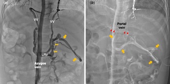
A 4-year-old girl was diagnosed with polysplenia syndrome, functional single right ventricle, pulmonary stenosis, and interrupted inferior caval vein with azygos continuation. She underwent total cavopulmonary shunt at 1 year of age. Since angiography performed before this surgery did not indicate a lower azygos venogram or superior mesenteric arteriogram, the existence of a portosystemic venous shunt was unclear. Cardiac catheterisation at the age of 2 years revealed a portosystemic venous shunt (Fig 1a and b), originating from the azygos vein via the left renal vein. A large number of aortopulmonary shunts were observed. We prioritised coil embolisation of the aortopulmonary shunt over closure of the portosystemic shunt before Fontan completion.
( a ) Azygos venogram (early phase) showing multiple veno-veno collateral veins and a portosystemic shunt (arrow). ( b ) Azygos venogram (late phase) shows a portosystemic shunt (arrow) and a portal vein (arrow head). VV: veno-veno collateral veins; LRV: left renal vein.

The patient underwent fenestrated Fontan-type surgery at 2 years and 9 months of age. Cardiac catheterisation performed 5 months after Fontan completion demonstrated an unchanged portosystemic shunt with stagnated bidirectional blood flow. Portal vein pressure was measured before and after balloon occlusion (both, 16 mm Hg). Serum ammonia and total bile acid levels were 84 μg/dL and 2.3 μmol/dL, respectively. We attempted coil embolisation of the portosystemic shunt a year later. However, it was unsuccessful because of the difficulty in delivering a microcatheter through the azygos vein to the distal portion of the portosystemic shunt. Pressures measured at this catheter session were as follows: azygos venous pressure 10 mmHg and hepatic venous pressure 11 mmHg.
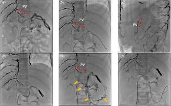
Seven months later, when the patient was aged 4 years, we reattempted coil embolisation of the portosystemic shunt using a 2.4-Fr steerable microcatheter (Leonis Mova, Sumitomo Bakelite, Tokyo, Japan). The patient’s body weight, height, and saturation at this point were 14.2 kg, 100.2 cm, and 91% (at room air), respectively. Serum ammonia and total bile acid levels before coil closure were 73 μg/dL and 27.3 μmol/dL, respectively. First, we performed angiography of the portosystemic venous shunt using a 5-Fr wedge pressure catheter with balloon inflation. It revealed a long and tortuous shunt heading toward the portal vein (Fig 2a). We then advanced a steerable microcatheter with a 0.016-inch guidewire (Meister®, Asahi Intec, Nagoya, Japan) to the distal portion of the portosystemic shunt (Fig 2b and c). After a selective angiogram confirmed its exact position inside the shunt, we retracted the microcatheter to the mid-portion of the shunt. Then, occlusion was achieved using detachable coils (MicrusFrame®S, Deltafill®, Galaxy G3, Johnson&Johnson NJ, USA; Azur 18, Azur CX, Terumo, Tokyo, Japan) (Fig 2d). Left renal venography after coil embolisation revealed a long and tortuous portosystemic shunt (Fig 2e). The steerable microcatheter was advanced distally to the shunt, and coil embolisation was achieved (Fig 2f). After coil embolisation of the portosystemic shunt, serum ammonia and total bile acid levels decreased to 32 μg/dL and 9.1 μmol/dL, respectively.
( a ) Left renal venogram with an inflated balloon of wedge pressure catheter showing a portosystemic shunt and a portal vein (arrow head). ( b , c ) Fluoroscopy shows a microcatheter with its tip reaching the portal vein (arrow head). B: frontal view. C: lateral view. ( d ) Fluoroscopy showing successful coil embolisation of the portosystemic shunt. ( e ) Left renal venography after coil embolisation shows another tortuous portosystemic venous shunt (arrow) and a portal vein (arrow head). ( f ) Fluoroscopy shows a microcatheter with its tip reaching the distal portion of the portosystemic shunt. PV: portal vein.

Discussion
Portosystemic venous shunts after the Fontan operation are sometimes very long and tortuous. Therefore, catheterisation and coil embolisation of these shunts could be challenging. To embolise these shunts, microcatheters should be advanced relatively distally from the shunt orifice. If the microcatheter tip is too proximal to the shunt, sufficient space for coil placement would not be secured. A steerable microcatheter can be used for hair-pin like vascular lesions and small-diameter targeted vessels. However, there is scarce literature on the use of a portosystemic shunt. Reference Soyama, Yoshida, Sakuhara, Morita, Abo and Kudo5 The steerable microcatheter tip can be easily advanced through the curves of the tortuous portosystemic shunt and positioned at any favourable point in the shunt, enabling effective coil embolisation.
Patients after a Fontan-type operation with a portosystemic shunt could present with hyperammonaemia and portal-systemic encephalopathy long after the Fontan operation. Reference Koteda, Suda, Kishimoto and Iemura2,Reference Nagata, Sakamoto and Fukuoka3 In our patient, serum ammonia and total bile acid elevations occurred in a relatively early post-Fontan period. After Fontan completion in patients with polysplenia and interrupted inferior caval vein, the hepatic vein pressure could increase and become equal to azygos or hemiazygos venous pressure. Reference Ikeda, Aotsuka, Nakajima and Sawada1 Generally, since hepatic congestion and fibrosis gradually advance, the transhepatic resistance increases. Consequently, portal venous blood flows in the opposite direction (to the azygos vein). The portosystemic shunt should be closed before portal venous flow refluxes to the azygos vein to prevent hyperammonaemia and portal-systemic encephalopathy.
The pathogenesis of portosystemic shunts could be either congenital or acquired. There are several reports regarding the association between congenital portosystemic shunts and polysplenia. Reference McElhinney, Marx and Newburger6–Reference Newman, Feinstein and Cohen8 After a bidirectional Glenn shunt in patients with polysplenia with interrupted inferior caval vein, the hepatic and portal veins remain as low-pressure systemic veins. Therefore, veno-veno collateral vessels from the azygos vein to the portal vein can occur. Reference Uemura, Yagihara, Hattori, Kawahira, Tsukano and Watanabe9 The portosystemic shunt, likely induced by the progression of hepatic cirrhosis, has been reported long after Fontal-type surgery. Reference Koteda, Suda, Kishimoto and Iemura2 The pathogenesis of the portosystemic shunt in our patient was not ascertained, as no azygos venogram was obtained before the bidirectional Glenn shunt. Therefore, these shunts might have occurred as venovenous collateral vessels after the bidirectional Glenn shunt due to its morphology and absence of the hypoplastic intrahepatic portal vein and hepatic cirrhosis. Based on these findings, patients with polysplenia with interrupted inferior caval vein and undergoing Fontan-type operation may have a higher incidence of portosystemic shunts. Therefore, efforts to detect portosystemic shunts should be made throughout the patient’s lifetime.
In conclusion, a portosystemic shunt occurring after the Fontan operation may induce hyperammonaemia in a relatively early phase and potentially lead to long-term portal-systemic encephalopathy. These shunts can be very long and tortuous, and coil embolisation could be challenging. Steerable microcatheters might be used for the treatment of these vessels.
Acknowledgement
None.
Financial support
This research received no specific grants from any funding agency, commercial, or not-for-profit sectors.
Conflicts of interest
None.
Ethical standards
The authors assert that all procedures contributing to this work comply with the ethical standards of the relevant national guidelines on human experimentation and the Helsinki Declaration of 1975, as revised in 2008. Informed consent for the publication of patient information was obtained from the patient’s caretaker.