A pulmonary sequestration is defined as a mass of non-functioning lung tissue that is supplied by an anomalous systemic artery. When associated with a functionally univentricular heart, the sequestration imposes a significant increased liability with respect to Fontan palliation, because the anomalous blood supply can result in high-output cardiac failure due to systemic ventricular volume overload, and the sequestered pulmonary tissue is at higher risk for recurrent pneumonia and abscess formation. Thus, it is reasonable to consider prophylactic lobectomy.Reference Adzick and Farmer 1
This study describes a 2-year-old girl with a functionally univentricular heart and pulmonary sequestration, who underwent right lower lobectomy, followed by a total cavopulmonary connection. The lobectomy appeared to contribute to the development of excellent Fontan haemodynamics. To our knowledge, this is the first report in a patient with a pulmonary sequestration associated with a functionally univentricular heart that describes the sequential evaluation of pulmonary haemodynamics both before and after lobectomy and after performing a total cavopulmonary connection.
Case report
A 3878 g female neonate born at 42 weeks of gestation, after in vitro fertilisation and embryo transfer, was transferred soon after birth to a children’s hospital because of cyanosis. She was diagnosed with double-inlet left ventricle with atrial situs solitus and normally related great vessels, patent ductus arteriosus, and pulmonary over-circulation. She underwent pulmonary artery banding and ligation of the patent ductus arteriosus at 21 days of age, and at 48 days of age she underwent an additional pulmonary artery banding to more adequately reduce excessive pulmonary blood flow. A bidirectional cavopulmonary connection was performed at 10 months of age.
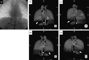
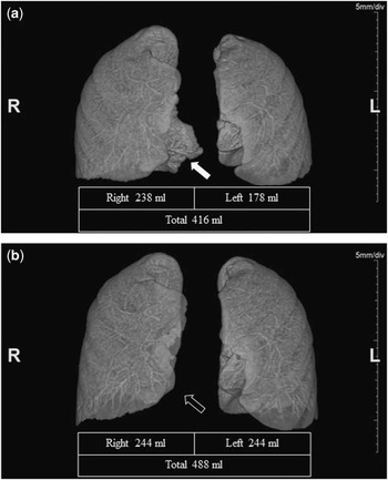
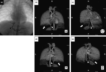
The patient was subsequently referred to our institution, where she underwent cardiac catheterisation at 23 months of age. Her mean pulmonary artery pressure was 12 mmHg, pulmonary vascular resistance 1.35 Wood units·m2, and left ventricular ejection fraction 74%. These haemodynamics seemed suitable for performing a total cavopulmonary connection, but the descending aortogram revealed that the right lower lobe was supplied by an anomalous systemic artery arising from the abdominal aorta (Fig 1a). A contrast-enhanced CT showed pulmonary sequestration of her right lower lobe with vascular supply from the abdominal aorta (Fig 1b-e), with estimated right, including the sequestered lobe, left, and total lung volumes of 238, 178, and 416 ml, respectively (Fig 2a).
( a ) Descending aortography and ( b–e ) three-dimensional contrast-enhanced computed tomography before lobectomy. Blood flow to the right lower lobe was supplied by an anomalous artery (arrowheads) from the abdominal aorta. Arrows show pulmonary sequestration of the right lower lobe.
Lung volumetric analysis by computed tomography ( a ) before and ( b ) after lobectomy. Right lung volume increased from 238 to 244 ml after lobectomy, despite resection of the right lower lobe (open arrow). Left lung volume also increased from 178 to 244 ml. The white solid arrow indicates the sequestered lobe before lobectomy.
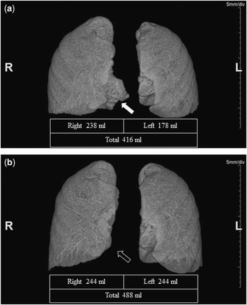
A right lower lobectomy was performed at 25 months of age to reduce the ventricular volume overload due to the anomalous blood supply and to prevent recurrent infections in the sequestered lobe. A cardiac catheterisation performed 36 days after the lobectomy documented a lower mean pulmonary artery pressure and pulmonary vascular resistance of 10 mmHg and 1.05 Wood units·m2, respectively. The pulmonary volumetric analysis by CT showed that her estimated right, left, and total lung volumes were 244, 244, and 488 ml, respectively (Fig 2b), demonstrating that the remaining lung tissue had increased in volume after the right lower lobectomy. Her pulmonary circulation was considered to be more favourable for maintaining a satisfactory Fontan circulation.
A total cavopulmonary connection with an extracardiac conduit was performed at 27 months of age. Although intravenous or oral prednisolone for persistent right pleural effusion was required for 11 days, the patient was discharged on the 29th postoperative day after a satisfactory postoperative course. A cardiac catheterisation performed 5 months after the total cavopulmonary connection demonstrated a mean central venous pressure of 13 mmHg, a mean pulmonary artery pressure of 12 mmHg, a pulmonary vascular resistance of 1.54 Wood units·m2, and a cardiac index of 3.22 l/minute/m2. Angiography in both the superior and inferior caval veins showed well-balanced blood flow to both the lungs. Quantitative evaluation of pulmonary blood flow distribution, based on vessel diameter and flow velocity measured by Doppler-tipped guidewire (FloWire, Volcano), showed a right-to-left pulmonary blood flow ratio of 51:49. She showed excellent Fontan physiology with well-balanced pulmonary circulation. Her clinical condition is good at 5 years of age.
Discussion
To achieve excellent Fontan physiology in this patient, it was necessary to definitively treat the pulmonary sequestration. It was also necessary to preserve ventricular function, by reducing the ventricular volume overload caused by the anomalous blood supply from the descending aorta to the sequestered lobe. Although percutaneous transcatheter embolisation of the anomalous artery was another therapeutic option, lobectomy was performed to prevent recurrent infections in the sequestered lobe. Transcatheter embolisation may be indicated in patients with anomalous systemic arterial supply to normal basal segments of the lung, a subtype of pulmonary sequestration with normal bronchial connections of the lower lobe. Lobectomy, however, may be superior even in these patients, because it may, as in this case, induce compensatory lung growth, including vascular growth, resulting in better pulmonary circulation with a rich pulmonary vascular bed and low pulmonary vascular resistance.
Compensatory lung growth, defined as an adaptive growth in the size of the remaining lung after pneumonectomy or lobectomy, has been described in many species including humans, particularly in children.Reference Rannels and Rannels 2 , Reference Laros and Westermann 3 Physical forces such as stretching or distortion of the remaining lung by overinflationReference Rannels 4 as well as retinoic acid,Reference Kaza, Kron and Kern 5 epidermal growth factor,Reference Kaza, Laubach and Kern 6 hepatocyte growth factor,Reference Sakamaki, Matsumoto, Mizuno, Miyoshi, Matsuda and Nakamura 7 and endothelial nitric oxide synthaseReference Leuwerke, Kaza, Tribble, Kron and Laubach 8 have been reported to be involved in the mechanism of this compensatory lung growth. Pulmonary vascular growth can also occur in the remaining lung after pneumonectomy or lobectomy. Evaluation of rats following left pneumonectomy showed pulmonary arterial growth in all lobes, with the greatest increase (76%) in the upper lobe.Reference Fernandez, Le Cras, Ruiz, Glover, Kron and Laubach 9
Assessments of pulmonary haemodynamics after pneumonectomy or lobectomy in 18 children (aged 6–16 years, mean 12 years) with bronchiectasis found that the mean pulmonary artery pressure remained normal, even after exercise, in the 14 children who underwent lobectomy, but exceeded 20 mmHg at rest or after exercise in all four children who underwent pneumonectomy or bilobectomy.Reference Vyhnalek, Wiedermann, Sery and Hamalova 10 Resection of a single lobe will likely induce compensatory lung growth, including vascular growth, in children, thereby maintaining more “normal” pulmonary haemodynamics.
In conclusion, lobectomy may be indicated in patients with pulmonary sequestration and a functionally univentricular heart to promote more optimal pulmonary haemodynamics and to reduce the chances of post-Fontan pneumonitis. There is evidence that after lobectomy there is a compensatory growth of existing lung tissue and pulmonary vasculature, thereby maintaining low pulmonary artery pressures and resistances as well as normal right, left, and total lung volumes.
Acknowledgements
Lobectomy was performed by Wataru Sumida (MD) and Yoshio Watanabe (MD). The authors thank Mitsuaki Sumi (RT) and Hiroyasu Iwasaki (RT) for their assistance in lung volumetric analysis and three-dimensional reconstruction of CT.
Financial Support
This research received no specific grant from any funding agency, commercial or not for-profit sectors.
Conflicts of Interest
None.