Introduction
Ca2+ ions are involved in the regulation of a variety of cellular processes such as muscle contraction, secretion, proliferation or cell death (Ref. Reference Berridge, Bootman and Roderick1). Therefore, there is a need of precise temporal and spatial control of Ca2+ fluxes and Ca2+ concentration within the cell. Indeed, Ca2+ concentration is tightly controlled in all eukaryotic cells by complex interactions among voltage sensors, Ca2+ transporters, Ca2+ channels, Ca2+ exchangers, Ca2+-binding proteins, ion pumps and ion exchangers (Refs. Reference Leong and MacLennan2, Reference Melzer, Herrmann-Frank and Luttgau3, Reference Murray4). This fine regulation of Ca2+ homeostasis allows this ion to act as one of the most important second messengers in signal transduction.
In skeletal muscle fibres, the status of the excitation–contraction–relaxation cycle is determined by the cytosolic Ca2+ levels: high Ca2+ levels are crucial for triggering muscle contraction, while low levels are critical for initiating muscle relaxation. Hence, a fast Ca2+-cycling mechanism and an efficient storage process are absolutely essential in muscle fibres. During normal contractions, local cytosolic Ca2+ concentration is greatly increased reaching up to 20 µM (Refs. Reference Baylor and Hollingworth5, Reference Chin and Allen6). In addition to these Ca2+ transients associated with muscle contraction, smaller but prolonged increases in intracellular Ca2+ levels associated with long-duration fatigue have been described (Ref. Reference Allen, Lamb and Westerblad7). Additionally, small but persistent increases in basal intracellular Ca2+ concentration likely occur during certain muscle diseases and in ageing (Ref. Reference Andersson8). Since changes in cytosolic Ca2+ levels are particularly frequent and diverse in the skeletal muscle, a tight control in Ca2+ homeostasis is most significant in this system. In this regard, small changes in the Ca2+ handling apparatus in the skeletal muscle might result in major pathophysiological consequences. Indeed, abnormal expression patterns of ion-regulatory proteins have been reported repeatedly in muscular dystrophy (Refs. Reference Alderton and Steinhardt9, Reference Culligan and Ohlendieck10, Reference Gailly11, Reference Gillis12, Reference Ruegg13).
Existing evidence implicates Ca2+ dysregulation as a common underlying event in the pathophysiology of muscular dystrophies, a group of diseases characterised by a primary degeneration of skeletal muscle tissue that results in progressive muscle weakness. This review examines Ca2+ handling in muscular dystrophies, with a special focus on dystrophinopathies, calpainopathies and myotonic muscular dystrophies. We also summarise recent advances in our understanding of Ca2+ ion cycling through the sarcolemma, the sarcoplasmic reticulum (SR) and mitochondria, and its involvement in the pathogenesis of these dystrophies. Finally, we discuss some of the clinical implications of these findings, and in particular, novel pharmacological therapeutic approaches for muscular dystrophies that target Ca2+ regulatory proteins.
Calcium handling in the skeletal muscle
Structure and physiology of the skeletal muscle
The skeletal muscle is under the control of the somatic nervous system through innervation of muscle fibres by motoneurons. Muscle fibres are long, cylindrical multinucleated cells that originate from the fusion of myoblasts during embryonic development and behave as a single unit. Each fibre contains several bundles of myofibrils, with inter-digitating myosin (thick) and actin (thin) filaments arranged longitudinally in repeating units known as sarcomeres. This particular arrangement of the cytoskeletal elements accounts for the banding pattern, characteristic of skeletal fibres. Actin filaments are bound to the Z line, which forms the borders of the sarcomere. At this location, deep invaginations of the sarcolemma (T-tubules) permit the conduction of the electrical impulses to the cell interior (Fig. 1).
Representation of calcium signalling dynamics in the muscle fibre. Signalling begins when sarcolemmal depolarisation reaches the T-tubules. In these structures, DHPR respond with a conformational change that activates closely apposed RyR1, resulting in Ca2+ release from the sarcoplasmic reticulum (SR). Ca2+ diffuses out to activate contraction by binding with Troponin C. Relaxation occurs as Ca2+ is returned into the SR by SERCA ATPase or is pumped out of the cell by NCX or PMCA. A portion of this Ca2+ reaches the mitochondria and driven by the negative membrane potential, it enters through MCU and LETM1 importers, where it stimulates the metabolism to provide the ATP required for maintaining contraction. Ca2+ can be released from mitochondria through NCLX exchanger and the mitochondrial PTP. Low SR Ca2+ levels are detected by STIM1 and activate SOCE, an extracellular Ca2+ influx through Orai1 or TRPC channels to refill intracellular Ca2+ stores.

A single motoneuron innervating one or more muscle fibres with similar functional properties constitutes a motor unit. A muscle is formed by many motor units, each one bringing its own specific contribution (Ref. Reference Schiaffino and Reggiani14). Motor units differ in the size of motoneurons and also in the type of innervated muscle fibres. They are divided into three categories: (a) slow; (b) fast fatigable; and (c) fast fatigue-resistant motor units (Ref. Reference Purves and Williams15).
(a) Slow motor units: Constituted by type I, slow twitch muscle fibres, are essential for sustained muscular contraction. Type I fibres contract very slowly and generate small force, but they are very resistant to fatigue. They contain high mitochondria density and they generate ATP by the aerobic metabolism.
(b) Fast fatigable motor units: Constituted by type IIB fibres, or type IIX in humans, are used for tasks that require large brief bursts of energy such as sprinting. Type IIB/IIX or fast glycolytic fibres have a fast contraction velocity but they fatigue easily. They contain few mitochondria and they generate ATP mainly by the anaerobic system.
(c) Fast fatigue-resistant motor units: Constituted by type IIA, fast twitch A fibres or fast oxidative fibres, which contain many mitochondria and, thus, can generate ATP by aerobic metabolism. They have a fast contraction velocity and they are resistant to fatigue, although not as much as type I fibres (Ref. Reference Scott, Stevens and Binder-Macleod16).
Some of the fibre-type differences in twitch kinetics are also likely due to differences in Ca2+ handling. Indeed, the amount of Ca2+ released in type II fibres is up to fourfold larger than in type I fibres, there is a faster decline of the calcium transient in type II fibres (Ref. Reference Schiaffino and Reggiani14) and the proportion of Ca2+ that binds troponin to activate contraction is substantially smaller (Ref. Reference Baylor and Hollingworth5). On the other hand, type I fibres show increased affinity for SR Ca2+ uptake, higher relative levels of SR Ca2+ loading and increased SR Ca2+ leakage compared with type II fibres (Refs. Reference Lamboley17, Reference Lamboley18). Muscle fibres display a high plasticity, being able to adapt to changing demands by modifying size or fibre-type composition (Ref. Reference Pette and Staron19). Specific types of muscle fibres are preferentially affected in different forms of muscular dystrophies. In Duchenne muscular dystrophy, for example, type IIB fibres are progressively replaced by type I and IIA fibres (Ref. Reference Webster20). Recently, we have also found in collaboration with Dr Spencer's group that slow fibres are preferentially involved in Limb-girdle muscular dystrophy-type 2A (LGMD2A) dystrophic patients (Ref. Reference Kramerova21). Interestingly, some very specific muscle types present in extra ocular muscles show great resistance to damage in Duchenne muscular dystrophy, partially due to an enhanced Ca2+ buffering (Ref. Reference Zeiger, Mitchell and Khurana22).
The Ca2+ cycle during excitation–contraction (E–C)
Skeletal muscle contraction is initiated by depolarisation of the sarcolemma in response to acetylcholine release from motoneurons. The sarcolemmal depolarisation propagates down the T-tubular system and activates dihydropyridine receptors (DHPRs) (Refs. Reference Baylor and Hollingworth5, Reference Stutzmann and Mattson23). The resulting conformational change of DHPRs activates closely apposed ryanodine receptors (RyRs), which are Ca2+ release channels localised in the SR membranes (Ref. Reference Franzini-Armstrong and Jorgensen24). As these release channels open, Ca2+ moves from the SR into the cytosol, driven by a steep Ca2+ concentration gradient. Afterwards, Ca2+ diffuses throughout the sarcomere and binds to various cytosolic constituents or Ca2+ buffers. Binding of Ca2+ to troponin triggers the contractile response of the myofibrils: Ca2+-bound troponin undergoes a conformational change that leads to the movement of tropomyosin, which unmasks myosin-binding sites on actin filaments and allows myosin and actin ATP-dependent cross-bridge cycling and shortening of the muscle. Lastly, relaxation is initiated by Ca2+ diffusion from the myofilaments to the cytosol and Ca2+ transfer back to the SR by the sarco/endoplasmic reticulum Ca2+ pump SERCA (Ref. Reference Periasamy and Kalyanasundaram25). The sarcolemmal Na+/Ca2+-exchanger (NCX) and the plasmalemmal Ca2+-ATPase (PMCA) are other mechanisms involved in relaxation, since they remove cytosolic Ca2+ to the extracellular space (Ref. Reference Sacchetto26). Figure 1 illustrates the major intracellular movements of Ca2+ taking place during E–C coupling in skeletal muscle fibres.
Cytosolic Ca2+ buffers
Cytosolic Ca2+ buffering is the rapid binding of cytosolic incoming Ca2+ ions to different cellular binding sites, and constitutes a critical process implicated in the precise regulation of Ca2+ signalling and homeostasis. Ca2+ buffers are cytosolic Ca2+-binding proteins that modulate intracellular Ca2+ transients by controlling diffusion of free Ca2+ ions within the cytosol. This process occurs at the sub-second scale, much faster than Ca2+ sequestration into intracellular stores. Concentration of Ca2+ buffers and other parameters, such as mobility and Ca2+-binding affinity, are important for determining their specific role in modulation of Ca2+ signalling (Ref. Reference Gilabert27).
In skeletal muscle fibres, the most significant Ca2+ buffers in order of binding occurrence in time are, first, troponin C is a component of thin filaments that allows interaction of actin and myosin and initiates contractile activation when its regulatory sites are bound to Ca2+. Different types of muscle fibres have specific versions of troponin C with different Ca2+-binding capacities. Second, ATP acts as a cytosolic mobile buffer for Ca2+ and Mg2+ (Ref. Reference Baylor and Hollingworth5). Then parvalbumin plays an important role in regulating the speed of relaxation in mice (Ref. Reference Schwaller28), although it is expressed at very low concentrations in human skeletal muscle (Ref. Reference Heizmann, Berchtold and Rowlerson29). Afterwards, Ca2+ binds to calmodulin, contributing to the regulation of contractile function via myosin light-chain kinase. It is also conducive to the activation of Ca2+-dependent signalling pathways involved in muscle gene regulation. Lastly, Ca2+ binds to SERCA pumps for Ca2+ sequestration into the SR.
Contribution of sarcolemma to Ca2+ homeostasis
In skeletal muscle, Ca2+ entry through the sarcolemma has been shown to play an important role in store repletion (Ref. Reference Kurebayashi and Ogawa30), limiting fatigue under conditions of extensive exercise (Ref. Reference Pan31), activation of NFAT transcription factor signalling (Refs. Reference Rosenberg32, Reference Stiber33) and muscle differentiation (Ref. Reference Darbellay34). The sarcolemma has a very low permeability to Ca2+ and a very particular organisation with the majority of the sarcolemma internalised as the tubular system, which allows for tightly regulated Ca2+ trans-sarcolemmal fluxes. Aside from voltage-gated channels, Ca2+ may enter the muscle fibre via SOCE or store-operated Ca2+ entry (Ref. Reference Kurebayashi and Ogawa30); ECCE, or excitation-coupled Ca2+ entry (Ref. Reference Cherednichenko35); SAC or stretch-activated cation channels (Ref. Reference Haws and Lansman36); SIC or stretch-inhibitable cation channels (Ref. Reference Suzuki37); and leak channels (Ref. Reference Hopf38). Previous studies have shown that dysregulation of Ca2+ entry may lead to severe muscle pathologies. In particular, several genetic mouse models of muscular dystrophy point towards an enhanced Ca2+ influx through SOCE or SAC that leads to Ca2+-dependent apoptosis and muscle degeneration (Refs. Reference Vandebrouck39, Reference Vandebrouck40). On the other hand, Ca2+ extrusion from cells may also play an important role in maintaining Ca2+ homeostasis. In muscle fibres, Ca2+ is pumped out of the cell by the Na+/Ca2+ exchangers NCX1-3 and the Ca2+-ATPase PMCA isoforms 1, 3 and 4, localised at the sarcolemma (Ref. Reference Schiaffino and Reggiani14).
Ca2+-handling proteins in the sarcoplasmic reticulum
The SR, the major reservoir of intracellular Ca2+ in the muscle fibre, is a highly specialised form of endoplasmic reticulum. It is organised as an extensive tubular network surrounding myofibrils with periodical dilated end sacs or terminal cisternae. A central T-tubule flanked on both sides by two terminal cisternae from the SR constitutes a triad, a highly specialised anatomical structure that is the basis of E–C coupling (Ref. Reference Al-Qusairi and Laporte41). In the SR, the balance between Ca2+ storage, release and reuptake is achieved through the coordinated action of three major types of Ca2+-handling proteins: luminal Ca2+-binding proteins, SR Ca2+ release channels and Ca2+-ATPase pumps for Ca2+ reuptake (Ref. Reference Rossi and Dirksen42). Ca2+-handling proteins are heterogeneously distributed within the SR, which results in functionally distinct subdomains where Ca2+ release and reuptake occur in the terminal cisternae and the longitudinal SR, respectively.
(a) Luminal Ca 2+-binding proteins: Within the SR lumen, a large proportion of Ca2+ is bound to Ca2+-binding proteins that perform a dual function as Ca2+ reservoir components and as endogenous regulators of Ca2+ fluxes. Luminal Ca2+-binding proteins help to reduce the concentration of free Ca2+ within the SR, which assists in the reuptake of Ca2+ to the SR. They determine normal and maximal Ca2+ levels in the SR, in combination with SERCA pump (Ref. Reference Murphy43). Intraluminal Ca2+ levels have been reported to range between high micromolar and low milimolar concentrations, thus, constituting a steep electrochemical gradient aimed at the cytosol. The most significant Ca2+-binding proteins in the SR are: calsequestrin (CSQ), which is a moderate-affinity, high-capacity Ca2+-binding proteins located in the terminal cisternae region; histidine-rich Ca2+-binding protein (HRC), which has been found to bind SERCA and triadin, and therefore, might be involved in the regulation of Ca2+ release by RyRs and sequestration (Refs. Reference Arvanitis44, Reference Lee45); sarcalumenin, a Ca2+-binding/shuttle protein localised in the longitudinal SR; and calreticulin. In particular, CSQ is involved in the SR Ca2+-loading capacity (Ref. Reference Murphy43), while also modulating activity of RyR channels (Ref. Reference Paolini46). In the skeletal muscle, CSQ1 is the main isoform, although CSQ2 is also expressed in type I fibres. In fact, CSQ1 has been found to both inhibit and activate RyR1 depending on its binding manner to RyR1 (Ref. Reference Beard, Wei and Dulhunty47). Recent work supports an essential role of CSQ1 in the mechanism of RyR1 channel closure and termination of SR Ca2+ release in mouse skeletal muscle (Ref. Reference Sztretye48).
(b) SR Ca 2+release channels: Activation of these channels results in Ca2+ release from SR, which contributes to elevation of cytosolic Ca2+ levels, whereas Ca2+ sequestration to SR assists termination of Ca2+ signals in the cytosol. RyRs, the largest known ion channels, are responsible for Ca2+ release from the sarco/endoplasmic reticulum and they also play a central role in the regulation of cytosolic and SR luminal Ca2+ levels. At least three isoforms are expressed in mammals, RyR1-3, being RyR1 the predominant isoform expressed in the skeletal muscle. RyR2 and RyR3 are expressed in cardiac and immature skeletal muscle, respectively (Refs. Reference Capes, Loaiza and Valdivia49, Reference Mackrill50, Reference Van Petegem51, Reference Mackrill52). RyR1 is essential for the E–C coupling in the skeletal muscle and its gating is strictly controlled by DHPR activation. In fact, Ca2+ release through RyR1 is the key determinant of muscle force since skeletal muscle contraction depends on Ca2+ released from the SR (Ref. Reference Allen, Lamb and Westerblad7). Cytosolic Ca2+ activates RyR1 in the low micromolar, but acts as an inhibitor in the low milimolar range. In addition, luminal Ca2+ levels are also able to modulate RyR1 activity, since previous reports show that high luminal Ca2+ levels enhance RyR1 responsiveness to cytosolic agonist (Ref. Reference Capes, Loaiza and Valdivia49).
Ca2+ release through RyR1 is also modulated by post-translational modifications and a vast variety of proteins and small molecules, both in the SR lumen and cytosol. In addition to Ca2+ and DHPR channels, other positive regulators of RyR1 activity in the cytosol are ATP, CaMKII and PKA. Calstabin-1 stabilises the closed stage of the channel and calmodulin has a biphasic effect on RyR1, functioning as an activator at the low cytosolic Ca2+ levels and as and inhibitor at the high cytosolic Ca2+ levels. On the luminal side, RyR1 interacts with the CSQ–junctin–triadin complex, which plays a dual role as luminal Ca2+ sensor and RyR activity modulator.
RyRs can be phosphorylated by several kinases, including CaMKII and PKA. A recent study has implicated PKA phosphorylation of RyR1 in skeletal muscle enhancement of contractile force (Ref. Reference Andersson53). However, PKA hyper-phosphorylation of RyR1 might lead to SR Ca2+ leak under prolonged pathological stress (Refs. Reference Ward54, Reference Reiken55). Finally, RyR1 activity can also be modulated by oxidative stress through modifications of cysteine thiol residues, such as S-nitrosylation and S-glutathionylation. Although the effect of RyR1 oxidative modifications may vary depending on which residue is modified, exposure of skeletal muscle to nitric oxide has been reported to increase RyR1 activity (Refs. Reference Capes, Loaiza and Valdivia49, Reference Van Petegem51). Interestingly, RyRs have been found to release Ca2+ spontaneously under conditions of luminal Ca2+ overload in a process known as SOICR or Store Overload-Induced Ca2+ Release (Refs. Reference Jiang56, Reference Palade, Mitchell and Fleischer57). This mechanism has been recently linked to cardiac and muscular disorders, such as malignant hyperthermia or catecholaminergic polymorphic ventricular tachycardia (Ref. Reference MacLennan and Chen58).
The other major SR Ca2+ release channels are the inositol 1,4,5-triphosphate receptors (IP3Rs). These Ca2+ channels are gated by the combined binding of IP3 and Ca2+, which initiates a slow wave of internal store Ca2+ release that often occurs at specific subcellular locations (Ref. Reference Marks59). Whereas in myoblasts IP3Rs have a major role regulating Ca2+ homeostasis during skeletal muscle development, the roles that IP3Rs may play in adult skeletal muscle are controversial. A recent study has not found any evidence of IP3R affecting global Ca2+ levels in adult mouse skeletal muscle (Ref. Reference Blaauw60). Conversely, a previous work supports a role of IP3Rs mainly at the neuromuscular junction (Ref. Reference Zayas, Groshong and Gomez61), and recent evidence indicates that crosstalk between IP3R1 and RyR1 underlies activation of stress-induced Ca2+ sparks in adult mice skeletal muscle (Ref. Reference Tjondrokoesoemo62).
(c) Ca 2+-ATPase pumps: Ca2+ reuptake into the SR after contraction relies on the Ca2+ ATPase pump SERCA, which actively transports Ca2+ from the cytosol to the SR against a large concentration gradient at the expense of ATP hydrolysis. Three genes localised on different chromosomes encode for SERCA1, 2 and 3 proteins, although isoform diversity is further increased by alternative splicing. Skeletal fast muscle fibres express SERCA1a exclusively, while SERCA2a is expressed in slow fibres and cardiac muscle. During development or regeneration, muscle fibres express the neonatal isoform SERCA1b (Refs. Reference Stutzmann and Mattson23, Reference Periasamy and Kalyanasundaram25). SERCA pumps are highly affected by changes in cell energetics and ATP supply, since they are, together with myosin ATPases, major ATP consumers (Ref. Reference Maack and O'Rourke63). SERCA pumps are also a substantial pathway for SR Ca2+ leakage and this leakage is increased by high cytosolic ADP levels (Ref. Reference Macdonald and Stephenson64). Low levels of free intraluminal Ca2+ are, thus, necessary so that Ca2+ leakage does not metabolically compromise muscle function (Ref. Reference Murphy43).
In addition to ATP, SERCA activity is modulated by several cytosolic and luminal proteins, and also by post-translational modifications. The endogenous inhibitory proteins phospholamban and sarcolipin modulate SERCA activity in slow and fast-twitch fibres, respectively (Ref. Reference MacLennan, Asahi and Tupling65). Phosphorylation by PKA or CaMKII, results in SERCA activation by dissociation of phospholamban and sarcolipin from the Ca2+ pump. N-glycosylation and other post-translational modifications such as glutathiolation have been found to affect SERCA activity in different ways: SERCA activity can be reversibly increased by oxidative modifications under normal or mild oxidative stress levels, but high oxidant levels present in some pathological conditions may further lead to irreversible SERCA inactivation and degradation (Refs. Reference Lancel66, Reference Vangheluwe67). This particularly susceptibility of SERCA to oxidative and nitrative modifications has led to the hypothesis of SERCA functioning as a sensor of cellular stress (Ref. Reference Bigelow68). Moreover, these data suggest a central role of SERCA in energy production and consumption through regulation of Ca2+ homeostasis.
Ca2+ in mitochondria
Mitochondria are located in close proximity to the SR in the skeletal muscle, and they account for about 15% of the cytosolic volume in oxidative fibres (Refs. Reference Franzini-Armstrong69, Reference Eisenberg70). There is a significant interplay between the mitochondria and the SR, through the mitochondria-associated SR membrane (MAM), which is a structural element essential for cell physiology and Ca2+ homeostasis (Refs. Reference Hajnoczky71, Reference Rizzuto72). Besides playing a central role in muscle bioenergetics, mitochondria are also able to store Ca2+ transiently and therefore, they contribute significantly to Ca2+ homeostasis in the muscle fibre. In fact, their ability to rapidly uptake Ca2+ for later release in response to different stimuli makes them very good cytosolic Ca2+ buffers (Ref. Reference Challet73). Ca2+ fluxes between mitochondria and SR are believed to help shape mitochondrial metabolism and ATP synthesis to the physiological demands of skeletal fibres (Ref. Reference Kavanagh, Ainscow and Brand74). At the same time, mitochondria have been found to modulate intracellular Ca2+ transients in skeletal muscle during E–C coupling (Ref. Reference Yi75). The outer mitochondrial membrane is permeable to Ca2+, unlike the inner mitochondrial membrane. Influx into the mitochondrial matrix is driven by the negative membrane potential and occurs through two types of importers: the low-affinity mitochondrial Ca2+ uniporter (MCU) (Ref. Reference Pizzo76) and a high-affinity mitochondrial Ca2+/H+ exchanger, LETM1. In skeletal muscle fibres, Ca2+ release from mitochondria can occur via a Na+/Ca2+ exchange protein (NCLX) or via Ca2+-induced–Ca2+-release pathways (Ref. Reference Pizzo76). Also, at high matrix Ca2+ levels mitochondria release Ca2+ through the permeability transition pore (PTP), a mechanism that can lead to cell death and might have an important role in muscular dystrophies (Refs. Reference Ascah77, Reference Fraysse78). Interestingly, in humans, mutations in the mitochondrial calcium uptake 1, a regulator of MCU, causes dysfunctional Ca2+ uptake and results in clinical and pathological features that overlap with those of mitochondrial myopathies, congenital core myopathies and muscular dystrophies (Ref. Reference Logan79).
Calcium dysregulation in muscular dystrophies
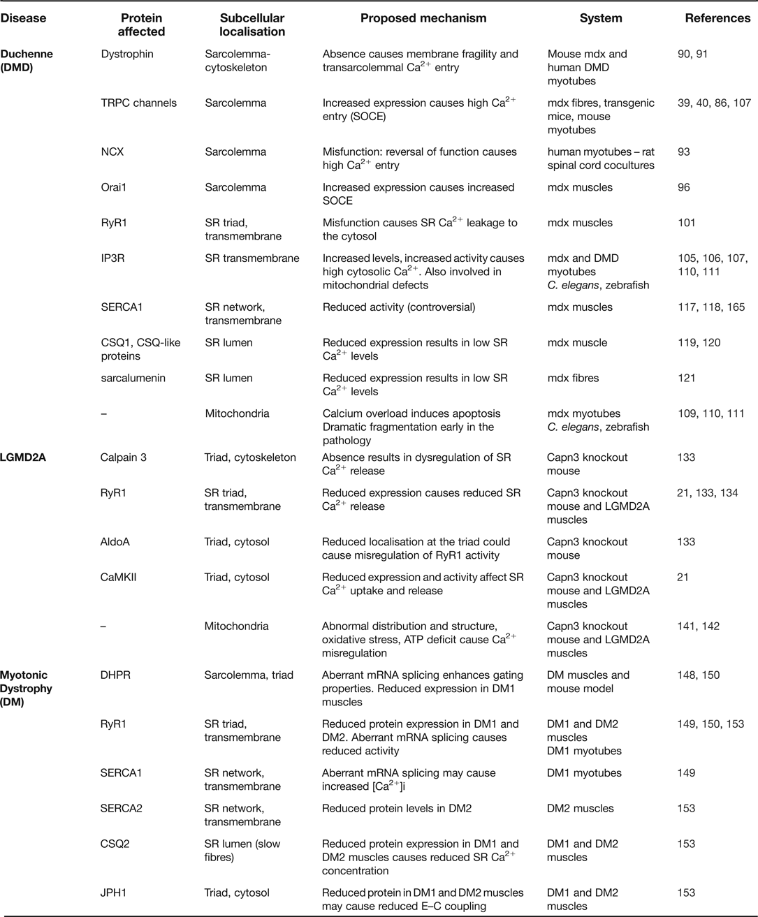
In this section, we will discuss current knowledge regarding Ca2+ homeostasis in dystrophinopathies, calpainopathies and myotonic muscular dystrophies. The main Ca2+-related pathways altered in these pathologies are summarised in Table 1.
Calcium-related mechanisms affected in muscular dystrophies
Dystrophinopathies
Duchenne muscular dystrophy (DMD) is an inherited X-linked neuromuscular disease that affects 1 in 3500 male births and it is characterised by severe and progressive skeletal muscle degeneration, weakness and premature death caused by respiratory failure and cardiac dysfunction (Ref. Reference Emery80). DMD results from deficiency in the structural protein dystrophin (Ref. Reference Hoffman, Brown and Kunkel81), a 427 kDa protein located beneath the sarcolemma whose major role is to protect and maintain muscle fibre integrity by linking intracellular cytoskeletal actin to the extracellular matrix. This is achieved through its association to a group of plasma membrane glycoproteins known as the dystrophin–glycoprotein complex (DGC) (Ref. Reference Blake82). Dystrophin is essential for sarcolemmal stability, and thus dystrophin-deficient muscle fibres are prone to endure recurrent membrane damage (Ref. Reference Petrof83). This sarcolemma fragility, however, does not fully explain the onset and progression of DMD and, up to date, the exact cause of muscle fibre death still remains unsolved. The pathological events most widely accepted to occur in DMD muscle fibres as a result of dystrophin deficiency include membrane fragility and increased basal intracellular Ca2+ levels, which leads to calpain activation, protein degradation, mitochondrial PTP opening and, ultimately, fibre death by necrosis (Refs. Reference Alderton and Steinhardt9, Reference Gailly11, Reference Ruegg13, Reference Allen84, Reference Bodensteiner and Engel85, Reference Millay86). Studies performed more than 30 years ago in muscle biopsies (Ref. Reference Bodensteiner and Engel85) and later on, in human foetuses and premature infants (Ref. Reference Bertorini87), showed increased Ca2+ accumulation in prenecrotic DMD fibres, which suggests that Ca2+ dysregulation is an early event in the pathophysiology of this disorder. Afterwards, many other studies have demonstrated increased intracellular Ca2+ levels in dystrophic muscles from DMD mouse models (mdx mice) and Duchenne patients compared to normal muscles (Refs. Reference Ruegg13, Reference Allen84, Reference Turner88, Reference Yeung89). Increased intracellular Ca2+ levels observed in dystrophin-deficient fibres is a complex process that involves, at least, trans-sarcolemmal Ca2+ fluxes, SR Ca2+ leakage and abnormal SR Ca2+ levels.
High cytosolic Ca2+ levels may result from enhanced Ca2+ influx through the dystrophin-deficient sarcolemma (Refs. Reference Fong90, Reference Franco and Lansman91). This abnormal influx seems to occur mostly through TRPC channels, a type of mechanosensitive voltage-independent Ca2+ channels with increased expression in dystrophic muscle fibres (Ref. Reference Vandebrouck40). Indeed, several groups have found that blockage of TRPC channels in dystrophic mice reduces the abnormal extracellular Ca2+ influx. Furthermore, overexpression of TRPC3 channels in normal mouse skeletal muscle enhances Ca2+ influx and results in a phenotype that closely resembles DGC-lacking dystrophic mice (Ref. Reference Millay86). TRPC channels can function as both, stretch-activated and store-operated channels, and therefore they are also involved in SOCE response, an influx of extracellular Ca2+ triggered by SR depletion of Ca2+ that is enhanced in dystrophic fibres (Ref. Reference Vandebrouck39). An increased SOCE associated with TRPC1 channels has been reported in dystrophic fibres, and remarkably, it can be restored to normal levels by inducing expression of minidystrophin, a smaller and partially functional dystrophin variant (Ref. Reference Vandebrouck92). Finally, enhanced Ca2+ influx in dystrophic fibres has also been reported to occur by reversal NCX sarcolemmal channels (Ref. Reference Deval93) and also by the Orai1/Stim SOCE pathway. In addition to TRPC channels, other proteins may also be responsible for increased SOCE in DMD, such as stromal interaction molecule 1 (STIM1), which acts as an SR Ca2+ sensor that can trigger SOCE through both TRPCs and Orai1 channels (Ref. Reference Lyfenko and Dirksen94). In this regard, it has been recently reported increased Orai1 (Refs. Reference Edwards95, Reference Zhao96) and STIM1 (Ref. Reference Edwards95) expression in dystrophic muscles from mdx mice. This indicates that enhanced activation of SOCE pathways may contribute to the disrupted Ca2+ homeostasis in DMD pathology. Several other proteins have been involved in this enhanced SOCE activity in dystrophin-deficient muscle myotubes, such as phospholipase C, protein kinase C (Ref. Reference Sabourin97), phospholipase A2 (iPLA2) and its derived metabolites (Ref. Reference Boittin98). In this regard, increased iPLA2 activity has been known for more than 10 years in DMD muscles (Ref. Reference Lindahl99). Therefore, specific inhibitors of these pathways may be of interest to reduce Ca2+ influx and subsequent degeneration of dystrophic muscle (Ref. Reference Ismail100).
Another source of elevated cytosolic Ca2+ levels may involve SR Ca2+ leakage through RyRs or IP3Rs. Regarding RyRs, several studies have pointed to abnormally high S-nitrosylation of RyR1 cysteine residues in mdx mice causing calstabin-1 depletion from the RyR complex, that lead to unstable and leaking RyR1 channels at resting conditions (Refs. Reference Bellinger101, Reference Brillantes102, Reference Bellinger103). These abnormal S-nitrosylation levels seem to be due to inducible nitric oxide synthase (iNOS), which was also found increased in dystrophic mdx muscles.
A recent study has found evidence of significant Ca2+ leakage through IP3Rs as well as through RyRs in dystrophic mdx myotubes contributing to the increased intracellular Ca2+ levels (Ref. Reference Altamirano104), which suggests that IP3R malfunction may also be involved in DMD Ca2+ mishandling. Indeed, previous studies have shown increased IP3R and IP3 levels, and enhanced IP3R-dependent Ca2+ transients in myotubes from DMD patients and mdx mice (Refs. Reference Liberona105, Reference Basset106, Reference Cardenas107). Moreover, induced expression of minidystrophin in dystrophin-deficient myotubes reverts the IP3R-dependent increase in Ca2+ release to normal levels (Ref. Reference Balghi108), which suggests an IP3R involvement in DMD impaired Ca2+ homeostasis. Finally, there seems to be a connection between IP3R and mitochondria that may play a relevant role in Ca2+ dysregulation. Previous work in mdx dystrophic myotubes has shown mitochondrial Ca2+ overload due to increased SR Ca2+ release (Ref. Reference Robert109). Interestingly, overexpression of Bcl-2 antiapoptotic protein in mdx myotubes prevents this mitochondrial Ca2+ overload by inhibiting IP3Rs (Ref. Reference Basset110). More recently, abnormal mitochondrial fragmentation has been observed in Caenorhabditis elegans and zebrafish models of DMD, where involvement of cytochrome c in muscle fibre death was demonstrated, acting in part through an interaction with IP3R (Ref. Reference Giacomotto111).
In DMD and in many other neuromuscular disorders such as sarcoglinopathies, desminopathies, LGMD2B muscular dystrophy and Pompe disease, the cardiac and respiratory muscles are affected and, consequently, there is an increased risk of respiratory and cardiac failure in these patients that leads to early mortality (Ref. Reference Allen84). Abnormal Ca2+ homeostasis and mitochondrial dysfunction have also been reported in these muscles. In particular, previous works in mdx cardiac myocytes have shown that stretch-activate channels contribute to abnormal Ca2+ influx (Ref. Reference Williams and Allen112), and excessive cytosolic Ca2+ signals induced by mechanical stress result in mitochondrial dysfunction (Ref. Reference Jung113). More recently, higher mitochondrial Ca2+ levels have been reported in cardiac dystrophic myocytes and activation of DHPR seems to contribute to abnormal mitochondrial Ca2+ handling (Ref. Reference Viola114). Evidence for early mitochondrial dysfunction in dystrophic cardiomyopathy is further supported by alterations in mitochondrial citric acid cycle-related parameters observed in ex vivo mdx hearts preceding overt cardiomyopathy (Ref. Reference Khairallah115).
Another controversial issue is whether SERCA expression or function is reduced, unchanged or increased in dystrophin-deficient fibres, with recent examples in the literature supporting all different possibilities (Refs. Reference Zhao96, Reference Robin, Berthier and Allard116, Reference Goonasekera117, Reference Gehrig118). Regardless the need for further studies to establish a clear understanding on this subject, the majority of the studies agree with a reduced SERCA activity in mdx muscles. In this line, nitrative stress could be causing a reduced SERCA activity in dystrophic fibres, since SERCA is highly sensitive to reactive nitrogen species (Ref. Reference Bigelow68) and high iNOS levels have been reported in mdx fibres (Ref. Reference Bellinger101).
Ca2+ binding proteins and decreased SR Ca2+ buffering have also been involved in the pathology of dystrophinopathies. In particular, in the SR lumen, reduction of CSQ1, CSQ-like proteins and sarcalumenin has been observed in mdx dystrophic muscles (Ref. Reference Culligan119, Reference Doran120, Reference Dowling, Doran and Ohlendieck121). A significant reduction of the SR Ca2+ buffering would have a direct negative impact on the Ca2+ reuptake capacity of SERCA, which in turn, would result in the increased cytosolic Ca2+ levels. Remarkably, less affected muscles in the mdx muscular dystrophy mouse model, such as the extraocular and intrinsic laryngeal muscles, exhibit higher expression levels of several Ca2+-handling proteins such as CSQ, calmodulin and/or SERCA (Refs. Reference Ferretti122, Reference Pertille123), which suggests that these proteins are playing a protective role in DMD pathology.
Calpainopathies
LGMDs are a large group of hereditary muscular dystrophies characterised by progressive proximal weakness and a predominant involvement of the scapula, pelvic girdle and trunk muscles without affecting cardiac or facial muscles (Ref. Reference Laval and Bushby124). Among the recessive forms of LGMDs, calpainopathy or LGMD2A is the most frequent muscular dystrophy with a prevalence of 82–100% in genetically isolated populations such as the Basque Country (Refs. Reference Saenz125, Reference Urtasun126). LGMD2A is caused by mutations in the gene encoding Calpain 3 (CAPN3), a non-lysosomal cysteine protease necessary for normal muscle function and regeneration (Refs. Reference Hauerslev127, Reference Richard128). The exact pathogenic mechanisms that underlie muscular dystrophic features caused by mutations in CAPN3 are still unclear, and in view of the lack of effective therapies available for LGMD2A patients, understanding these pathological mechanisms is needed in order to advance in the design of potential treatments.
In skeletal muscle CAPN3 is present in different subcellular locations including myofibrils, membrane and cytoplasm. Most of the CAPN3 is bound within the contractile filament network, where it remains in its inactive state through its association with titin, a giant protein that connects the Z line to the M line in the sarcomere (Refs. Reference Beckmann and Spencer129, Reference Murphy and Lamb130). Absence of CAPN3 from mouse and human muscles results in sarcomere disorganisation (Refs. Reference Hauerslev127, Reference Kramerova131), which suggests that CAPN3 is involved in sarcomere repair and maintenance and may also be acting as a sensor of its integrity and functionality (Ref. Reference Beckmann and Spencer129). Remarkably, sarcomere alignment is preserved in mice expressing an inactive form of CAPN3 (Capn3cs/cs mice), indicating that the structural role of CAPN3 and not the enzymatic one is involved in sarcomeric remodelling (Ref. Reference Ojima132).
In the membranes, CAPN3 is localised in the triad protein complex and plays an essential role in preserving its structure and function. CAPN3 has been found to interact with RyR1 (Ref. Reference Ojima132), the main SR Ca2+ release channel, and regulate Ca2+ release from SR (Ref. Reference Kramerova133). CAPN3 has also been found to interact with the Ca2+-handling protein CSQ by immunoprecipitation analysis (Ref. Reference Ojima132). This interaction is proposed to occur by means of connecting proteins such as triadin, since CSQ and CAPN3 are localised in different cellular compartments. In CAPN3 knockout mice (C3KO, Capn3-/-), reduced expression of RyR1 is concomitant with the reduced SR Ca2+ release (Refs. Reference Kramerova133, Reference Dayanithi134). Also, it has been previously reported slower Ca2+ reuptake into SR after SR Ca2+ depletion in CAPN3 mice, although the basis of these finding needs further analysis (Ref. Reference Dayanithi134). However, loss of CAPN3 proteolytic activity in Capn3cs/cs mice does not affect Ca2+ homeostasis (Ref. Reference Ojima132), which indicates that the structural role of CAPN3 is a key for maintenance of Ca2+ homeostasis. In this line, we have recently shown in a collaboration study that muscles from LGMD2A patients with deficient CAPN3 also expressed lower RyR1 protein levels. Moreover, we found a correlation between CAPN3 and RyR1 expression levels that supports a structural function of CAPN3 in the preservation of the triad protein complex (Ref. Reference Kramerova21).
Another CAPN3-binding partner in the triad complex is aldolase A (AldoA) (Ref. Reference Kramerova133), a glycolytic enzyme that may modulate Ca2+ handling in the muscle fibre through multiple mechanisms, including direct regulation of RyR activity by intermediates and products of glycolysis, and modulation of SERCA activity through local changes of glycolytically derived ATP (Ref. Reference Kockskamper, Zima and Blatter135). In the skeletal muscle, AldoA directly interacts with RyR1 and in vitro it has been shown to modulate RyR1 activity (Refs. Reference Kramerova133, Reference Seo136). Interestingly, CAPN3 knockout mice show decreased levels of AldoA in the sarcomere due to impaired recruitment of AldoA to the triads (Ref. Reference Kramerova133). Remarkably, mutations in the gene encoding AldoA result in severe skeletal muscle damage in humans (Refs. Reference Kreuder137, Reference Yao138).
Ca2+ calmodulin kinase II (CaMKII) is another triad component that is modulated by CAPN3. In this regard, our group has found that CaMKII signalling is compromised in CAPN3 knockout mice and, thus, we propose that impaired Ca2+-mediated signalling and weakened muscle adaptation are pathogenic mechanisms likely operating in CAPN3-deficient muscles (Ref. Reference Kramerova21). CaMKII is a multifunctional protein activated by muscle contraction-induced Ca2+ increase that regulates several Ca2+-handling proteins (Ref. Reference Allen, Lamb and Westerblad7). In slow twitch skeletal fibres, CaMKII may increase SERCA2 activity through direct phosphorylation or by phosphorylation of its inhibitory protein phospholamban (Refs. Reference Vangheluwe67, Reference Sacchetto139). On the other hand, CaMKII is also able to modulate Ca2+ release through phosphorylation of related proteins, such as triadin, which inhibits RyR1 activity when unphosphorylated (Ref. Reference Rose140). Thus, decreased CaMKII signalling levels, comparable to the ones observed in CAPN3 deficient muscles, may result in reduction of SR Ca2+ release during contractions as well as reduced Ca2+ reuptake by SERCA. However, this hypothesis needs verification through experimental analysis.
Finally, there seems to be a connection between CAPN3 and mitochondrial function, since LGMD2A patients and CAPN3 knockout mice show abnormal distribution and structure of mitochondria in skeletal fibres (Ref. Reference Kramerova, Beckmann and Spencer141). Moreover, CAPN3-deficient muscles show ATP production deficits and increase oxidative stress levels (Ref. Reference Kramerova142). Accordingly, muscles with higher percentage of slow twitch fibres, which have higher oxidative metabolism, are the most affected in CAPN3 knockout mice (Refs. Reference Kramerova21, Reference Kramerova142). These mitochondrial abnormalities are likely associated to a severe Ca2+ dysregulation in CAPN3-deficient muscle fibres, although future experiments are still needed in order to elucidate the pathogenic mechanisms involved in the mitochondrial dysfunction observed in LGMD2A.
Myotonic dystrophy
Myotonic dystrophy type 1 (DM1), the most frequent adult form of muscular dystrophy (Ref. Reference Harper143), is an inherited autosomal dominant disease caused by an unstable expansion of the CTG triplet repeat located in the 3′ untranslated region of DMPK gene that codes for myotonic dystrophy protein kinase. This gene is located in the long arm of chromosome 19 and it is predominantly expressed in skeletal muscle (Ref. Reference Brook144). DM2 form is a rare disease associated with a CCTG tetranucleotide repeat expansion, located in the first intron of the zinc finger protein 9 (ZNF9) gene in chromosome 3. Thus, both DM1 and DM2 originate from long non-coding repeat expansions, which cause a similar chronic, slowly progressing, multisystemic disease with a dominant inheritance pattern. Expanded mRNA molecules in the cell nucleus affect expression and/or re-allocation of RNA-binding proteins and lead to alteration in RNA processing (Refs. Reference Dansithong145, Reference Philips, Timchenko and Cooper146, Reference Timchenko147). The multi-systemic nature of both diseases is thought to be caused by aberrant splicing affecting a number of proteins involved in multiple cellular processes. It has recently been shown that myotonic dystrophy is associated with deregulated alternative splicing of the gene CACNA1S that encodes for DHPR alpha 1S subunit (DHPRα1s), a voltage sensor that plays a central role in E–C coupling (Ref. Reference Tang148). In DM1 and DM2, skipping of CACNA1S exon 29 is enhanced, which increases DHPRα1s channel conductance and voltage sensitivity. The long-term consequences are very likely harmful to muscle, especially when DHPR missplicing is combined with deregulated alternative splicing of other genes related to Ca2+-handling or E–C coupling such as the ones encoding chloride channel type 1, SERCA and RyR1 (Refs. Reference Kimura149, Reference Santoro150). This would likely lead to a chronic Ca2+ overload, a common scenario occurring in other forms of muscular dystrophies. In fact, several studies have reported that Ca2+-signalling pathways are consistently affected in DM muscles, DM myotubes and mouse models of this disease (Refs. Reference Benders151, Reference Jacobs152, Reference Vihola153). These findings suggest that the combined effect of misregulated splicing of several genes involved in Ca2+ regulation and E–C coupling contributes to muscle degeneration in myotonic dystrophy. In particular, a recent study performed on human DM samples demonstrates abnormal expression of mRNA and proteins involved in muscle Ca2+ release, reuptake, storage and signalling, such as junctophilin-1 (JPH1), SERCA2, RyR1, CSQ2 and DHPR (Ref. Reference Vihola153). In DM muscles they found reduced protein levels of JPH1, but not mRNA, which is consistent with a general dysregulation of Ca2+ homeostasis since JPH1 undergoes Ca2+-dependent proteolysis when intracellular Ca2+ levels are elevated for a sustained period (Refs. Reference Murphy154, Reference Toral-Ojeda, Aldanondo and Vallejo-Illarramendi155). Interestingly, in most cases mRNA levels of Ca2+-handling molecules were found increased while the protein levels were decreased, which suggests that the translational block may be one of the underlying mechanisms of DM.
Therapeutic targeting of mitochondria and calcium homeostasis in muscular dystrophies
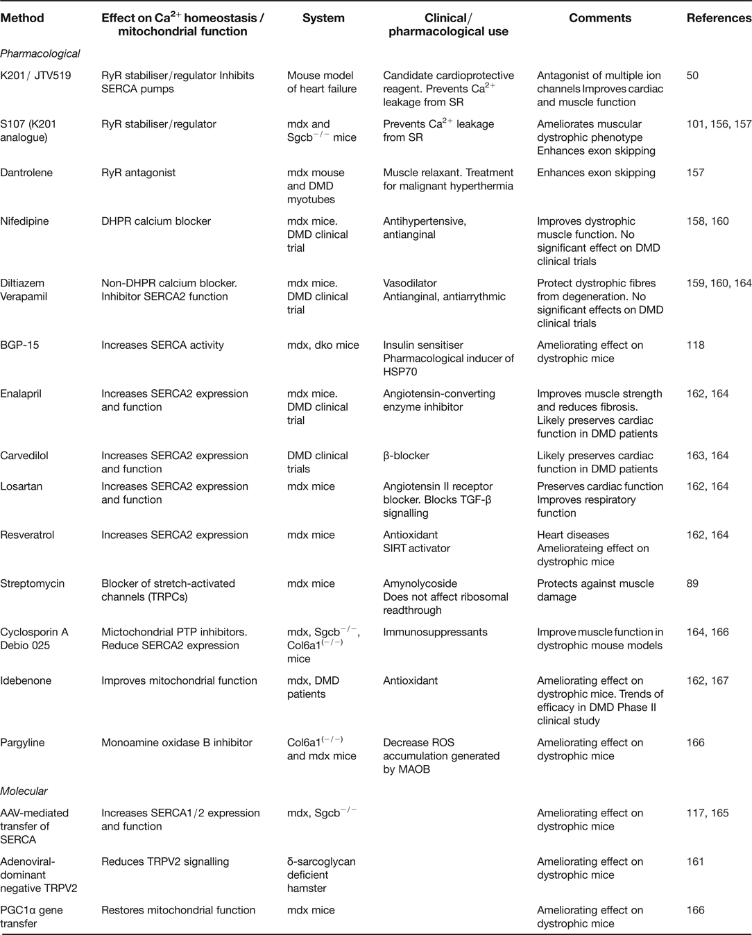
Over the years, several pharmacological and molecular therapies that target Ca2+ homeostasis and/or mitochondrial function have been proposed as potential treatments for muscular dystrophies (summarised in Table 2). Different studies have demonstrated beneficial effects of RyR stabilisers on different animal models of muscular dystrophy and heart failure (Refs. Reference Mackrill50, Reference Bellinger101, Reference Andersson156). In particular, treatment of mouse models of Duchenne and LGMD2E muscular dystrophy with the RyR stabiliser S107 results in reduced RyR1 nitrosylation, reversion of calstabin-1 depletion and an overall improvement of muscle function and exercise performance (Refs. Reference Bellinger101, Reference Andersson156). Interestingly, a recent study has demonstrated that S107, as well as the RyR inhibitor dantrolene, synergise with antisense-mediated exon-skipping therapies and increases muscle function in mdx mice and human DMD myotubes (Ref. Reference Kendall157). Also, several Ca2+ channel blockers such as nifedipine, diltiazem and verapamil have shown to improve muscle structure and function in dystrophic mice (Refs. Reference Altamirano158, Reference Matsumura159), but they have failed to prove significant beneficial effects on DMD patients in clinical trials (Ref. Reference Phillips and Quinlivan160). Likewise, pharmacological or molecular blockage of stretch-activated channels (TRPC, TRPV2) has also shown a beneficial effect on several dystrophic models (Refs. Reference Yeung89, Reference Iwata161).
Treatments targeting calcium homeostasis and mitochondrial function in muscular dystrophies
Angiotensin II-receptor blockers, β-blockers and angiotensin-converting-enzyme inhibitors have shown beneficial effects for the treatment of cardiomyopathies in clinical trials and animal models (Refs. Reference Finsterer and Cripe162, Reference Kwon163). Interestingly, some pharmacological agents from these groups have also been reported to increase SERCA2 expression and/or function, and therefore they are potential enhancers of cardiac Ca2+ homeostasis (Ref. Reference Shareef, Anwer and Poizat164). In this line, different strategies comprising overexpression of SERCA1 or SERCA2 in skeletal muscle of dystrophic mice, such as treatment with BGP-15, or AAV-mediated gene transfer have consistently shown a significant improvement in the dystrophic phenotype (Refs. Reference Goonasekera117, Reference Gehrig118, Reference Morine165), which further supports that reduced SERCA activity is a contributing factor in the pathology of muscular dystrophies.
Finally, several therapeutic approaches targeting mitochondrial dysfunction have been shown to improve muscle function in different dystrophic mouse models. These include treatment with cyclosporine A and cyclophilin D inhibitors (Debio 025) for impaired mitochondrial PTP opening, treatment with pargyline for excessive accumulation of reactive oxygen species or overexpression of peroxisome proliferator-activated receptor γ coactivator 1-gene α (PGC1 α) for mitochondrial dysfunction in postnecrotic dystrophic muscles (Ref. Reference Katsetos, Koutzaki and Melvin166). In this line, an antioxidant that improves mitochondrial respiratory function, idebenone, has shown protection of cardiac and skeletal muscle in mdx mice and trends of efficacy in a Phase II study with DMD patients (Ref. Reference Buyse167).
Conclusions and future address
The full extent to which intracellular Ca2+ signalling influences the physiopathology of primary and secondary muscle diseases is only beginning to be understood. In the forthcoming years, future technological advances, including new powerful Ca2+ indicators are needed in order to accelerate and target-specific Ca2+-signalling pathways within cells. Some promising genetically encoded Ca2+ indicators from invertebrates or algae are already under development and will likely provide us with new clues to understanding the role of Ca2+ in degenerative processes. Since these indicators are both, incorporated at the genome level and translated as fluorescent proteins that modify their emission intensity as a function of Ca2+ levels, they can be used for long-term experiments over days to months and they can also be introduced into in vivo animal models to test Ca2+ activity over a lifetime (Refs. Reference Kotlikoff168, Reference Mank169). Furthermore, other imaging technologies such as fluorescence resonance energy transfer-based (FRET) analysis can demonstrate Ca2+-dependent responses at the single protein level (Ref. Reference Rochefort and Konnerth170). In the near future, these developments will contribute to the design of new experiments with potential applications in basic science and translational research studies (Ref. Reference Schoenenberger, Scharer and Oertner171).
The target of these research lines in the field of muscular dystrophies should be focused in the first place on disorders where Ca2+ homeostasis seems to play a central role, such as in malignant hyperthermia, central core disease or arrythmogenic cardiomyopathies (Ref. Reference Mackrill50). Nevertheless, as awareness of Ca2+-signalling dysregulation in other muscular diseases progresses, such as LGMD2A, attention should be addressed to specific elements of intracellular Ca2+-signalling cascades in these entities. From a therapeutical point of view, some progress in these areas is already taking place: the DHPR blocker nifedipine and the SR leaky-channel blockers dantrolene and S-107, for instance, have been successfully tested in animal models of muscular dystrophy (Refs. Reference Bellinger101, Reference Andersson156, Reference Altamirano158). It is very likely that whatever achievements may come from these on-going research projects, they will be useful for several other muscular disorders sharing similar underlying Ca2+ pathological mechanisms.
Financial support
Research in our laboratories is supported by the Instituto Carlos III (A.V.-I., grant number PI11/01499), Gobierno Vasco (A.V.-I., grant numbers SAIO12-PR12BN002, 2010111089), Marie Curie International Reintegration Grant (A.V.-I., grant number IRG256512) and Fundación de Estudios Neurológicos ILUNDAIN. I.T.-O. and G.A. are funded by PhD fellowships from Gobierno Vasco.
Conflicts of interest
None.