1. Introduction
Although dermochelyid turtles are currently represented by only a single living species, the leatherback turtle Dermochelys coriacea (Vandelli, Reference Vandelli1761), they have an extensive fossil record that mirrors a complex evolution (Wood et al. Reference Wood, Johnson-Gove, Gaffney and Maley1996). Despite the fact that D. coriacea has a very short evolutionary history inscribed in its mtDNA (<1 Ma; Dutton et al. Reference Dutton, Bowen, Owens, Barragan and Davis1999), the earliest definite dermochelyids are known from the Late Paleocene to Early Eocene (Nielsen, Reference Nielsen1959; Tong et al. Reference Tong, Buffetaut, Thomas, Roger, Halawani, Memesh and Lebret1999). Besides the Mesozoic Mesodermochelys Hirayama & Chitoku, Reference Hirayama and Chitoku1996 (Late Cretaceous, Japan) and Corsochelys Zangerl, Reference Zangerl1960 (Late Cretaceous, North America), at least seven Cenozoic fossil dermochelyid genera can be identified (Danilov, Reference Danilov and Fritz2005; Bever & Joyce, Reference Bever, Joyce and Fritz2005): (1) Arabemys crassiscutata Tong et al. Reference Tong, Buffetaut, Thomas, Roger, Halawani, Memesh and Lebret1999 (Paleocene–Eocene, Saudi Arabia), (2) Cosmochelys Andrews, Reference Andrews1919 (Eocene, Africa), (3) Egyptemys Wood et al. Reference Wood, Johnson-Gove, Gaffney and Maley1996 (Eocene, Africa), (4) Eosphargis Lydekker, Reference Lydekker1889 (Eocene, Europe), (5) Cardiochelys Moody, Reference Moody1993 (Oligocene, Europe), (6) Natemys Wood et al. Reference Wood, Johnson-Gove, Gaffney and Maley1996 (Oligocene, Peru) and (7) Psephophorus Meyer, Reference Meyer1847 (mainly Miocene, Europe and possibly North America). The two Mesozoic forms are closely related to dermochelyids according to Hirayama & Chitoku (Reference Hirayama and Chitoku1996) and Hirayama (Reference Hirayama, Callaway and Nicholls1997); however, the relationships of these taxa await reassessment in a more comprehensive phylogenetic analysis. It is apparent that the basal members (e.g. Eosphargis spp.) still retained neural, costal and peripheral bones and otherwise lacked an ‘epithecal’ shell (Nielsen, Reference Nielsen1963; see also Karl & Lindow, Reference Karl and Lindow2010). Conversely, in D. coriacea the primary ‘thecal’ shell, with the exception of the nuchal bone, is largely reduced. Instead a secondary ‘epithecal’ mosaic of dermal elements, the ossicles, embedded in the thick fatty dermis, is exceptionally developed (e.g. Hay, Reference Hay1898; Versluys, Reference Versluys1913; Deraniyagala, Reference Deraniyagala1930; Zangerl, Reference Zangerl1939, Reference Zangerl, Gans, Bellairs and Parsons1969; Rieppel, Reference Rieppel2001; Bever & Joyce, Reference Bever, Joyce and Fritz2005; Frazier, Gramentz & Fritz, Reference Frazier, Gramentz, Fritz and Fritz2005). Seven longitudinal ridges run along the carapace, and the unique secondary carapace consists of thousands of ‘epithecal’ polygonal ossicles (e.g. Gervais, Reference Gervais1872). The ‘thecal’ elements of the plastron are still present, but only as thin bony rod-like elements. After Brongersma (Reference Brongersma1969), the plastron is referred to as being additionally covered in isolated rows of small ‘epithecal’ platelets that coincide with six plastral ridges (see also Bever & Joyce, Reference Bever, Joyce and Fritz2005).
For several decades, it has been assumed that Dermochelys evolved from the extinct taxon Psephophorus Meyer, Reference Meyer1847 from the middle Miocene Studienka Formation of Devínska Nová Ves, close to Bratislava, Slovakia. However, according to the last comprehensive revision of the taxonomy and phylogeny of fossil Dermochelyidae (Wood et al. Reference Wood, Johnson-Gove, Gaffney and Maley1996), Dermochelys belongs to a clade that is the sister group to P. polygonus (i.e. an unresolved trichotomy with ‘Psephophorus’ calvertensis and a yet undescribed specimen from the Eocene Selma Formation of Alabama, USA). Wood et al. (Reference Wood, Johnson-Gove, Gaffney and Maley1996) concluded that most of the species previously referred to Psephophorus are not assignable to this genus, which is represented by a single species, Psephophorus polygonus Meyer, Reference Meyer1847. They distinguished Dermochelys and Psephophorus on the basis of several characters. Particularly diagnostic for Psephophorus should be the fact that longitudinal ridges are not apically pointed in cross-section, and are expressed only on the external surface of the carapace ossicles, their visceral surface being flat. This contrasts with the condition in Dermochelys, where the osteoderms that comprise the longitudinal ridges are tectiform, pointed in cross-section and expressed also on the ventral surface of the shell. Moreover, ossicles of Dermochelys vary greatly in size but not in Psephophorus.
In the revision by Wood et al. (Reference Wood, Johnson-Gove, Gaffney and Maley1996), the morphology of the type species, Psephophorus polygonus Meyer, Reference Meyer1847, was based only on what is considered to be the neotype (NHMW 2011/0330/0001) as it is doubtful whether Meyer's specimens, which he did not figure, will ever become identifiable. In fact, the history of the definition of this species has not been straightforward and several inaccuracies, related mostly to the nature of the type material and its whereabouts, have been progressively disseminated in the literature.
Meyer (Reference Meyer1846) first briefly mentioned some isolated ossicles from Slovakia that he tentatively referred to an unidentified dasipodid mammal, without naming any new taxon. Then, the year after, he examined a drawing of a slab coming from the Slovakian locality Neudorf an der March (= Dévényújfalu, now Devínska Nová Ves), containing ossicles in connection, and on the basis of it – without studying the real fossil – confirmed the previous referral to a mammal, briefly describing the new taxon Psephophorus polygonus Meyer, Reference Meyer1847. Regrettably, he did not publish the drawing and therefore we only know that the slab contained about 70 ossicles, some of which formed a ridge. As already summarized by Seeley (Reference Seeley1880, p. 406), who associated for the first time P. polygonus to chelonians, the slab was originally in Bratislava but was later acquired by the Museum of the Imperial Geological Survey in Vienna, which also obtained a second slab ‘containing a larger portion of the same carapace’ that fitted onto the first (see also von Hauer, Reference Hauer1868, Reference Hauer1870). This second slab is the only one of the two that is currently available: it is displayed at the Naturhistorisches Museum Wien (NHMW 2011/0330/0001) and is considered the neotype of P. polygonus (Wood et al. Reference Wood, Johnson-Gove, Gaffney and Maley1996). Seeley (Reference Seeley1880) described in detail both slabs and figured them along with a vertebra that he referred to the same taxon. He reported the presence of a fragment of a rib and five fragmentary vertebrae, the best preserved of which, the one figured, ‘is chelonian in its characters’ (Seeley, Reference Seeley1880, p. 410). On the basis of the description and the figure, it is not actually possible to confirm the identification of the vertebrae as chelonian, and possibly because of this, vertebrae were not mentioned in subsequent accounts (i.e. Wood et al. Reference Wood, Johnson-Gove, Gaffney and Maley1996; Gemel & Rauscher, Reference Gemel and Rauscher2000). Wood et al. (Reference Wood, Johnson-Gove, Gaffney and Maley1996, p. 279) wrote that ‘the type and only specimen of Psephophorus polygonus is housed in the collections of the Natural History Museum in Vienna’ but it is known that other shell remains were found in the same locality (Abel, Reference Abel1919; Szalai, Reference Szalai1934; Młynarski, Reference Młynarski1966). In particular, Abel (Reference Abel1919) described and figured isolated ossicles from Dévényújfalu (the Hungarian name for Devínska Nová Ves) that were deposited at the Geologische Bundesanstalt in Vienna, at the Institute of Palaeontology of Vienna University and in the Mineral and Palaeontological collections of the Hungarian National Museum in Budapest. Młynarski (Reference Młynarski1966) later published catalogue numbers and a brief comment on the available remains stored in Budapest.
In order to provide a sound basis for future analyses of fossil dermochelyids and to further our understanding of the relationship among the fossil taxa and the only living relative, D. coriacea, we here summarize the current location of the remains of P. polygonus from the type locality that are available in public collections. On the basis of this material, we re-evaluate the gross morphology of the ossicles of P. polygonus and describe for the first time their microstructure, both of which are compared with those of the ossicles of extant D. coriacea.
Institutional abbreviations. GBA – Geologische Bundesanstalt, Vienna, Austria; IPUW – Institut für Paläontologie an der Universität Wien, Vienna, Austria; MDHC – Massimo Delfino Herpetological Collection, deposited at Università di Torino, Italy; MTM – Magyar Természettudományi Múzeum, Budapest, Hungary; NHMW – Naturhistorisches Museum Wien, Vienna, Austria; PIMUZ – Paläontologisches Institut und Museum, Universität Zürich, Zürich, Switzerland; QM – Queensland Museum, Brisbane, Queensland, Australia.
2. Systematic palaeontology
Order TESTUDINES Batsch, Reference Batsch1788
Family Dermochelyidae Fitzinger, Reference Fitzinger1843
Genus Psephophorus Meyer, Reference Meyer1847
Diagnosis. As for the only species ascribed to this genus by Wood et al. (Reference Wood, Johnson-Gove, Gaffney and Maley1996).
Psephophorus polygonus Meyer, Reference Meyer1847
Neotype. NHMW 2011/0330/0001: a slab containing ridge and non-ridge ossicles (Wood et al. Reference Wood, Johnson-Gove, Gaffney and Maley1996).
Topotype material. GBA 2008/93/1: 13 ossicles; IPUW 6224/1–5: five ossicles; MTM V 61.1216: 37 ossicles, 28 of which are almost complete (six from a ridge); all the ossicles are isolated except six (three from a ridge) that have been joined in a major shell fragment and two that constitute a minor one; MTM V.61.1215: 46 ossicles (two from the ridge); MTM V.61.1218: 17 ossicles (two from the ridge); MTM V.61.1219: a slab broken into two pieces containing 37 connected ossicles (all except ten are broken into two along their horizontal plane); NHMW 1848/0015/a–z, aa–aq: 43 ossicles; NHMW 1857/0019/0010a–e: five ossicles; NHMW 1857/0028/0006a–b: two ossicles; NHMW 1859/0005/0125a–b: two ossicles; NHMW 1868/0008/0004: one ossicle; NHMW 2011/01/0149/001–002: two ossicles; NHMW 2011/0150/001–2: two ossicles; NHMW 2011/0151/0001–006: six ossicles (one from a ridge); NHMW 2011/0152/0001–0013: 13 ossicles; NHMW 2011/137/0001–0010: ten ossicles (three from a ridge).
Type locality. Devínska Nová Ves, close to Bratislava (Slovakia). The locality reported on the label of the materials in the collections of the MTM is Dévényujfalu/Neudorf (the Hungarian and German names for Devínska Nová Ves, respectively), upper layers of Sandberg Hill. It is noteworthy that the same locality was sometimes called Neudorf an der March in the German literature (= Neudorf at the Morava river). The labels of the isolated ossicles at the NHMW also indicate Sandberg (Neudorf an der March), whereas the material at the GBA is known to come from Neudorf an der March but more precise collection data are unavailable. Based on the labels at the MTM and NHMW together with the sandstone matrix of MTM V.61.1219, we assume that all known material of P. polygonus from the type locality of Devínska Nová Ves comes from a single site and horizon (Sandberg). This is supported by the presence of very similar matrix in the neotype specimen (NHMW 2011/0330/0001) and in MTM V.61.1219. Sandberg is a sandstone hill just at the southern tip of Devínska Nová Ves (longitude: 17°00′, latitude: 48°12′). The succession, classified as the Sandberg Member of the Studienka Formation and interpreted as the shallow marine marginal facies of the latter, consists of predominately clastic sediments, including sand, clayey sand, calcareous sandstone, limestone, breccia and gravel. During the middle Miocene, Sandberg was part of an archipelago or peninsula in the Central Paratethys Sea as testified by a rich vertebrate and invertebrate marine and terrestrial fauna (Švargovský, Reference Švagrovský, Papp, Cicha, Seneš and Steininger1978; Baráth, Nagy & Kováč, Reference Baráth, Nagy and Kováč1994; Sabol & Holec, Reference Sabol and Holec2002; Holec & Emry, Reference Holec and Emry2003; Kováč et al. Reference Kováč, Baráth, Harzhauser, Hlavatý and Hudáčková2004).
Age. According to molluscan and foraminiferal biostratigraphy, the age of the fossiliferous layers of Sandberg at Devínska Nová Ves is middle Miocene (Late Badenian or earliest Langhian). They represent the faciostratotype of the Bulimina–Bolivina Zone (Švargovský, Reference Švagrovský1974, Reference Švagrovský, Papp, Cicha, Seneš and Steininger1978; Holec & Emry, Reference Holec and Emry2003; Kováč et al. Reference Kováč, Baráth, Harzhauser, Hlavatý and Hudáčková2004). The large terrestrial mammal assemblage is typical of the Astaracian faunal unit that corresponds to basal MN6–MN7/8? (Sabol & Holec, Reference Sabol and Holec2002).
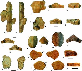
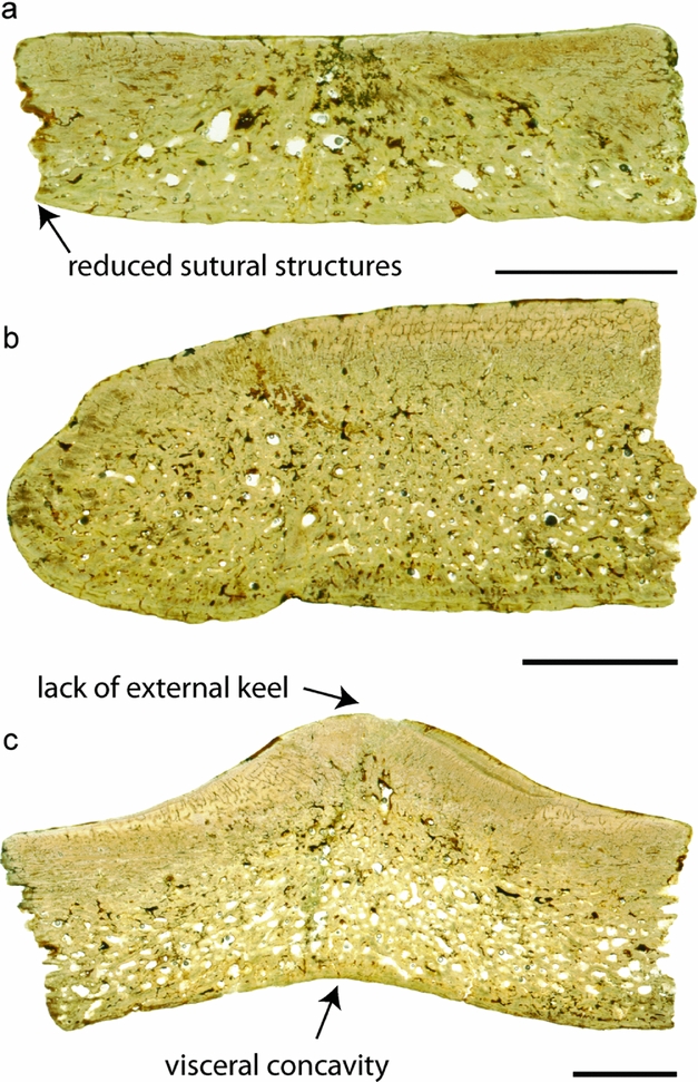
Diagnosis. Because only shell fragments are known from the type locality, this diagnosis is based only on the morphology and arrangement of the ossicles forming the shell. P. polygonus differs from all the other dermochelyid turtles provided with an ‘epithecal’ shell formed by several small bony ossicles based on the following combination of characters: thick ossicles; ossicles not greatly varying in size and shape; ossicle edges with reduced sutural structures; ossicles not arranged in the ‘sunflower’ pattern typical of Natemys; largest ossicles arranged in several longitudinal ridges; ridge-forming ossicles often viscerally flat (ridge expressed only on the external surface) and dorsally arched in cross-section, but in some cases viscerally concave (the ridge is also expressed on the visceral surface) and tectiform in cross-section; ridge ossicles characterized by variable thicknesses but without evident external keels (present in Dermochelys; compare Fig. 1 with Fig. 2); presence of peaks and valleys on the longitudinal ridges; peaks and bottom of the valleys corresponding to the sutures among ossicles; neighbouring longitudinal ridges separated by at least five ossicles (which are 13 on average in the remains from Southern Italy; Chesi et al. Reference Chesi, Delfino, Varola and Rook2007).
Ossicles of Psephophorus polygonus Meyer, Reference Meyer1847 from Devínska Nová Ves (Slovakia). (a–d) External, lateral and extremities views of the ossicles forming a longitudinal ridge (MTM V 61.1216/a); note that in lateral view the edge of the ridge is approximately straight and that in anterior and posterior views the visceral surface of the carapace corresponding to the keel does not markedly follow the external surface. (e–g) External, extremity and lateral views of a ridge ossicle (MTM V 61.1216/b); note that the ventral surface is nearly flat and that in lateral view the thickness of the ossicle is not uniform. (h–j) Idem (MTM V 61.1216/c). (k, l) Ossicle with a marked ventral concavity and a clear tectiform shape in cross-section; external and extremity views, respectively (MTM V 61.1216/d). (m, n) Idem (NHMW 2011/0151/0001). (o) Large isolated ossicle coming from the region between two ridges; external view (MTM V 61.1216/e). (p) Pair of ossicles from the same region; external view (MTM V 61.1216/f). (q–s) Flat polygonal ossicle (NHMW 1848/0015/0001al) with rounded margin in external, visceral and lateral view. (t–v) Flat polygonal ossicle (NHMW 1848/0015/0001am) in external, visceral and lateral view. Arrows indicate planes of sectioning for histology. Scale bars equal 10 mm.

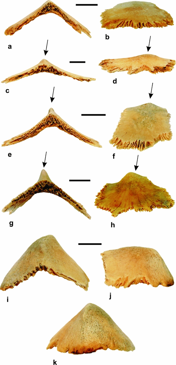
Ridge ossicles of extant Dermochelys coriacea MDHC 336 showing the variability in the development of the apical keel (indicated by the arrow). (a, b) Keel absent. (c, d) Keel very low. (e, f) Keel moderately developed. (g, h) Keel well developed. (i, k) Posterior ridge ossicles, devoid of any keel, characterized by being posterodorsally convex, having sutures only on the anterior and visceral surfaces. (a, c, e, g, i) Anterior view. (b, d, f, h, j) Left lateral view. (k) Posterior view. Note that all the ridge ossicles are ventrally concave (despite considerable variation in the degree of concavity) and that they are very thin, with a modest increase in the thickness corresponding to the dorsal apex. Scale bars equal 10 mm.

From a histological perspective, P. polygonus differs at least from D. coriacea in having shell ossicles that retain the diploe structure with an internal cortical coarse fibrous meshwork throughout ontogeny (the ossicles of D. coriacea do not have the internal cortex).
3. Gross morphology
The following description deals with the isolated ossicles and the poorly preserved slab MTM V.61.1219 available in the four above-mentioned collections and does not take into consideration the slab NHMW 2011/0330/0001 that is thought to be the neotype of P. polygonus (for its description see Seeley, Reference Seeley1880; Wood et al. Reference Wood, Johnson-Gove, Gaffney and Maley1996).
None of the isolated ossicles here described is characterized by a ‘radiating pattern’ (Seeley, Reference Seeley1880; Wood et al. Reference Wood, Johnson-Gove, Gaffney and Maley1996) on the external surface, whereas many have a fibrous visceral surface usually hosting few small foramina (usually from two to five). Among the ten well-preserved ossicles in the slab MTM V.61.1219, three show a weak radiating decoration that is comparable to what is seen in the neotype of P. polygonus. It is apparent that this pattern was subsequently obscured by erosion on the ‘epithecal’ portions that suffered more intense transportation and fell apart into isolated ossicles. The edge of the lateral surfaces of both ridge and flat ossicles do not have sharply serrated sutural margins as in Dermochelys, being rather smooth and straight. However, irregular sutural surfaces, with variably expressed smaller denticles, are visible on the lateral sides of the ossicles.
Six ossicles in the MTM collections (MTM V 61.1216/a) form a major shell fragment that is 94.2 mm long and 51.9 mm wide (Fig. 1a–d); it is composed of three ossicles forming a ridge and three lateral ossicles fitting with them. The ossicles of the ridge are distinctly elongated (32.0 × 21.0 mm; 29.0 × 19.0 mm; 32.0 × 22.0 mm) and larger than the surrounding elements. Owing to absence of informative characters, it is not possible to identify the anterior and posterior edges, but the designation of an external and a visceral surface is obvious. The outer surface of the ridge has a wide convexity (about 140°) and it is apically rounded (Fig. 1c, d). If seen in lateral view, the ridge is approximately linear (Fig. 1b), without the distinctive peak and valleys typical of the neotype of P. polygonus (Wood et al. Reference Wood, Johnson-Gove, Gaffney and Maley1996; Chesi et al. Reference Chesi, Delfino, Varola and Rook2007). The visceral surface of the ridge ossicles is nearly flat, having only a weak concavity that is amplified by the ossicles lateral to the ridge in Figure 1c, d. The ridge ossicles are slightly thicker at the centre (at the extremities of the shell fragment: 14.1 and 13.0 mm) than at the lateral edges (12.1 and 11.1 mm, respectively).
Several other isolated ossicles at the MTM were clearly part of a ridge. Most of them are congruent with the descriptions provided by Wood et al. (Reference Wood, Johnson-Gove, Gaffney and Maley1996). Two of the largest, MTM V 61.1216/b,c (Fig. 1e–j), are not distinctly elongated (37.5 × 35.3 mm; 34.6 × 43.2 mm) but are asymmetrically thickened: the thickness varies by about 3 mm at the two sides of the main axis of the ridge (13.8 and 10.7 mm in one case, 13.1 and 10.2 mm in the other); the external profile is therefore slightly undulating in lateral view (Fig. 1g, j). As in the case of the ridge ossicles previously described, the ventral surface is approximately flat or only very slightly concave (Fig. 1f, i).
Other ridge ossicles are not congruent with the descriptions provided by Wood et al. (Reference Wood, Johnson-Gove, Gaffney and Maley1996). MTM V 61.1216 (Fig. 1k, l) is characterized by a tectiform profile, a markedly convex external surface (about 90°) and a strikingly concave visceral surface; the thickness at the centre (15.1 mm) is about double that at the edges (8.5 mm). This element is not completely preserved since its lateral surface is broken off at its base.
A similar condition is shown by NHMW 2011/0151/0001 (Fig. 1m, n). It is rather large, 45.1 mm long and 39.2 mm wide, and characterized by a markedly concave ventral surface even if the thickness of the ossicles is 6.9 mm along the edge parallel to the axis of the ridge and 12.1 mm at the extremity of the ridge. The angle between the external sides is 117° whereas the one corresponding to the visceral sides of the ossicles is 145°. In cross-section, the ossicle is tectiform and not arched.
In the four collections, all the isolated ossicles originally located between the longitudinal ridges of the shell (examples in Fig. 1o, v) are approximately flat, variably shaped and sized (from 40.7 × 24.3 mm to 16.8 × 8.7 mm) and with thicknesses varying from 19.2 to 4.8 mm.
4. Microstructure
4.a. Materials
4.a.1. Psephophorus polygonus
Three ossicles from the type locality of P. polygonus were obtained for histological study, including one ridge ossicle (MTM V 61.1216/c, Fig. 1h–j) and two ossicles of the flat polygonal type (NHMW 1848/0015/0001al, Fig. 1q–s, and NHMW 1848/0015/0001am, Fig. 1t–v). The thicker flat ossicle (NHMW 1848/0015/0001al) has a maximum thickness of 10.0 mm and shows one rounded margin without sutures, maybe representing a marginal ossicle of the carapace. In the ridge ossicle (MTM V 61.1216/c), the ridge extends only to about half of the maximum length of the specimen (presumably representing the anteroposterior axis of the element); thus, it might represent the first or the last ossicle in a series of ridge ossicles in the carapace. The external bone surface is smooth in all three specimens. Only in the thinner flat ossicle (NHMW 1848/0015/0001am; maximum thickness of 5.0 mm) and the ridge ossicle (thickness ranging between 10.0 mm at the margins and 12.0 mm below the ridge) are a few foramina visible on the visceral bone surface.
4.a.2. Dermochelys coriacea
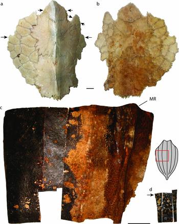
Several associated ossicles, including six irregularly formed flat polygonal ossicles and two ridge ossicles of a carapace fragment of a presumably juvenile to subadult specimen (PIMUZ A/III 1288 collected along the Atlantic coast of northern Africa) of D. coriacea were sampled (Fig. 3a, b). The carapace fragment was completely devoid of soft tissue, so the interdigitating sutures between the ossicles were well visible. The external bone surfaces of both ossicles were rugose, with the ridge ossicles also showing some faint striation radiating from the ossicle centre towards the margins. The visceral bone surfaces were also slightly rugose and several foramina were scattered over the surface of the ossicles. The thickness of the ossicles varied between 1.5 mm (usually the flat ossicles) and up to 4.0 and 5.0 mm on each ridge ossicle. As noted by Deraniyagala (Reference Deraniyagala1939), ossification of ossicles starts during the first year of life below the ridges (ossicles larger in outer ridges, smaller in the more medially situated ones), and then spreads towards the areas between ridges. In an animal of 662 days, so less than two years of age, he further noted that the ‘epithecal’ carapace has almost fully developed (see also Frazier, Gramentz & Fritz, Reference Frazier, Gramentz, Fritz and Fritz2005, p. 298, table 29). We here assume that the specimens used for sectioning derived from an individual that was thus at least two years old, because the carapace fragment shows well-developed and well-sutured ossicles both of ridge and intra-ridge areas. A more accurate age determination of the material is not possible owing to the lack of growth marks (see below).
Carapace fragments of Dermochelys coriacea sampled in the study. (a, b) Carapace fragment (PIMUZ A/III 1288) in external (a) and visceral view (b), respectively. Note size and shape differences in the flat polygonal ossicles lateral to the ridge ossicles, as well as the presence of an anomalous, elongated, extra ossicle which is not seen in MDHC 336 (Fig. 2). Arrows indicate planes of sectioning for histology. (c, d) Left part of the carapace of an adult specimen (QM J73979, cranial towards the top of image). The position of the removed part is indicated by the red box on the small sketch of the overall carapace. (d) Portions of carapace that were removed from the larger piece for histological sectioning. The sectioned ridge ossicle shown in Figures 6 and 7 derived from the slim rectangular portion in the upper left. Plane of sectioning is indicated by black arrow. Scale bars equal 10 mm in (a, b) and 10 cm in (c, d). Abbreviation: MR – medial ridge.

Additionally, ossicles from three wild-caught specimens of D. coriacea from southeastern Queensland were sampled between 2004 and 2006 by three of the authors (SM, TF and SWS): a flat, polygonal ossicle from a small adult male (QM J81592) and a ridge ossicle from a large adult (QM J73979; Fig. 3c, d). Because these ossicles were cut out of the integument, the margins of the plates were not sampled. Portions of the carapace of a hatchling specimen (QM J58751) from a failed nest were also examined in order to cover a wide range of ontogenetic development.
The young adult specimen (QM J81592; male, maximum curved carapace length 163.5 cm) was found in a shark net off the Gold Coast, southeastern Queensland, on 25 September 2004. After collection, a portion of carapace was removed and kept on ice for few days before it was preserved in 90% ethanol. The exact location and orientation of the portion of carapace that was removed was not recorded, but it is assumed to have come from the caudal left side of the animal. The ossicle that was sampled from this portion of carapace is situated just below the transition between the external cuticle and the lipid-rich fibrous tissue of the dermis, where the dermal fibre bundles trend sub-parallel to the external surface of the integument. A thin layer of dermal connective tissue thus separates the ossicle from the epidermal tissue. In cross-section, the layer of horizontally arranged dermal fibre bundles is followed internally by a thick, adipose-rich layer of integumentary tissue, where fibre bundles are more loosely arranged. The fibre bundles extend in several directions, with a slight dominance of fibre bundles that extend roughly diagonally towards the external surface of the integument.
The adult specimen (QM J73979; sex unknown, total length recorded as 200 cm, assumed to be curved carapace length but not specified) was found drowned in a fishing net in Moreton Bay, just off Skirmish Point, Bribie Island, southeastern Queensland, sometime during 2004. The carcass was taken ashore and left for several months, during which time portions of the carapace became desiccated. The extremely high lipid content of the integument prevented processing of good sections during this period. During the desiccation process, the ‘epithecal’ (secondary) shell locally separated from the ribs. Histological samples were subsequently taken from a ridge ossicle on the left side of the desiccated carapace (Fig. 3c, d). The sampled ridge ossicle is tectiform in cross-section. Because the bone was dissected out of the surrounding integumentary tissue, its connection with the dermal tissue is not recorded.
The hatchling specimen (QM J58751; sex unknown, maximum carapace length 8.2 cm) from a failed nest, thought to be at Wreck Rock, between Gladstone and Bundaberg, in the Wide Bay–Burnett region of southeastern Queensland, was gutted and fixed in formalin, and then kept in 70% alcohol. Histological slides from this specimen were based on a transverse section through a caudal portion of the carapace, dorsal to a rib and extending from the lateral margin to 3 mm past the median osteoderm ridge. Because the hatchling specimen shows largely unossified soft tissue integumentary structures, i.e. subcutical adipose tissue, and the formation of the ribs and vertebral column only, with no ‘epithecal’ ossicles (which is again in order with the developmental timing presented by Deraniyagala, Reference Deraniyagala1939, see above), it was excluded from the following Results and Discussion sections.
4.b. Histological sampling
The sampling of the ossicles followed standard procedures for petrographic thin-sections. The slides were studied using a Leica DM2500 M composite microscope mounted with a Leica DFC 420 C digital camera.
4.c. Results
4.c.1. Psephophorus polygonus
All three ossicles sampled show similar histological features, thus warranting their description in one section. Differences tied to variation in overall shape between the flat polygonal and the ridge ossicles are pointed out where necessary. Although all three specimens are quite compact bones, they still present a diploe structure (Fig. 4), with external and internal cortices surrounding a cancellous core. On the other hand, it is extremely difficult to differentiate between the tissue present in the cancellous core and that of the internal cortex, because both tissues share histological features.
Overview of thin-sections of the ossicles of Psephophorus polygonus in normal transmitted light. Note thickness of ossicles and the diploe structure that is retained in all three specimens. (a) Flat polygonal ossicle (NHMW 1848/0015/0001am). (b) Flat polygonal ossicle (NHMW 1848/0015/0001al). (c) Ridge ossicle (MTM V 61.1216/c). Scale bars equal 5 mm.

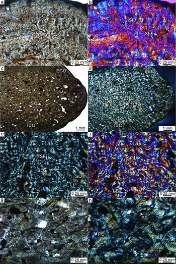
External cortex. The external surface of the ossicles is even, disturbed only by the insertion of small foramina. The cortex itself is composed of interwoven structural fibre bundles, in which the bundles are mostly homogeneous in size and diameter, with a slight dominance of fibre bundles that extend at high angles to the external bone surface (Fig. 5a, b). The bone tissue is vascularized by an extensive network of primary vascular canals, scattered primary osteons and few secondary osteons. The amount of secondary osteons increases towards the internal cancellous bone. Bone cell lacunae are round or slightly flattened and elongated and appear scattered or clustered. In the lamellar bone of the secondary osteons, the cell lacunae are flattened and elongated and are aligned along the secondary bone lamellae. The ridge ossicle shows patches in the cortex, in which the typical interwoven structure is abruptly offset by a more organized lamellar structure reminiscent of parallel-fibred bone tissue. Although growth marks are visible in all three specimens, it is only in these more organized areas of the external cortex of the ridge ossicle that they are most pronounced (although not continuous). In NHMW 1848/0015/0001al, the external cortical bone is continuous all along the rounded margin, grading into the internal cortical tissue at the visceral side of the ossicle (Fig. 5c, d).
Histology of the ossicles of Psephophorus polygonus. Images in (a, c, g) normal transmitted light, (d, e, h) polarized light, and (b, f) in polarized light with additional lambda compensator. (a, b) The external cortex of ossicle NHMW 1848/0015/0001al is composed of interwoven structural fibre bundles vascularized by a reticular network of primary vascular canals. Sharpey's fibres point perpendicular to the external bone surface. (c, d) Rounded margin of ossicle NHMW 1848/0015/0001al. Note how the external cortical bone continues onward to the visceral side of the ossicle. (e, f) Interior core area of ossicle NHMW 1848/0015/0001al showing interwoven structural fibre bundles and numerous secondary osteons. (g, h) Close-up of the irregular meshwork of mainly longitudinally sectioned coarse fibre bundles of the internal cortex of ossicle NHMW 1848/0015/0001am. The tissue is vascularized by simple primary vascular canals. Abbreviations: ECO – external cortex; ICO – internal cortex; ISF – interwoven structural fibre bundles; LB – lamellar bone; PC – primary vascular canals; ShF – Sharpey's fibres; SO – secondary osteons.

Cancellous bone and internal cortex. The interior core of the ossicles is more compact (Fig. 5e, f) in the flat polygonal ossicles than in the ridge ossicle, with larger and longer trabeculae being completely absent. Even the ridge ossicle shows only a few trabeculae. The tissue is predominantly still primary and of a coarse cancellous type. Remodelling of the bone appears to be restricted to scattered secondary osteons and few larger erosion cavities. Internally, the tissue increasingly becomes coarsely fibrous, with the internal cortex consisting completely of a meshwork of coarse fibre bundles (Fig. 5g, h). Although the coarse bundles form a three-dimensional meshwork, the whole tissue is dominated by fibre bundles extending from the growth centre of the ossicle (at the plate centre close to the external bone surface) towards the internal bone surface in a fan-shaped pattern. Growth marks are also faintly seen in the internal cortical bone.
Sutures. The sutural zones are narrow with short bony protrusions and sockets. The bone tissue adjacent to the sutures is similar to the tissue described for the external cortex, but shows more signs of remodelling. Sharpey's fibres that extend perpendicular to the margin dominate the sutural bone tissue.
4.c.2. Dermochelys coriacea
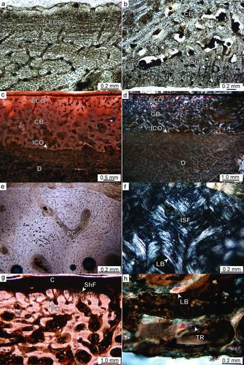
All sampled ossicles of D. coriacea are similar in bone microstructure, so they will be described in one section (Fig. 6), with peculiarities being pointed out if applicable. The ridge ossicles of the presumably juvenile to subadult specimen (PIMUZ A/III 1288) show a diploe structure, with external and visceral cortices framing an interior cancellous centre. In some of the flat polygonal ossicles, the interior core is often restricted to a thin area of vascular primary bone. Noteworthy, one of the ridge ossicles of the juvenile to subadult specimen revealed a small internal (visceral) ‘keel’, caused by a local thickening of the cancellous bone core below the external ridge. The bony ossicle of the young adult specimen (QM J81592) comprises a layer of external cortical bone and an internal layer of cancellous bone, with an internal cortical layer of compact bone being almost absent. The ossicle of the adult specimen (QM J73979), marginally thicker than that of the young adult specimen, is composed of two layers only, a more compact external layer and a vascular internal layer; an internal cortical layer is not visible.
Histology of the ossicles of Dermochelys coriacea. Images (a–c, e, g) in normal transmitted light and (d, f, h) in polarized light. (a) External cortex of the flank of smaller ridge ossicle of PIMUZ A/III 1288. Note extensive vascular system and scalloped external bone surface. (b) Interior core area and internal cortical bone of smaller ridge ossicle of PIMUZ A/III 1288. (c, d) Ossicle of young adult specimen (QM J81592) still embedded within the integument. Note remnant of internal cortex of the ossicle, as well as the soft tissue structure of the dermis. (e, f) Close-up of the external cortex of the ossicle of QM J81592. (g) Close-up of the external cortex and strongly remodelled interior core area of the ossicle of the adult specimen (QM J73979). (h) Close-up of the strongly remodelled internal area of the ossicle of the adult specimen (QM J73979) leading to complete absence of compact bone. Abbreviations: C – epidermal cuticle; CB – cancellous bone; D – dermis, ECO – external cortex; ICO – internal cortex; ISF – interwoven structural fibre bundles; LB – lamellar bone; PC – primary vascular canals; ShF – Sharpey's fibres; TR – trabeculae.

External cortex. In all specimens (e.g. Fig. 6a, e, f), the external cortex consists of interwoven structural fibre bundles. The bone tissue is highly vascularized with an extensive reticular network of primary vascular canals and scattered primary osteons. The external bone surface is rough and scalloped with deep foramina inserting into the cortical bone (Fig. 6a, e, g). Especially in the older individuals, Sharpey's fibres are locally found as thick and dark fibrous strands inserting at high angles (Fig. 6g) into the external-most layers of the cortex, whereas they are less conspicuous in the juvenile to subadult specimen. In the ridge ossicle of the adult individual, the cortical bone itself resembles only a spongy meshwork of trabecular struts, with the large vascular intertrabecular cavities being filled with brown adipose tissue. In the juvenile to subadult specimen, the external-most cortical layer usually does not show bone cell lacunae, followed by a second layer in which the cell lacunae are small and round and usually devoid of canaliculi. Only the deeper cortical layers then also house larger flattened osteocyte lacunae with canaliculi. This stratification of the cortex was not observed in the two older specimens. Furthermore, growth marks (i.e. lines of arrested growth) were not observed in any of the samples.
Cancellous bone. The internal cancellous bone comprises trabeculae and scattered secondary osteons (Fig. 6b–d, g). Interstitial primary bone is still present within the lamellar bone walls of many trabecular struts and trabecular branching areas. Other trabeculae, especially in the ridge ossicle, are at various stages of remodelling, resulting in secondary trabeculae composed solely of lamellar bone. Bone cell lacunae in the lamellar bone are flattened and elongated, while they are more round and plump in interstitial areas. The intertrabecular cavities are also filled with brown adipose tissue. Where the adipose tissue desiccated, small empty vascular spaces appeared.
Internal cortex. The ossicles of the presumably juvenile to subadult specimen all showed a thin internal cortex (Fig. 6b), with the cortical tissue being composed of parallel-fibred bone. The tissue is vascularized by primary vascular canals extending predominantly anteroposteriorly (parallel to the trend of the ridge). Abundant coarse Sharpey's fibres insert into the internal cortex perpendicularly or at moderate angles, especially in the flanks of the ridge ossicles. In the young adult specimen (QM J81592), only remnants of this internal compact cortical layer are still visible, with other parts being already completely cancellous (Fig. 6c, d). Unfortunately, most of the structural details in this specimen are obscured here because of the high fat content of the adjacent deeper dermal tissue. The cortex is completely remodelled in the adult ossicle (Fig. 6h).
Sutures. The internal structure of sutural areas could only be studied in the purported juvenile to subadult specimen (PIMUZ A/III 1288). As indicated already by the irregular shape of the ossicles in external view, the bones show wide sutural areas in cross-section with long protrusions and sockets. The bone tissue again is similar to that of the external cortex, with Sharpey's fibres inserting perpendicularly into the bone tissue.
5. Discussion
Wood et al. (Reference Wood, Johnson-Gove, Gaffney and Maley1996) identified about 20 characters for the analysis of the phylogeny of dermochelyid turtles. The evaluation of some of them, such as the number of ossicles between ridges, is restricted to the fossils where portions of shell (with ossicles) are preserved in connection (i.e. in vivo). Conversely, other characters can be better evaluated on isolated ossicles. The type locality materials of P. polygonus listed and described herein have been overlooked for several decades by researchers who concentrated only on the in vivo ossicles preserved on the slab displayed at the Naturhistorisches Museum Wien, NHMW 2011/0330/0001 (among others Wood et al. Reference Wood, Johnson-Gove, Gaffney and Maley1996; Chesi et al. Reference Chesi, Delfino, Varola and Rook2007). On the basis of the isolated ossicles here described, as well as those of the neotype, it is possible to re-evaluate the morphological differences between the extinct P. polygonus and the extant D. coriacea.
5.a. Macro-morphological differences between Psephophorus polygonus and Dermochelys coriacea
The main differences can be summarized as follows.
According to Wood et al. (Reference Wood, Johnson-Gove, Gaffney and Maley1996), P. polygonus has ridge ossicles that are viscerally flat, whereas the ridge ossicles of D. coriacea are characterized by a concave visceral surface (see Wood et al. Reference Wood, Johnson-Gove, Gaffney and Maley1996, fig. 19c, e). Such morphology has been only partly confirmed by our material. Some ridge ossicles of P. polygonus are actually viscerally flat, unlike those of D. coriacea (compare Fig. 1c, d, f, i with Fig. 2a, c, e, g). However, two isolated ridge ossicles (MTM V 61.1216/d and NHMW 2011/0151/001) are characterized by a distinct visceral concavity (Fig. 1l, n). Even if the viscerally concave ossicles clearly do not belong to D. coriacea because, among other factors, of their thickness, such polymorphism requires a reconsideration of the phylogenetic analysis of the dermochelyid taxa performed by Wood et al. (Reference Wood, Johnson-Gove, Gaffney and Maley1996), which was based on very few characters, some of which are correlated with each other or are phylogenetically uninformative. It is worth noting that an unnamed Eocene dermochelyid from Alabama (USNM 23699) has ossicles similarly concave viscerally, but (at least from the only figured specimen; Wood et al. Reference Wood, Johnson-Gove, Gaffney and Maley1996, fig. 14) it is characterized by a nearly uniform thickness of the ossicles. In P. polygonus, the thickness of the ridge ossicles significantly decreases laterally.
Although the carapaces of both P. polygonus and D. coriacea have ridges that are not straight but undulated, the nature of the undulations is quite different. The peaks and valleys on the ridges of P. polygonus correspond to the sutures between contiguous ossicles so that each convexity is formed by two contiguous ossicles with its apex – the peak – corresponding to the suture between them. The asymmetrical thickening of the isolated ossicles described above (see Fig. 1g, j) obviously contributes to the undulating profile. Conversely, in D. coriacea there is no significant asymmetrical thickening and each peak usually corresponds to a keel on the external surface of a single osteoderm. As in P. polygonus, the bottom of a valley corresponds to the suture between two ossicles. The expression of the keel on the ridge ossicles of D. coriacea can be quite variable, from non-existent as shown in Figure 2a, b, to distinctly developed, as in the case of the ossicle shown in Figure 2g, h. A particular case of undulation is the one occurring in the posterior region of the carapace (Fig. 2i–k), where the external edges of the ridge ossicles do not have any sort of keel, but are so markedly convex that the posterior edges of the ossicles do not host any sutures (which are limited to the anterior and ventral edges).
All the ossicles, either from the ridge or from the area between the ridges, are proportionally much thicker in P. polygonus than in D. coriacea (compare for example Fig. 1f, n, s, v with Fig. 2a, c, e, g). Probably because of the difference in thickness and (considering the density) weight, the sutures of the ossicles of P. polygonus are not as developed as those of D. coriacea (compare for example Fig. 1e, f with Fig. 2e, f), which appears to need relatively deeply interdigitating sutures to keep the thin and light ossicles in firm contact with each other. The ossicles of P. polygonus are also not as variable in size as those of D. coriacea, which shows a remarkable difference between the large ridge ossicles and the much smaller flat ossicles (compare Fig. 1 with Fig. 3).
Some of the differences in carapace morphology observed between P. polygonus and D. coriacea may relate to diving capability. D. coriacea routinely dive to depths of around 300 m (Houghton et al. Reference Houghton, Doyle, Davenport, Wilson and Hays2008), but have been recorded going as deep as 1280 m (Doyle et al. Reference Doyle, Houghton, O'Súilleabháin, Hobson, Marnell, Davenport and Hays2008). It is very likely that a rigid shell would collapse or fracture during such deep dives, but the carapace of D. coriacea is thought to be able to deform under pressure, returning to its original shape upon ascent (Spotila, Reference Spotila2004). If such deformation does occur, this flexibility appears to be the result, at least in part, of the reduction of the ‘thecal’ elements and the configuration and structure of the osteoderms and ossicles of which the secondary (‘epithecal’) carapace is comprised. The osteoderms and ossicles are much thinner and internally more vascularized within the external cortex and cancellous core areas, particularly in mature individuals, relative to the fused ‘thecal’ plates that make up the carapace and plastron of other extant sea turtles, and their small size and interdigitating sutural connections, along with their lack of direct connectivity with the ribs and girdle elements, appear to facilitate this flexibility.
The thicker, denser and proportionately larger osteoderms and ossicles of P. polygonus, with their straighter sutural margins, are very likely to have resulted in a more rigid carapace than in D. coriacea. Assuming that diving ability in D. coriacea correlates in part with the size, shape, thickness and configuration of the carapace osteoderms and ossicles, it seems likely that, while probably a capable diver relative to extant sea turtles, P. polygonus would not have been able to reach the depths that D. coriacea can. As initially observed by Wood et al. (Reference Wood, Johnson-Gove, Gaffney and Maley1996), within the various clades of Dermochelyidae there is a clear evolutionary trend for a decrease in the thickness, size and density of the osteoderms and ossicles that compose the secondary (‘epithecal’) carapace. Our observations of the osteoderms and ossicles of P. polygonus support this trend, and indicate that many of the morphological changes that have occurred within the Dermochelyidae relate to an improved capacity for deep dives.
5.b. Histological properties of Psephophorus polygonus and Dermochelys coriacea
All ossicles of P. polygonus shared similar histological structures (e.g. diploe structure, growth marks in the external cortex, coarse fibrous meshwork of the internal cortex) although there were also aspects that appear to be linked to the overall shape of the ossicles (flat v. ridge ossicles). As such, the abrupt change from interwoven to more parallel-fibred organization in the external cortex was only visible at the flanks of the ridge ossicle.
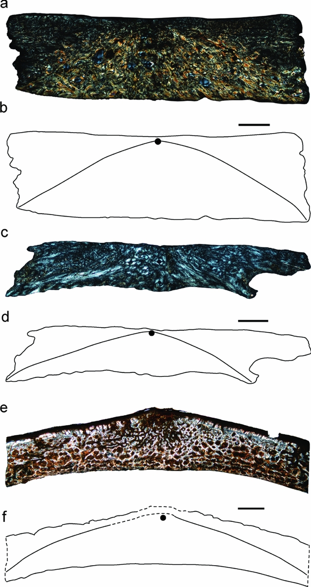
Independent of shape, the growth centre was situated close to the external bone surface at the ossicle centres in P. polygonus, as well as in D. coriacea ossicles (Fig. 7). On the other hand, ontogenetic changes became apparent in the histology of the D. coriacea ossicles such as the loss of internal cortical bone (i.e. the parallel-fibred bone tissue), increased vascularization of the external cortex and cancellous core areas, as well as an increased rugosity of the external bone surface with increased age, all of which clearly distinguish them from ossicles of P. polygonus.
Comparison of histological sections of flat polygonal and ridge ossicles of Psephophorus polygonus and Dermochelys coriacea, with interpretative sketches of the distribution of external and marginal bone tissue versus interior core and internal bone tissue of the specimens sampled. Growth centres of the plates are indicated by a black spot in the sketches. Images in (a, c, e) in polarized light. (a, b) Flat polygonal ossicle (NHMW 1848/0015/0001am) of P. polygonus. (c, d) Flat polygonal ossicle (PIMUZ A/III 1288) of carapace fragment of juvenile to subadult specimen of D. coriacea. (e, f) Ridge ossicle of the adult specimen of D. coriacea (QM J73979). Note that in this specimen, the cuticle is still attached to the ossicle. The flat polygonal ossicles as well as the ridge ossicle show growth centres located at the plate centres close to the external bone surface. Scale bars equal 2 mm in (a, b) and (e, f), and 1 mm in (c, d).

The ossicles of dermochelyid turtles, as exemplified by NHMW 1848/0015/0001am of P. polygonus or the thin flat ossicles of the presumably juvenile to subadult D. coriacea (PIMUZ A/III 1288), show growth centres situated at the plate centres just interior to the external bone surface (Fig. 7). These result in a peculiar wedge-like histological morphogenesis of the ossicles, in which the external cortical bone tissue is thinnest at the ossicle centre and expands towards the margins. In this aspect, dermochelyid ossicles differ from those of other lineages of amniotes with dermal elements in the trunk. In the latter, dermal elements are composed largely of flat polygonal ossicles or platelets. In the dermal ossicles of cyamodontoid placodonts (Sauropterygia), glyptodont xenarthrans (Mammalia), as well as thyreophoran dinosaurs and titanosaurian sauropod dinosaurs, the growth centres are usually either situated centrally within the cancellous core or closer to the internal compact bone layer (e.g. Westphal, Reference Westphal, Bellairs and Cox1976; Ricqlès et al. Reference Ricqlès, Pereda Suberbiola, Gasparini and Olivero2001; Hill, Reference Hill2006; Vickaryous & Hall, Reference Vickaryous and Hall2006; Scheyer, Reference Scheyer2007; Cerda & Powell, Reference Cerda and Powell2010), resulting in a more hourglass-shaped pattern. Accordingly, although fundamental functional aspects of the morphogenesis of polygonal carapace plates in general (e.g. Westphal, Reference Westphal, Bellairs and Cox1976) appear to apply also to dermochelyid ossicles, the growth centres of developing dermochelyid ossicles appear to lie at a much shallower level within the dermis close to the epidermal–dermal boundary, than in other amniote lineages.
5.c. Palaeobiogeography and taxonomic remarks
The genus Psephophorus has been considered a wastebasket taxon (Wood et al. Reference Wood, Johnson-Gove, Gaffney and Maley1996) and a few species have been erected from isolated ossicles, small portions of shells and humeri collected from sediments deposited in coastal settings worldwide (see among others Moody, Reference Moody, Callaway and Nicholls1997; Karl, Reference Karl2002). However, according to Wood et al. (Reference Wood, Johnson-Gove, Gaffney and Maley1996), the only remains clearly referable to the species P. polygonus were the ones from the type locality, and the results of their phylogenetic analysis suggested the splitting of Psephophorus into many genera. One of the consequences of this split has been the restriction of the range of Psephophorus from nearly worldwide to an area as specific as the type locality. The subsequent referral of large portions of a dermochelyid shell from the middle Miocene of Southern Italy to P. polygonus (Delfino, Reference Delfino2002; Chesi et al. Reference Chesi, Delfino, Varola and Rook2007) considerably expanded the range of this species to a larger sector of the Tethys.
However, despite evident macro- and microscopic differences in the shell architecture, P. polygonus is, in general terms, morphologically very close to D. coriacea (but we note that they are both significantly different from all the other marine turtles). It is thus tempting to assume that, perhaps with the exception of a capacity for extremely deep dives, the ecology of P. polygonus was similar to that of D. coriacea, which is the most cosmopolitan extant turtle species, ranging throughout the Pacific, Atlantic and Indian oceans (e.g. Pritchard, Reference Pritchard1980), occurring in cool waters up to 69° north and down to 47° south (Frazier, Gramentz & Fritz, Reference Frazier, Gramentz, Fritz and Fritz2005). Although the shell of P. polygonus was more massive than that of D. coriacea (which has thin ossicles without the internal cortex), the overall morphology is so similar that it seems likely that P. polygonus was similarly far ranging, such that its remains may one day turn up outside of the Tethys. Even if all the Psephophorus fossils known so far come from sediments deposited in coastal settings (therefore with a relative high energy that does not help the undisturbed preservation of large carcasses), this does not necessarily mean that Psephophorus inhabited only coastal waters. Despite the fact that Dermochelys is clearly a true pelagic turtle, its fossil record is virtually non-existent (Lapparent de Broin, Reference Lapparent de Broin2001), being limited to very few archaeological localities (Frazier, Reference Frazier2005; M. D. unpub. obs).
Moreover, the large size of Psephophorus, which was at least comparable to that of extant Dermochelys (see Chesi et al. Reference Chesi, Delfino, Varola and Rook2007), could demonstrate it was a gigantotherm. Given that the capacity for gigantothermy is also a key factor behind the worldwide distribution of Dermochelys (in the Dermochelyidae it can be traced back at least to the Middle Eocene; Albright et al. Reference Albright, Woodburne, Case and Chaney2003), it further supports our supposition that P. polygonus may have had a much broader geographic distribution than its present fossils indicate.
Future revisions of Miocene Psephophorus-like fossils, as well the collection of new materials, will help in clarifying these issues. In particular, it should be possible to test if ‘Psephophorus’ calvertensis Palmer, Reference Palmer1909 from the middle Miocene Calvert Formation of Maryland and ‘Psephophorus’ californiensis (Gilmore, Reference Gilmore1937) from the middle Miocene Temblor Formation of California, are not only conspecific, as recently suggested by Lynch & Parham (Reference Lynch and Parham2003), but also if they are junior synonyms of P. polygonus. If so, the range of P. polygonus would encompass both the Atlantic and the Pacific oceans, mirroring the distribution of the extant D. coriacea.
According to Wood et al. (Reference Wood, Johnson-Gove, Gaffney and Maley1996), ‘P.’ calvertensis differs from P. polygonus in only two characters: the carapacial ridges are also expressed on the visceral surface of the ossicles (the visceral surface of the ridge ossicles is concave; character 10) and the ridges are tectiform in cross-section (character 11). However, as shown in Figure 1n, the ossicles of P. polygonus can also have at the same time a concave visceral surface and a tectiform shape in cross-section. As such, ‘P.’ calvertensis and P. polygonus could be synonymous, something that should be verified in the future by direct revision of the material in question.
Acknowledgements
Thanks to H. Furrer (PIMUZ), M. Gasparik (HMNH), M. Harzhauser (NHMW) and U. Göhlich (NHMW) for assisting us while studying the fossil materials, for providing specimens for destructive sampling and relevant information about the fossil remains and their origin. U. Göhlich also discussed the chronological allocation of the fossil material. L. Kordos (Geological Institute, Budapest) helped in solving the toponymic synonymy. B. Villier (Torino) provided technical assistance for some photographs. A. Amey and P. Couper (Queensland Museum) are to be thanked for helping with the collection of QM J73979, and permitted the sampling of QM J81592 and QM J58751 for histological work. This work was supported by the Swiss National Science Foundation (T.M.S., Grant no. 31003A_127053/1), the Spanish Ministerio de Economia y Competitividad (CGL2011-28681), the Italian MIUR PRIN 2009MSSS9L_002 (to G. Pavia, Torino) and the Synthesys Programme (M.D., AT-TAF-1281 and HU-TAF-1894). M. Delfino's research was originally developed at Università di Firenze, thanks to the support of MIUR PRIN (to E. Abbate) and Fondi di Ateneo (to L. Rook).