Introduction
Nasal septal perforations are full-thickness anatomical defects of the nasal septum with loss of cartilage or bone and mucosa. Most patients are asymptomatic. When symptomatic, patients present with epistaxis, nasal obstruction, crusting and whistling. Small perforations are associated with whistling. Large perforations, particularly anterior ones, cause nasal airflow turbulence resulting in dryness and crusting within the nasal cavity, epistaxis and reduced olfaction.Reference Kridel and Delaney1 Patients who fail conservative measures or septal button insertion may be considered for surgical closure.
Surgical repair of nasal septal perforations is challenging. Many techniques have been described, with most comprising local flaps with or without interposition grafts. These include transplantation of septal cartilage, inferior turbinate flaps, interposition grafts using tragal cartilage and temporalis fascia, acellular human dermal interposition allograft, oral mucosal flaps, and two-staged procedures with tissue expanders. Both closed and open techniques have been described. Reported results vary from 70 to 100 per cent in terms of success.Reference Ribeiro and da Silva2–Reference Dosen and Haye5 Intranasal mucosal advancement flaps with interposition grafts give the best physiological outcome and long-term success rates.Reference Kridel and Delaney1
We report septal perforation closure results and symptom control using the previously reported technique of bilateral superior and inferior nasal mucosal advancement flaps with interposition graft.Reference Wong and Raghavan6 We also compare the results of using porcine acellular collagen versus autologous septal cartilage or bone as an interposition graft as well as the benefits of reduction rhinoplasty for closing perforations larger than 15 mm.
Materials and methods
All patients with symptomatic nasal septal perforations who were operated on between November 2005 and April 2018 were included. If the aetiology could not be identified during history taking, patients underwent investigation including testing for cytoplasmic anti-neutrophilic cytoplasmic antibody and angiotensin converting enzyme. If these blood markers did not identify a cause, biopsy of the septal perforation margin was taken. All patients underwent surgical nasal septal perforation repair by the senior author (UR).
Procedure
An open rhinoplasty approach under general anaesthesia was used for all patients. Peri-operative prophylactic antibiotics were administered. Our preferences were co-amoxiclav or clarithromycin (if penicillin allergic). Two per cent lidocaine with 1:80 000 adrenaline was injected into the nasal septum and dorsum to provide hydrodissection and vasoconstriction. Further vasoconstriction was provided by neurosurgical patties soaked in 2 ml of 10 per cent cocaine with 1 ml of 1:10 000 adrenaline placed in both nasal cavities for 15 minutes. Using rigid endoscopy and a sterile ruler, the horizontal and vertical dimensions of the nasal septal perforation were measured (Figure 1).
Images showing measurements of the (a) horizontal and (b) vertical dimensions of the septal perforation taken at the start of surgery.

An inverted V-shaped transcolumellar incision was performed, and the soft tissue envelope was elevated as per the standard open rhinoplasty approach. The caudal septum was exposed by separating the medial crura. The upper lateral cartilages were dissected from the dorsal septum with the mucoperichondrial flap left attached to the dorsal edge of the upper lateral cartilage. Any available nasal septal cartilage and bone was preserved. Septal deviation requiring correction was addressed and reconstructed using conchal or costal cartilage if required. Mucoperichondrial flaps were raised bilaterally around the perforation, and the perforation margins were separated (Figure 2). The inferior part of each flap was elevated along the floor of the nose, inferior meatus and inferior aspect of the inferior turbinate, leaving it attached along the medial border of the inferior turbinate (Figure 3). If the vertical dimension of the perforation was greater than 10 mm, the superior part of the septal mucoperichondrial flap was dissected from the inferior aspect of the upper lateral cartilage (Figure 4). The lateral extent of this dissection provided adequate length for a tension-free approximation of perforation margins. If additional inferior flap length was required, an incision could be made along the medial border of the inferior turbinate to convert the inferior flap into a bipedicled flap. In our series, we did not require the use of a bipedicled flap.
(a) Image and (b) diagram of mucoperichondrial flaps raised around the perforation.

(a) Diagram and (b) image of dissection of the inferior flap along the floor of the nose and inferior meatus.

(a) Diagram and (b) image of dissection of the superior flap from the inferior aspect of the upper lateral cartilage.

After sufficiently mobilising the flaps, the perforation margins were approximated without tension and sutured with vertical mattress sutures using 4-0 polydioxanone with a reverse cutting circular needle and with the knots facing the nasal cavity (Figure 5). If autologous septal cartilage or bone was available, this was positioned between the mucoperichondrial flaps. Alternatively, acellular porcine collagen was used (Permacol®; Figure 6). The upper lateral cartilages were supported with spreader grafts and sutured to the dorsal septum. A tongue-in-groove technique was required to reposition the tip and to counter the traction exerted by the superior flap on the upper lateral cartilage. If the vertical length of the perforation was greater than 20 mm, a reduction rhinoplasty was performed to reduce the dorsum. This left excess mucoperichondrium in the superior flap to approximate the edges of the perforation.
Approximation and suturing of the flaps with vertical mattress sutures following sufficient mobilisation showing (a) diagram with arrows showing the margins of the perforation before approximation, (b) diagram with arrows showing the margins of the perforation after approximation and (c) intra-operative image of the approximated margins without tension.

(a) Diagram and (b) image of use of an acellular porcine collagen interposition graft between the mucoperichondrial flaps.

Soft Silastic® nasal splints were secured in each nasal cavity with a single 3-0 silk suture anteriorly, without compressing the superior and inferior flaps. The trans-columellar incision was closed with 6-0 prolene sutures and marginal incisions with 5-0 vicryl sutures. SteristripsTM and a nasal splint were applied on the dorsum. Internal and external splints and Steristrips were removed one week post-operatively in clinic. Oral antibiotics were continued for a week and saline nasal douching was continued for six weeks.
During the immediate post-operative period, the inferior flap was seen stretching across the nasal cavity from the medial border of inferior turbinate to the septum and the superior flap was seen blunting the nasal valve angle. The position of these flaps reverted back to the normal position within four weeks.
Assessment of symptoms
Patients rated their symptom severity using a visual analogue scale (VAS) from 0 (no symptoms) to 10 (maximal symptoms) for whistling, epistaxis, nasal obstruction and crusting pre-operatively and at six months and one year post-operatively. Patients who were followed up longer than this were asked to repeat this at their final follow-up visit.
Statistical analysis
The paired t-test was used to calculate p-values for the pre- and post-operative VAS scores.
Literature review
The Cochrane Central Register of Controlled Trials (Cochrane Library, 2011), Medline (1946–1 June 2018) and Embase (1974–1 June 2018) were interrogated. Two reviewers independently selected studies and extracted data. Disagreements were resolved by discussion.
The search terms were: ‘nasal septal perforation’, ‘nasal septum’, ‘repair’, ‘surgery’, ‘surgical’ and ‘closure’. We included papers in all languages reporting results of nasal septal perforation repair.
Results
Our case series
A total of 159 patients underwent surgical closure of nasal septal perforations during the study period. Five patients were lost to follow up; therefore 154 patients were recruited to the study. There were 102 males and 52 females. Mean age was 47 years (range, 21–76 years). The aetiology of perforation was previous nasal surgery in 62 patients (40 per cent), cocaine abuse in 23 patients (15 per cent), perforation secondary to cautery in 6 patients (4 per cent) and idiopathic aetiology in 63 patients (41 per cent). Mean perforation dimensions were 24 mm vertically (range, 5–35 mm) and 29 mm horizontally (range, 5–43 mm). Mean follow up was 76 months (range, 12–127 months).
A total of 42 patients (27 per cent) had a vertical measurement of less than 10 mm and required bilateral inferior flaps alone. A total of 112 patients (73 per cent) required bilateral superior and inferior flaps. Reduction rhinoplasty was required in 21 patients (14 per cent) where the vertical diameter of the perforation was over 20 mm. A total of 107 patients (69 per cent) had acellular porcine collagen, and 47 (31 per cent) had autologous septal cartilage or bone used as an interposition graft.
Our surgical technique achieved an overall success of 95 per cent (146 patients) in closing the septal perforation. Of the 107 patients who received acellular porcine collagen, the perforation was not closed in 5 patients (4.7 per cent), giving a success rate of 95.3 per cent. Two of these patients, with 35 × 35 mm and 30 mm × 35 mm vertical and horizontal diameter perforations following previous septal surgery, re-perforated and remained symptomatic despite reduction rhinoplasty, bilateral superior and inferior flaps, and acellular porcine collagen interposition graft. These patients continued to report crusting, epistaxis and nasal obstruction. A further 2 patients who received acellular porcine collagen had perforations measuring 23 × 20 mm and 25 × 22 mm following previous septal surgery. They had undergone previous attempts to close the perforation in another centre. Superior and inferior flaps were raised and closed without tension on the left side. However, on the right side the flaps were atrophic and scarred. It was not possible to approximate the perforation edges without tension. These patients re-perforated and remained symptomatic with nasal blockage and crusting. The final patient in the failed acellular porcine collagen group had a 15 × 15 mm perforation but failed to heal completely resulting in a 5 × 5 mm residual perforation when seen at 6 months’ follow up in the post-operative period. This patient achieved complete symptom control post-operatively.
Three patients (6.3 per cent) of the 47 who received autologous septal tissue also did not obtain closure of the perforation, giving a success rate of 94.7 per cent. Their perforations were 18 × 15 mm, 15 × 15 mm and 15 × 10 mm horizontally and vertically, respectively. They received crushed septal cartilage because only a small piece of intact cartilage could be harvested safely. Pre-operatively, both patients refused to have acellular porcine collagen or to use conchal cartilage. Both repairs failed completely, and the patients remained symptomatic. One of these patients developed neuralgic pain, which was controlled with amitriptyline. One patient in this group had a 20 × 20 mm perforation and also had crushed cartilage, but the perforation healed partially leaving a 10 × 5 mm perforation towards the posterior part of the original perforation. This patient was completely asymptomatic.
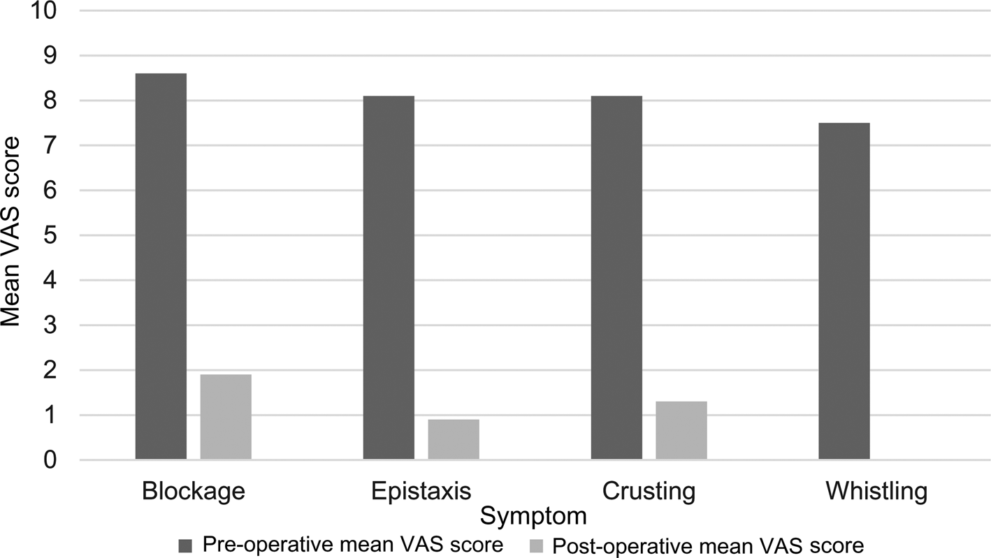
In this study, 130 patients complained of nasal blockage, 102 of crusting and 98 of epistaxis pre-operatively. Post-operatively, 139 of 154 patients were asymptomatic (90 per cent). Persisting symptoms were a feeling of blockage, epistaxis and crusting. There were statistically significant differences between the pre- and post-operative mean VAS scores for epistaxis (p < 0.001), crusting (p < 0.001), whistling (p < 0.001) and nasal obstruction (p < 0.001; Table 1 and Figure 7).
Pre- and post-operative mean visual analogue scale (VAS) scores for nasal symptoms.

Pre- and post-operative VAS scores for nasal symptoms

VAS = visual analogue scale; SD = standard deviation
Literature search
A total of 52 papersReference Kridel and Delaney1–Reference Hong, Mutsumay and Jin52 were extracted during the literature search (Table 2), describing techniques for nasal septal perforation repair along with their closure results, involving 1227 patients (mean, 23 patients per study; range 1–258). Thirty-seven papers (69 per cent) made some assessment of nasal symptoms. Follow-up periods ranged from 3 months to 6 years. Reported closure rates ranged from 66 to 100 per cent.
Results of literature review: nasal septal perforation closure techniques and outcomes

AP = ; NA = not available; ResT150 = total resistance
Discussion
Many techniques have been described for the surgical repair of nasal septal perforations. The reported outcomes are variable, with no single technique consistently favoured. We have presented an open rhinoplasty technique of bilateral superior and inferior mucosal advancement flaps with the use of autologous septal cartilage or bone or acellular porcine collagen. Tension-free everted closure ensured good healing, and an open septorhinoplasty approach was used to give better access.
In the 52Reference Kridel and Delaney1–Reference Hong, Mutsumay and Jin52 studies we reviewed (Table 2), unilateral or bilateral local mucosal flaps were used more commonly, usually advancement or bipedicled flaps, with an autologous interposition graft, commonly cartilage, bone or temporalis fascia. Local mucosal flaps alone were used in 22 studies, and 18 used local mucosal flaps with either allograft or xenograft interposition. Free grafts were used in five studies. Inferior turbinate flaps were used in six studies, and a middle turbinate flap in one study, either alone or with local mucosal flap. The remaining study enlarged the perforation by removing the posterior border of the perforation.
Large septal perforations, revision cases and iatrogenic septal perforations from previous nasal surgery are challenging when there is minimal septal cartilage or bone available. Tissue may be harvested from a secondary donor site, such as conchal cartilage. However, this results in further donor site morbidity. Moreover, the conchal cartilage is cup shaped and introduces a septal deviation. Non-autologous options have been described, such as the use of a human acellular dermal graft used by Kridel and Delaney.Reference Kridel and Delaney1
The size of the perforation is an important consideration in closure technique and outcomes. We have found the vertical dimension to be the deciding factor in nasal septal perforation surgical closure. Of the 51 studies we looked at, only 4 mentioned the vertical dimension. Kridel and DelanyReference Kridel and Delaney1 mentioned vertical diameters from 13 to 30 mm. Taskin et al.Reference Taskin, Yigit and Sisman17 treated vertical diameters of 20 to 27 mm and MansourReference Mansour20 described closure of perforations with vertical diameters of 5 to 15 mm. Wong et al.Reference Wong and Raghavan6 closed perforations with vertical diameters from 10 to 37 mm. Various reports have been published where rhinoplasty was combined with closure of nasal septal perforation to reduce the size of the perforation, thereby facilitating closure.Reference Kridel and Delaney1,Reference Foda and Magdy51,Reference Hong, Mutsumay and Jin52
Development of post-operative nasal obstruction was a frequently reported complication in the studies identified. Dosen and HayeReference Dosen and Haye5 reported that four patients developed post-operative nasal vestibular stenosis, a narrowing caused by healing of the nasal mucosa in the surgically advanced position. Ribeiro and da SilvaReference Ribeiro and da Silva2 reported that 37.9 per cent of patients complained of nasal obstruction in the initial 2 months post-operatively, which improved spontaneously. Two patients had long-term stenosis of the nasal valves bilaterally, requiring surgical correction. Both Dosen and HayeReference Dosen and Haye5 and Ribeiro and da SilvaReference Ribeiro and da Silva2 used closed techniques. Dosen and HayeReference Dosen and Haye5 reported extending the transfixion incision laterally along the floor of the vestibule at the nasal sill to the insertion of the inferior turbinate. This may have been the site of scarring that led to the complication of vestibular stenosis.
Nasal septal perforation repair is technically challenging surgery with no standardised technique, and the skill of the surgeon is important in the outcome. This may explain different complication rates between studies, despite theoretically similar techniques. The learning curve in septal perforation repair is highlighted by the reduction in re-perforation rates for a single operating surgeon; for example in the study by Dosen and Haye it was 47 per cent in the first 5 years of the study and 2 per cent in the latter 5 years.Reference Dosen and Haye5
We have described septal perforation closure results and symptom control using the previously reported technique of bilateral superior and inferior nasal mucosal advancement flaps with interposition graft.Reference Wong and Raghavan6 This technique uses autologous septal cartilage or bone when available (31 per cent) or acellular porcine collagen, a xenograft (69 per cent). This method avoids the need to harvest tissue from a secondary donor site (e.g. conchal cartilage) and consequent donor site morbidity. Porcine collagen is an easily available, robust material that is resistant to collagenase enzymes and is acellular and therefore non-immunogenic. It needs little preparation and is ready to use out of the packet.
We found the vertical dimension to be the deciding factor in nasal septal perforation surgical closure. Those less than 10 mm can be closed with an inferior flap alone. Any perforation larger than 10 mm vertically requires superior and inferior flaps. When the vertical diameter of perforations is 20 mm or more, the nasal dorsum can be lowered to release more mucosa for superior flaps, improving approximation of perforation margins. In our series, this was performed in 21 patients.
A further point of importance is the need for the edges of the flap to be sutured tension free to reduce the rate of failure. We used local mucosal advancement flaps on both sides, rather than bipedicled or rotation flaps, to ensure better vascularity. Tension-free closure was assured by mobilising superior and inferior mucosal advancement flaps bilaterally, if required. In addition, the perforation edges were sutured from the mucosal side with vertical mattress sutures to ensure everted edges and better healing.
The better access accorded by an open rhinoplasty technique allows the surgeon to release the maximum amount of mucosa and avoids distortion of tissues seen during an endonasal approach. Open rhinoplasty improves access to the posterior part of the perforations, particularly when the horizontal diameter is long. Endoscopic approach limits access and denies binocular vision.Reference Kridel and Delaney1 In addition, use of an open rhinoplasty technique allows any co-existent external nasal deformity or septal deviation to be corrected simultaneously.
Approximation of flaps may exert forces on the tip complex, thereby distorting it. This was reduced using the tongue-in-groove technique. If the lateral crura appeared to be displaced despite using the tongue-in-groove technique, cephalic trimming was performed to separate the lateral crura at the scroll region.Reference Kridel and Delaney1
Although we used a similar mucosal advancement technique to that described in the literature,Reference Dosen and Haye5 we did not experience post-operative nasal vestibular stenosis in our series despite the perforations extending up to 43 mm. We hypothesised that this may be because of our use of an open technique, which allows better access and more precise raising of the mucosal flaps without the need for multiple incisions. A concern raised about our technique was the possibility of the nasal passage narrowing because of the flaps raised. Immediately post-operatively, the nasal passage appeared narrowed because the inferior flap was raised from the floor and the superior flap was dissected off the undersurface of the upper lateral cartilage. However, when reviewed four weeks post-operatively, the flaps returned to their original positions. Three months post-operatively, it was difficult to see any evidence that the flaps were raised.
The reported closure rates among the 52 studies ranged from 66 to 100 per cent. A total of 26 studies (51 per cent) reported closure rates of 90 per cent or better. There may be a degree of publication bias, with poorer closure rates less likely to be reported.
In our series, the main symptoms were nasal blockage (84 per cent), crusting (66 per cent), epistaxis (64 per cent) and whistling (15 per cent) (Table 1). Our overall symptom control was 90 per cent which is comparable with the figure of 91.1 per cent reported by Kridel and Delaney.Reference Kridel and Delaney1 A total of 139 patients had complete symptom control (90 per cent). There were statistically significant differences between the pre- and post-operative mean VAS scores for epistaxis (p < 0.001), crusting (p < 0.001), whistling (p < 0.001) and nasal obstruction (p < 0.001).
• Surgical closure of nasal septal perforations is technically challenging and there is no standardised method
• This study describes a technique involving mucosal advancement flaps and autologous or acellular porcine interposition grafts
• The long-term closure rate and symptom control were assessed
• Using this technique, complete closure of septal perforations was achieved in 95 percent of patients
• Using this technique, 90 per cent of patients had complete symptom control
• Surgical closure of nasal septal perforation with mucosal advancement flaps and an interposition graft is a reliable technique with excellent long-term outcomes
There were re-perforations for eight cases in our series, all during the first six months. Five of these cases received acellular porcine collagen, of whom 2 had large initial perforations measuring up to 35 mm in the vertical diameters, 2 were revision cases where the flaps were difficult to raise because the mucosa was atrophic and scarred, and 1 failed to heal completely leaving a 5 × 5 mm asymptomatic perforation. Three patients refused acellular porcine collagen and received crushed septal cartilage as only a small amount of septal cartilage could be harvested safely.
A major strength of this work was the large number of patients included and the long follow-up period. This work was limited by the lack of objective nasal flow measurements, such as peak inspiratory nasal flow. VAS sores were used to assess whistling, epistaxis, nasal obstruction and crusting. Although these were not validated patient-reported outcome measures, the same score was used pre-operatively and at 6 and 12 months post-operatively so the results are likely to be comparable.
Conclusion
No single technique is universally accepted as the ‘gold standard’ method for nasal septal perforation repair. We present an open rhinoplasty technique of bilateral superior and inferior mucosal advancement flaps for larger perforations and inferior flaps for smaller ones. Good interposition grafts are required for healing of the repaired site. When available, patients’ own septal cartilage or bone can be used. Alternatively, acellular porcine collagen is a good alternative. Tension-free everted closure ensures good healing. An open septorhinoplasty approach gives better access. The vertical dimension is the limiting factor and if bigger than 20 mm a dorsal reduction rhinoplasty is required.
Acknowledgements
We would like to thank Dr Jane Dyer for her input into the statistical analysis.
Competing interests
None declared