Implications
There is scarce information regarding the role, importance and diversity of yeast in the rumen of dairy cattle. The internal transcribed spacer (ITS) approach allows the profiling and identification of ruminal yeast in a simple, fast and sensitive way that could provide new insights into the diversity and ecology of many different groups of fungi. This approach could be an important tool in rumen microbiome studies.
Introduction
The rumen allows the development and interactions of a wide variety of microorganisms (Mendes et al., Reference Mendes, Robson, Flávia, Cláudio, Castro and Rosa2012), which are able to adapt to the rumen anaerobic conditions, temperature and pH (Fonty and Chaucheyras-Durand, Reference Fonty and Chaucheyras-Durand2006). In addition, the composition and proportion of ruminal microorganisms are influenced by external factors such as diet, feeding frequency, age (Denman and McSweeney, Reference Denman and McSweeney2006) and have an important function in feed digestion (Oliveira et al., Reference Osinska-Jaroszuk, Jarosz-Wilkołazka, Jaroszuk-Sciseł, Szałapata, Nowak, Jaszek, Ozimek and Majewska2014).
In the rumen inhabit yeasts, which are a phylogenetically diverse group of unicellular fungi, and belong to the phylum Ascomycota (de Barros Lopes et al., Reference Bellemain, Carlsen, Brochmann, Coissac, Taberlet and Kauserud1998) and Basidiomycota (Osinska-Jaroszuk et al., Reference Oliveira, Robson, Silva, Alves, Castro, Ferreira, Rosa and Rodrigues2015); however, these microorganisms have received little attention from researchers working on ruminal microbiology.
Clarke and Di Menna (Reference de Barros Lopes, Soden, Martens, Henschke and Langridge1961) and Lund (Reference Lund1974) were pioneers in the study of these microorganisms in bovine through traditional techniques. Traditional methods for yeast identification are based on cellular morphology and reactions on fermentation protocols (Kurtzman and Robnett, Reference Kurtzman and Robnett1998). These procedures are difficult and time-consuming (Villa-Carvajal et al., Reference Villa-Carvajal, Querol and Belloch2006), and can be ambiguous due to strain variability of the microorganisms (Kurtzman and Robnett, Reference Kurtzman and Robnett1998). For these reasons, identification of yeast species by phenotypic characteristics has been largely replaced by DNA-based methods (Kurtzman, Reference Kurtzman2006) because they are quick, sensitive and specific (Hayashi et al., Reference Hayashi, Sugita, Hata, Katsuda, Zhang, Kihu, Sugawara, Ozawa, Matsubara, Ando, Obayashi, Itoh, Yabusaki, Kudo, Yamamoto, Koiwa, Oshida, Tagawa and Kawai2013). The ITS is a PCR approach validated to be useful for analysis of fungal diversity (Bellemain et al., Reference Carvalho, Ávila, Miguel, Pinto, Santos and Schwan2010). ITS is a non-coding ribosomal DNA (rDNA) spacer region located between the 18S and 28S ribosomal RNA (rRNA) genes (Sirohi et al., Reference Sirohi, Choudhury, Puniya, Singh, Dagar and Singh2013), and comprises ITS1/ITS2 intergenic sequences with highly conserved 5.8 rRNA in between (Zhang et al., Reference Zhang, Yuan, Yang, Huang and Huang2015). These regions were confirmed to be the most suitable marker genes for all classes of fungi providing complementary phylogenetic information (Koetschan et al., Reference Koetschan, Kittelmann, Lu, Al-Halbouni, Jarvis, Müller, Wolf and Janssen2014), and even useful for the detection and identification of fungal pathogens (Iwen et al., Reference Iwen, Freifeld, Sigler and Tarantolo2005). Moreover, ITS is a DNA region with high variation among closely related species due to its fast rate of evolution (Schoch et al., Reference Schoch, Seifert, Huhndorf, Robert, Spouge, Levesque and Chen2012) providing a molecular basis for determining phylogenetic relationships (Hayashi et al., Reference Hayashi, Sugita, Hata, Katsuda, Zhang, Kihu, Sugawara, Ozawa, Matsubara, Ando, Obayashi, Itoh, Yabusaki, Kudo, Yamamoto, Koiwa, Oshida, Tagawa and Kawai2013).
Due to the scarce information regarding the role, importance and diversity of yeast in the rumen of dairy cattle, the aim of this study was to use the ITS region to generate ruminal yeast profile and to identify ruminal yeast in dairy cows.
Material and methods
Animals, diet and management
The study was conducted at the Estación Experimental Pirque, Santiago, Chile (33°38'28''S, 70°34'27''W). Animals were handled following approved guidelines of the Animal Care and Use Committee of the Pontificia Universidad Católica de Chile. Three non-lactating, non-pregnant Holstein cows (785.5±37.3 kg of BW; with rumen cannulae no. 3C; Bar Diamond Inc., Boise, ID, USA) were used. Cows were individually fed a total mixed ration (TMR) at a fixed rate once daily (0930 h). Animals were housed in individual stalls (2.4×6 m) and had continuous access to water. The diet was formulated according to the NRC (2001) with a 56 : 44 forage : concentrate ratio to meet the requirements of a dry cow in the last trimester of gestation consuming 10 kg dry matter (DM) daily. A mixer wagon was used to mix forage (17% of alfalfa hay and 18% of corn silage of DM) and concentrate (10% of high-moisture corn, 34% of soybean hulls and 19% of wheat bran of DM). Briefly, the chemical composition (g/kg DM) of the diet was as follows: 514 of DM, 173 of CP, 342 of NDF, 200 of ADF and 19 of lignin. Cows were fed the diet for 21 days.
Samples
On day 21, whole ruminal contents were hand-collected from the anterior, dorsal and mid-ventral regions of the rumen at 0900 h (2 h after feeding) and were squeezed through three layers of cheesecloth. A quantity of 10 ml of residual ruminal fluid was immediately used to determine pH (PP-201; GOnDO Electronic, Taipei, Taiwan), 10 ml were kept for NH3–N analysis and another 10 ml were preserved for volatile fatty acid (VFA) determination by adding 1 ml of 25% metaphosphoric acid. Samples were frozen (−20°C) for later analysis. Samples of ruminal fluid were analyzed for VFA using a gas chromatograph (GC-2010; Shimadzu Scientific Instruments AOC-20s, Columbia, MD, USA) equipped with a 30 m wall-coated open-tubular fused-silica capillary column (Stabilwax-DA; 30 m×0.32 mm i.d., 0.25 µm film thickness; Restek, Bellefonte, PA, USA). Oven temperature was programmed for 145°C for 2 min and then increased from 145°C to 220°C at 4°C/min. The injector and flame ionization detector were maintained at 250°C and 300°C, respectively. Following pH determination, the strained ruminal fluid was centrifuged for 10 min at 3000×g at room temperature, discarding the supernatant and conserving the residue on ice – with later storage at 20°C – for subsequent DNA extraction.
DNA extraction
Before DNA extraction, samples of ruminal digesta were weighed (240±12 µg) and deposited in 1.5 Eppendorf tubes. For preparation and homogenization, 300 µl of phosphate-buffered saline solution was added to the samples. Subsequently, tubes were incubated for 30 min at 37°C with lysozyme (1 μg/µl) and then for 30 min at 37°C with proteinase K (0.1 mg/ml). DNA was extracted by using the PowerSoil DNA Isolation Kit (MO BIO Laboratories Inc.,Carlsbad, CA, USA) following the manufacturer’s guidelines.
PCR amplification of internal transcribed spacer region
The ribosomal ITS region was amplified from genomic DNA by two consecutive PCR reactions, using primers ITF1 (5'-TCCGTAGGTGAACCTGCGG-3') and ITF4 (5'-TCCTCCGCTTATTGATATGC-3'). For the first amplification, the ITS region was amplified in 40 µl of reaction volume and the PCR conditions included 30 cycles, with initial denaturation at 94°C for 5 min, followed by denaturation at 94°C for 30 s, annealing at 55°C for 30 s and extension at 72°C for 1 min. A blank sample was used to ensure that there were no PCR artifacts. The amplified PCR products were separated by electrophoresis in 5% polyacrylamide gels in 1× TBE (tris-borate-EDTA) buffer at 150 V for 50 min and visualized by 30 min of incubation with SYBR Green at room temperature (Molecular Probes, Eugene, OR, USA). The band sizes were estimated by comparison against 100-bp DNA ladders and excised from the gel on a UV-transilluminator using a sterile scalpel as described by Trabal et al. (Reference Trabal, Mazón-Suástegui, Vázquez-Juárez, Asencio-Valle, Morales-Bojórquez and Romero2012). The resulting slices were placed separately in 0.6 ml Eppendorf tube with 50 µl of nuclease-free water and incubated overnight at 4°C for elution of the DNA. Subsequently, 1 µl of each eluted band was used for re-amplification under the same conditions described above. To confirm the amplification, amplicons were visualized by electrophoresis in acrylamide gel under the same electrophoretic conditions previously described, and by silver staining method. Afterward, the amplicons were sent to Macrogen Inc. (Rockville, MD, USA) for sequencing service with both primers: ITF1 and ITF4.
Sequence and phylogenetic analysis
The obtained sequences were examined using the BLAST tool from the UNITE database (https://unite.ut.ee/) in order to find the sequence homology with the fungal sequences available in this database. Four UNITE reference sequences from phylum Ascomycota and Zygomycota were used in the phylogenetic analysis. These sequences included Millerozyma (SH201340.07FU), Rhizomucor (SH220342.07FU), Hyphopichia (SH203994.07FU) and Pichia (SH199823.07FU). The phylum Neocallimastigomycota reference sequence was used as an outgroup. The evolutionary distances were computed using the maximum likelihood method. The analysis involved 16 nucleotide sequences and all positions containing gaps were eliminated. Phylogenetic analysis was performed using the Molecular Evolutionary Genetics Analysis (MEGA6) software version 6.0.
Results and discussion
Ruminal fermentation parameters
The ruminal microbial composition and environmental parameters may vary depending on the diet composition (Monteils et al., Reference Monteils, Rey, Cauquil, Troegeler-Meynadier, Silberberg and Combes2011). In this study, ruminal fermentation parameters were measured to provide data on the rumen environment at the time when ruminal digesta was collected for molecular analysis. Those parameters averaged 6.9±0.01 for pH, 13.3±1.2 mg/dl for NH3–N, 57.5±6.3 mmol/l for total VFA, whereas molar proportions (mol/100 ml) for VFA were as follows: 63.8±0.1 for acetate, 22.7±0.2 for propionate, 11.2±0.1 for butyrate and 2.4±0.1 for valerate. The effect of the diet on the fermentation pattern is well known and the values obtained in the present study are similar to those reported for non-lactating cows fed corn-silage-based diets (Peyrat et al., Reference Peyrat, Baumont, Le Morvan and Nozière2016) and a diet with a 50 : 50 forage : concentrate ratio based on corn silage, alfalfa hay and soybean meal (Shahmoradi et al., Reference Shahmoradi, Alikhani and Riasi2016).
PCR amplification of internal transcribed spacer region
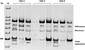
PCR amplification rendered product sizes between 460 and 750 bp (Figure 1). A total of 11 ITS sequences were obtained from those bands named in Figure 1. Ruminal yeast profile consisted mostly of four to six bands, hence potentially, four to six yeast genera. These profiles can be considered as equivalent to ruminal yeast microbiota profiles, as each ITS band may correspond to a particular yeast genus (Ide et al., Reference Ide, Miyazaki, Maki and Kobayashi2010).
Electrophoretic separation profiles of internal transcribed spacer (ITS1) region of ruminal yeast from dairy cows fed a diet with a 56 : 44 forage : concentrate ratio. Lines 1 to 3=ruminal fluid from cow 1; Lines 4 to 6=ruminal fluid from cow 2; Lines 7 to 9=ruminal fluid from cow 3; M=100-bp DNA marker.

An important part in this study is the PAGE adaptations that allowed an appropriate separation of the bands. Another comparative advantage of the approach used in this study is that we were able to obtain amplicons avoiding the use of purification and cloning kits. Consequently, this approach becomes an easy and economical alternative technique for ruminal yeast identification.
Identification of ruminal yeast
ITS bands were sequenced and the identification of the bands corresponded to the genera Millerozyma, Rhizomucor, Hyphopichia and Pichia. A more detailed information about the resulting sequence similarity searches against the records of the database UNITE are shown in Table 1. In this table, each ITS band is described based on the observed size of the band and the percentage of identity according to the UNITE RefSeq (sequences of the eluted amplicons).
UNITE reference sequence results for ruminal yeast

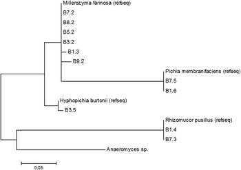
In this study, the phylogenetic analysis was performed using MEGA6 software comparing the retrieved sequences with the RefSeq for each yeast genus. The resulting maximum likelihood tree (Figure 2) showed the identity of ITS bands for Rhizomucor and Pichia RefSeq, in contrast with a more diverse genetic composition observed for Millerozyma and Hyphopichia.
Phylogenetic tree constructed from internal transcribed spacer sequences of ruminal yeast from dairy cows and reference sequence from UNITE. The evolutionary history was inferred using the maximum likelihood method. Evolutionary analyses were performed using MEGA6 software version 6.0.

Fungi are the second largest kingdom of eukaryotic life and several DNA regions were evaluated as potential DNA barcodes for this important biological group. For yeasts, the D1/D2 region of large-subunit rDNA was adopted for characterizing species long before the concept of DNA barcoding was promoted (Schoch et al., Reference Schoch, Seifert, Huhndorf, Robert, Spouge, Levesque and Chen2012). It had superior species resolution in some taxonomic groups, such as the early diverging lineages and the Ascomycete yeasts, but was otherwise slightly inferior to the ITS. Therefore, ITS has been suggested as the primary fungal barcode marker to the Consortium for the Barcode of Life (Schoch et al., Reference Schoch, Seifert, Huhndorf, Robert, Spouge, Levesque and Chen2012).
In previous studies, researchers have reported the predominance of the genus Pichia in the rumen. Lund (Reference Lund1974) identified Pichia kudriavzevii (previously named Candida krusei) as the most frequent species in cows fed with different diets that included grass, grass silage, or a mix of beet and straw plus molasses and concentrates. Abrao et al. (Reference Abrao, Freitas, Duarte, Geraseev, Barreto, Medeiros and Rosa2011) described Pichia membranaefaciens as the predominant ruminal yeast in goats through mycological cultures and micromorphological analysis. Similarly, Mendes et al. (Reference Mendes, Robson, Flávia, Cláudio, Castro and Rosa2012) reported P. kudriavzevii as the most abundant yeast in isolates from ruminal samples from dairy cattle fed tropical forages. The genus Pichia was also isolated from the rumen of dairy cows (Sirisan and Pattarajinda, Reference Sirisan and Pattarajinda2011; Sirisan et al., Reference Sirisan, Pattarajinda, Vichitphan and Leesing2013) using a PCR approach to amplify the D1/D2 domain of 26S rDNA.
The knowledge of the function exerted by yeast supplementation in the rumen has been the focus of several studies, for example, Yuan et al. (Reference Yuan, Liang, Muckey, Mendonça, Hulbert, Elrod and Bradford2015) studied the effect of yeast supplementation on milk production, feeding behavior and metabolism in dairy cows. They proposed that yeast removed the oxygen in the rumen, promoting anaerobic microorganisms’ development, and therefore enhance fiber digestion. The positive effects of yeast in the ruminal environment could also be due to the interaction of yeast with the rumen microorganisms, stimulating certain microbial population with its consequent effect (Sirisan et al., Reference Sirisan, Pattarajinda, Vichitphan and Leesing2013; Marrero et al., Reference Marrero, Castillo, Ruiz, Burrola and Angulo2015). Mendes et al. (Reference Mendes, Robson, Flávia, Cláudio, Castro and Rosa2012) suggested that yeasts are able to degrade simple carbohydrates, which could favor the growth of yeast populations, and also contribute to the regulation of ruminal pH. Both live and dead yeast (Saccharomyces) are used as feed additives for dairy cows (Salvati et al., Reference Salvati, Morais Júnior, Melo, Vilela, Cardoso, Aronovich, Pereira and Pereira2015); however, their precise role in improving rumen function is still unclear. Because yeast is involved in the oxygen utilization, fiber digestion and pH regulation (Salvati et al., Reference Salvati, Morais Júnior, Melo, Vilela, Cardoso, Aronovich, Pereira and Pereira2015) in the rumen, data from this study are relevant for a wide range of scientists, animal nutritionists and enterprises. Despite the fact that the effects of dietary supplementation of ruminants with yeast has been extensively studied, there is little information on the identification, quantification and function of these fungi in the rumen. In this regard, a striking feature of this study was that we were able to identify Millerozyma, Hyphopichia and Rhizomucor (mold), which have not been previously described in the rumen microbial community.
The yeast found in this study may be present in ingested feed; however, we did not evaluate that. A recent publication (Henderson et al., Reference Henderson, Cox, Ganesh, Jonker and Young2015) reported that rumen microbial community composition varies with diet and host, but a core microbiome can be found across a wide geographical range. Thus, ruminal yeast could be influenced by both diet and host. Jensen et al. (Reference Jensen, Olsen and Aalbaek1994) have established that the gastrointestinal tract is the main portal of entry for these fungi to the rumen. For example, Pichia manshurica was discovered in silage corn and non-fermented TMR under different air exposure, being predominant in low and high moisture levels (Carvalho et al., Reference Clarke and Di Menna2014; Hao et al., Reference Hao, Wang, Ning, Yang and Xu2015). The species Hyphopichia burtonii have been isolated from corn, wheat and rice (Limtong et al., Reference Limtong, Kaewwichian, Am-In, Boonmak, Jindamorakot, Yongmanitchai, Srisuk, Kawasaki and Nakase2010) and also described as food spoilage organism (Groenewald and Smith, Reference Groenewald and Smith2010). The presence of this yeast in different feedstuffs could explain the presence in the rumen through its ingestion from the diet. According to Lund’s (Reference Lund1974) hypothesis, yeast could be introduced to the rumen from feed and water, more recently, Henderson et al. (Reference Henderson, Cox, Ganesh, Jonker and Young2015) studied the microbial community composition of the ruminant and proposed that rumen microbes’ differences are attributed to diet. This assumption can be important for future studies on rumen microbiology and consider the analysis of yeast profile in diets and feedstuffs to confirm this hypothesis.
Finally, there are some aspects that need to be considered when using the ITS approach, for example, the ITS results can be a preliminary step for the quantification of yeast species in rumen. In future studies, the use of quantitative PCR with the same primers used in this study could provide more accurate results if the objective is to determine the total concentration of ruminal yeast. In addition, findings from this study showed that the ITS approach is not accurate enough to discern among yeast species and therefore this molecular technique is more suitable to study genera. In addition, it is recommended to use reference sequences from GenBank RefSeq or UNITE repositories, which are databases with validated sequences to avoid misleading or incorrect sequences.
Conclusion
In conclusion, the ITS approach used in this study allowed the identification of ruminal yeast in an easier, faster and economic procedure, and could provide new insights into the diversity and ecology of many different groups of fungi. In this study, the use of ITS allowed the identification of Millerozyma and Hyphopichia (phylum Ascomycota), which have not been previously described in ruminants.
Acknowledgments
This study was funded by a research grant from FONDECYT 11121142 and 1140734 (Fondo Nacional de Desarrollo Científico y Tecnológico, Chile). The authors would like to thank Juanita Clavijo, Jorge Manzor and Pamela Alvarez for their technical assistance and Fundación Agro U.C. for the animal facilities and assistance in obtaining research data.